 ムービー
ムービー コントローラー


+ データを開く

- 基本情報
基本情報
| 登録情報 | データベース: PDB / ID: 2bgz | ||||||
|---|---|---|---|---|---|---|---|
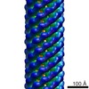
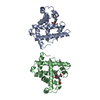
| タイトル | ATOMIC MODEL OF THE BACTERIAL FLAGELLAR BASED ON DOCKING AN X-RAY DERIVED HOOK STRUCTURE INTO AN EM MAP. | ||||||
 要素 要素 | FLAGELLAR HOOK PROTEIN FLGE | ||||||
 キーワード キーワード | MOTOR PROTEIN / BACTERIAL MOTIILITY / BACTERIAL FLAGELLAR HOOK / FLAGELLUM | ||||||
| 機能・相同性 |  機能・相同性情報 機能・相同性情報bacterial-type flagellum basal body / bacterial-type flagellum-dependent swarming motility 類似検索 - 分子機能 | ||||||
| 生物種 |  SALMONELLA TYPHIMURIUM (サルモネラ菌) SALMONELLA TYPHIMURIUM (サルモネラ菌) | ||||||
| 手法 | 電子顕微鏡法 / 電子線結晶学 / クライオ電子顕微鏡法 / 解像度: 9 Å | ||||||
 データ登録者 データ登録者 | Shaikh, T.R. / Thomas, D.R. / Chen, J.Z. / Samatey, F.A. / Matsunami, H. / Imada, K. / Namba, K. / Derosier, D.J. | ||||||
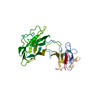
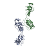
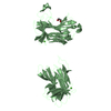
 引用 引用 | ジャーナル: Proc Natl Acad Sci U S A / 年: 2005 タイトル: A partial atomic structure for the flagellar hook of Salmonella typhimurium. 著者: Tanvir R Shaikh / Dennis R Thomas / James Z Chen / Fadel A Samatey / Hideyuki Matsunami / Katsumi Imada / Keiichi Namba / David J Derosier /  要旨: The axial proteins of the bacterial flagellum function as a drive shaft, universal joint, and propeller driven by the flagellar rotary motor; they also form the putative protein export channel. The N- ...The axial proteins of the bacterial flagellum function as a drive shaft, universal joint, and propeller driven by the flagellar rotary motor; they also form the putative protein export channel. The N- and C-terminal sequences of the eight axial proteins were predicted to form interlocking alpha-domains generating an axial tube. We report on an approximately 1-nm resolution map of the hook from Salmonella typhimurium, which reveals such a tube made from interdigitated, 1-nm rod-like densities similar to those seen in maps of the filament. Atomic models for the two outer domains of the hook subunit were docked into the corresponding outermost features of the map. The N and C termini of the hook subunit fragment are positioned next to each other and face toward the axis of the hook. The placement of these termini would permit the residues missing in the fragment to form the rod-like features that form the core domain of the hook. We also fit the hook atomic model to an approximately 2-nm resolution map of the hook from Caulobacter crescentus. The hook protein sequence from C. crescentus is largely homologous to that of S. typhimurium except for a large insertion (20 kDa). According to difference maps and our fitting, this insertion is found on the outer surface of the hook, consistent with our modeling of the hook. #1: ジャーナル: Nature / 年: 2004タイトル: Structure of the bacterial flagellar hook and implication for the molecular universal joint mechanism. 著者: Fadel A Samatey / Hideyuki Matsunami / Katsumi Imada / Shigehiro Nagashima / Tanvir R Shaikh / Dennis R Thomas / James Z Chen / David J Derosier / Akio Kitao / Keiichi Namba / 要旨: The bacterial flagellum is a motile organelle, and the flagellar hook is a short, highly curved tubular structure that connects the flagellar motor to the long filament acting as a helical propeller. ...The bacterial flagellum is a motile organelle, and the flagellar hook is a short, highly curved tubular structure that connects the flagellar motor to the long filament acting as a helical propeller. The hook is made of about 120 copies of a single protein, FlgE, and its function as a nano-sized universal joint is essential for dynamic and efficient bacterial motility and taxis. It transmits the motor torque to the helical propeller over a wide range of its orientation for swimming and tumbling. Here we report a partial atomic model of the hook obtained by X-ray crystallography of FlgE31, a major proteolytic fragment of FlgE lacking unfolded terminal regions, and by electron cryomicroscopy and three-dimensional helical image reconstruction of the hook. The model reveals the intricate molecular interactions and a plausible switching mechanism for the hook to be flexible in bending but rigid against twisting for its universal joint function. | ||||||
| 履歴 |
| ||||||
| Remark 700 | SHEET THE SHEET STRUCTURE OF THIS MOLECULE IS BIFURCATED. IN ORDER TO REPRESENT THIS FEATURE IN ... SHEET THE SHEET STRUCTURE OF THIS MOLECULE IS BIFURCATED. IN ORDER TO REPRESENT THIS FEATURE IN THE SHEET RECORDS BELOW, TWO SHEETS ARE DEFINED. |
- 構造の表示
構造の表示
| ムービー |
ムービービューア |
|---|---|
| 構造ビューア | 分子: MolmilJmol/JSmol |
- ダウンロードとリンク
-ダウンロード
| PDBx/mmCIF形式 | 2bgz.cif.gz | 65.5 KB | 表示 | PDBx/mmCIF形式 |
|---|---|---|---|---|
| PDB形式 | pdb2bgz.ent.gz | 47.6 KB | 表示 | PDB形式 |
| PDBx/mmJSON形式 | 2bgz.json.gz | ツリー表示 | PDBx/mmJSON形式 | |
| その他 |  その他のダウンロード その他のダウンロード |
-検証レポート
| 文書・要旨 | 2bgz_validation.pdf.gz | 760.4 KB | 表示 | wwPDB検証レポート |
|---|---|---|---|---|
| 文書・詳細版 | 2bgz_full_validation.pdf.gz | 767.9 KB | 表示 | |
| XML形式データ | 2bgz_validation.xml.gz | 15.2 KB | 表示 | |
| CIF形式データ | 2bgz_validation.cif.gz | 21.2 KB | 表示 | |
| アーカイブディレクトリ | https://data.pdbj.org/pub/pdb/validation_reports/bg/2bgzftp://data.pdbj.org/pub/pdb/validation_reports/bg/2bgz | HTTPS FTP |
-関連構造データ


-リンク
 PDBj
PDBj
- 集合体
集合体
| 登録構造単位 | 
|
|---|---|
| 1 |
|
-要素
| #1: タンパク質 | 分子量: 31335.084 Da / 分子数: 1 / 断片: FLGE31, RESIDUES 71-369 / 由来タイプ: 組換発現 詳細: FIT OF CRYSTAL STRUCTURE PDB ID 1WLG TO AN EM AMAP OF S. TYPHIMURIUM POLYHOOK 由来: (組換発現) SALMONELLA TYPHIMURIUM (サルモネラ菌)発現宿主: |
|---|
-実験情報
-実験
| 実験 | 手法: 電子顕微鏡法 |
|---|---|
| EM実験 | 試料の集合状態: 2D ARRAY / 3次元再構成法: 電子線結晶学 |
- 試料調製
試料調製
| 構成要素 | 名称: POLYHOOK / タイプ: COMPLEX |
|---|---|
| 緩衝液 | 名称: 10 MM TRIS/HCL,5MM EDTA,0. 1%V/V TRITON X-100 / pH: 8 / 詳細: 10 MM TRIS/HCL,5MM EDTA,0. 1%V/V TRITON X-100 |
| 試料 | 包埋: NO / シャドウイング: NO / 染色: NO / 凍結: YES |
| 試料支持 | 詳細: HOLEY CARBON |
| 急速凍結 | 装置: HOMEMADE PLUNGER / 凍結剤: ETHANE 詳細: SAMPLES PREPARED AT 4 DEG C PLUNGED INTO LIQUID ETHANE |
- 電子顕微鏡撮影
| 顕微鏡 | モデル: FEI/PHILIPS CM200FEG 詳細: DATA FROM 262 INDIVIDUAL SEGMENTS OF HOOK IN FINAL MAP. |
|---|---|
| 電子銃 | 電子線源:  FIELD EMISSION GUN / 加速電圧: 200 kV / 照射モード: FLOOD BEAM FIELD EMISSION GUN / 加速電圧: 200 kV / 照射モード: FLOOD BEAM |
| 電子レンズ | モード: BRIGHT FIELD / 倍率(公称値): 66000 X / 最大 デフォーカス(公称値): 2700 nm / 最小 デフォーカス(公称値): 1300 nm / Cs: 2 mm |
| 撮影 | 電子線照射量: 10 e/Å2 / フィルム・検出器のモデル: KODAK SO-163 FILM |
| 放射波長 | 相対比: 1 |
- 解析
解析
| EMソフトウェア |
| ||||||||||||
|---|---|---|---|---|---|---|---|---|---|---|---|---|---|
| 3次元再構成 | 解像度: 9 Å / 解像度の算出法: DIFFRACTION PATTERN/LAYERLINES 詳細: HELICAL SYMMETRY OPERATORS CAN BE CALCULATED USING RISE PER SUBUNIT 4.226475A ROTATION PER SUBUNIT 23.48758 DEG. DATA USED IN REFINEMENT. RESOLUTION RANGE HIGH 12A, RESOLUTION RANGE LOW 200A, ...詳細: HELICAL SYMMETRY OPERATORS CAN BE CALCULATED USING RISE PER SUBUNIT 4.226475A ROTATION PER SUBUNIT 23.48758 DEG. DATA USED IN REFINEMENT. RESOLUTION RANGE HIGH 12A, RESOLUTION RANGE LOW 200A, DATA CUTOFF (SIGMA(F)) 1, COMPLETENESS FOR RANGE 100(%) NUMBER OF REFLECTIONS 84. PROTEIN ATOMS 2156 対称性のタイプ: HELICAL | ||||||||||||
| 原子モデル構築 | プロトコル: OTHER / 詳細: METHOD--RECIPROCAL SPACE FITTING IN URO | ||||||||||||
| 精密化 | 最高解像度: 9 Å | ||||||||||||
| 精密化ステップ | サイクル: LAST / 最高解像度: 12 Å
|
