 Movie
Movie Controller

[English] 日本語
 Yorodumi
- PDB-29xz: Cryo-EM structure of Rhodobacter capsulatus cytochrome bc1 dimer ... -
+ Open data

- Basic information
| Entry | Database: PDB / ID: 29xz | |||||||||
|---|---|---|---|---|---|---|---|---|---|---|
| Title | Cryo-EM structure of Rhodobacter capsulatus cytochrome bc1 dimer at 2.16 A resolution with water molecules and quinone in the quinone reduction site | |||||||||
 Components Components |
| |||||||||
 Keywords Keywords | ELECTRON TRANSPORT / oxidoreductuse / proton pumping / quinol oxidation / respiration | |||||||||
| Function / homology |  Function and homology information Function and homology informationrespiratory chain complex III / quinol-cytochrome-c reductase / quinol-cytochrome-c reductase activity / respiratory electron transport chain / 2 iron, 2 sulfur cluster binding / electron transfer activity / oxidoreductase activity / heme binding / metal ion binding / plasma membrane Similarity search - Function | |||||||||
| Biological species |  Rhodobacter capsulatus SB 1003 (bacteria) Rhodobacter capsulatus SB 1003 (bacteria) | |||||||||
| Method | ELECTRON MICROSCOPY / single particle reconstruction / cryo EM / Resolution: 2.16 Å | |||||||||
 Authors Authors | Pietras, R. / Mielecki, B. / Wojcik-Augustyn, A. / Sarewicz, M. / Jaciuk, M. / Koziej, L. / Glatt, S. / Osyczka, A. | |||||||||
| Funding support |  Poland, 2items Poland, 2items
| |||||||||
 Citation Citation | Journal: Proc Natl Acad Sci U S A / Year: 2026 Title: Distinct ubiquinone binding at the oxidation and reduction sites of cytochrome bc. Authors: Rafał Pietras / Anna Wójcik-Augustyn / Bohun Mielecki / Marcin Sarewicz / Marcin Jaciuk / Łukasz Koziej / Sebastian Glatt / Artur Osyczka /  Abstract: The function of cytochrome bc, a widespread energy-conserving enzyme, requires the coordinated activity of two quinone-binding sites (Q catalyzing oxidation of ubiquinol and Q catalyzing reduction of ...The function of cytochrome bc, a widespread energy-conserving enzyme, requires the coordinated activity of two quinone-binding sites (Q catalyzing oxidation of ubiquinol and Q catalyzing reduction of ubiquinone). The operation of Q, but not Q, involves large-scale movement of the head domain of iron-sulfur protein (ISP-HD). How the respective sites accommodate quinone molecules for efficient catalysis remains elusive. Here, we present high-resolution cryoelectron microscopy structures of bacterial cytochrome bc with native ubiquinone molecules in various states. They show that the quinone headgroup occupies a catalytically competent position in Q only when the ISP-HD interacts with cytochrome b. When the ISP-HD does not interact with this subunit, quinone is present in the hydrophobic groove, however its headgroup is prevented from reaching the catalytic cavity by steric hindrance. In this state, the position of quinone headgroup is clearly not fixed. In contrast, all structures show Q in the same state with a well-resolved and catalytically competent quinone headgroup, but with its tail not fixed. These distinctly different ubiquinone binding modes for Q and Q secure the smooth operation of cytochrome bc. | |||||||||
| History |
|
- Structure visualization
| Structure viewer | Molecule: MolmilJmol/JSmol |
|---|
- Downloads & links
-Download
| PDBx/mmCIF format | 29xz.cif.gz | 363.7 KB | Display | PDBx/mmCIF format |
|---|---|---|---|---|
| PDB format | pdb29xz.ent.gz | 291.1 KB | Display | PDB format |
| PDBx/mmJSON format | 29xz.json.gz | Tree view | PDBx/mmJSON format | |
| Others |  Other downloads Other downloads |
-Validation report
| Arichive directory | https://data.pdbj.org/pub/pdb/validation_reports/9x/29xzftp://data.pdbj.org/pub/pdb/validation_reports/9x/29xz | HTTPS FTP |
|---|
-Related structure data
| Related structure data |  57440MC  29yaC  29ybC  29ycC  29ydC  29yeC M: map data used to model this data C: citing same article ( |
|---|---|
| Similar structure data |


-Links
 PDBj
PDBj
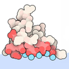

- Assembly
| Deposited unit | 
|
|---|---|
| 1 |
|
-Components
-Protein , 3 types, 6 molecules ADBECF
| #1: Protein | Mass: 20333.914 Da / Num. of mol.: 2 Source method: isolated from a genetically manipulated source Source: (gene. exp.) Rhodobacter capsulatus SB 1003 (bacteria)Gene: petA, fbcF, RCAP_rcc02768 / Production host: Rhodobacter capsulatus (bacteria) / References: UniProt: D5ANZ2, quinol-cytochrome-c reductase#2: Protein | Mass: 50297.418 Da / Num. of mol.: 2 Source method: isolated from a genetically manipulated source Details: affinity tag at C-terminus (StrepTag II) Source: (gene. exp.) Rhodobacter capsulatus SB 1003 (bacteria)Gene: petB, cytB, RCAP_rcc02769 / Production host: Rhodobacter capsulatus (bacteria) / References: UniProt: D5ANZ3#3: Protein | Mass: 28321.121 Da / Num. of mol.: 2 Source method: isolated from a genetically manipulated source Source: (gene. exp.) Rhodobacter capsulatus SB 1003 (bacteria)Gene: petC, RCAP_rcc02770 / Production host: Rhodobacter capsulatus (bacteria) / References: UniProt: D5ANZ4 |
|---|
-Non-polymers , 6 types, 195 molecules 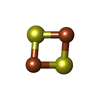
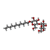
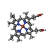
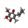
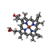

| #4: Chemical | 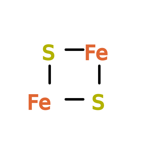 Mass: 175.820 Da / Num. of mol.: 2 / Source method: obtained synthetically / Formula: Fe2S2 / Feature type: SUBJECT OF INVESTIGATION Mass: 175.820 Da / Num. of mol.: 2 / Source method: obtained synthetically / Formula: Fe2S2 / Feature type: SUBJECT OF INVESTIGATION#5: Chemical | ChemComp-UMQ / 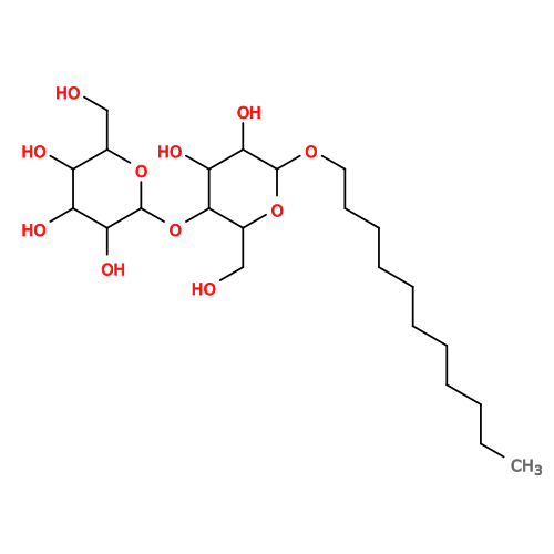 Mass: 496.589 Da / Num. of mol.: 8 / Source method: obtained synthetically / Formula: C23H44O11 / Comment: detergent*YM Mass: 496.589 Da / Num. of mol.: 8 / Source method: obtained synthetically / Formula: C23H44O11 / Comment: detergent*YM#6: Chemical | ChemComp-HEM / 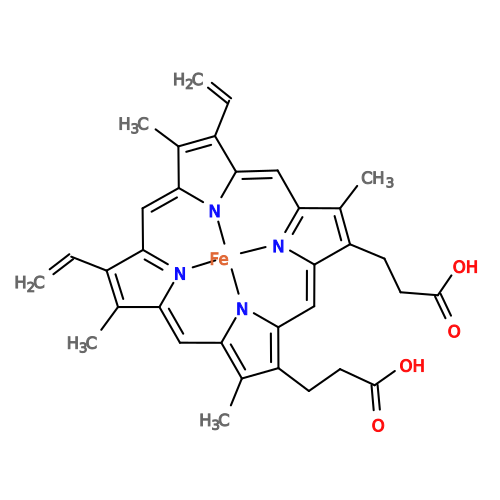 Mass: 616.487 Da / Num. of mol.: 4 / Source method: obtained synthetically / Formula: C34H32FeN4O4 / Feature type: SUBJECT OF INVESTIGATION Mass: 616.487 Da / Num. of mol.: 4 / Source method: obtained synthetically / Formula: C34H32FeN4O4 / Feature type: SUBJECT OF INVESTIGATION#7: Chemical | 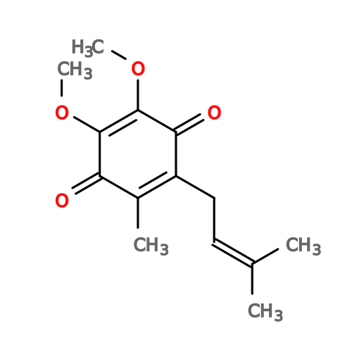 Mass: 250.290 Da / Num. of mol.: 2 / Source method: obtained synthetically / Formula: C14H18O4 / Feature type: SUBJECT OF INVESTIGATION Mass: 250.290 Da / Num. of mol.: 2 / Source method: obtained synthetically / Formula: C14H18O4 / Feature type: SUBJECT OF INVESTIGATION#8: Chemical |  Mass: 620.519 Da / Num. of mol.: 2 / Source method: obtained synthetically / Formula: C34H36FeN4O4 / Feature type: SUBJECT OF INVESTIGATION Mass: 620.519 Da / Num. of mol.: 2 / Source method: obtained synthetically / Formula: C34H36FeN4O4 / Feature type: SUBJECT OF INVESTIGATION#9: Water | ChemComp-HOH / | Mass: 18.015 Da / Num. of mol.: 177 / Source method: isolated from a natural source / Formula: H2O |
|---|
-Details
| Has ligand of interest | Y |
|---|---|
| Has protein modification | Y |
-Experimental details
-Experiment
| Experiment | Method: ELECTRON MICROSCOPY |
|---|---|
| EM experiment | Aggregation state: PARTICLE / 3D reconstruction method: single particle reconstruction |
- Sample preparation
| Component | Name: native dimeric cytochrome bc1 complex / Type: COMPLEX / Entity ID: #1-#3 / Source: RECOMBINANT |
|---|---|
| Molecular weight | Value: 0.20 MDa / Experimental value: NO |
| Source (natural) | Organism: Rhodobacter capsulatus SB 1003 (bacteria) |
| Source (recombinant) | Organism: Rhodobacter capsulatus (bacteria) |
| Buffer solution | pH: 8 / Details: 50mM bicine, 100mM KCl, 1mM EDTA, 0.8mM UDM |
| Specimen | Conc.: 6 mg/ml / Embedding applied: NO / Shadowing applied: NO / Staining applied: NO / Vitrification applied: YES |
| Specimen support | Grid material: COPPER / Grid mesh size: 200 divisions/in. / Grid type: Quantifoil R2/1 |
| Vitrification | Instrument: FEI VITROBOT MARK IV / Cryogen name: ETHANE / Humidity: 100 % / Chamber temperature: 277 K |
- Electron microscopy imaging
| Experimental equipment |  Model: Titan Krios / Image courtesy: FEI Company |
|---|---|
| Microscopy | Model: TFS KRIOS |
| Electron gun | Electron source:  FIELD EMISSION GUN / Accelerating voltage: 300 kV / Illumination mode: FLOOD BEAM FIELD EMISSION GUN / Accelerating voltage: 300 kV / Illumination mode: FLOOD BEAM |
| Electron lens | Mode: BRIGHT FIELD / Nominal magnification: 105000 X / Nominal defocus max: 2100 nm / Nominal defocus min: 900 nm / Cs: 2.7 mm / C2 aperture diameter: 50 µm |
| Specimen holder | Specimen holder model: FEI TITAN KRIOS AUTOGRID HOLDER |
| Image recording | Electron dose: 40.22 e/Å2 / Film or detector model: GATAN K3 BIOQUANTUM (6k x 4k) / Num. of grids imaged: 1 / Num. of real images: 8883 |
| EM imaging optics | Energyfilter name: GIF Bioquantum / Energyfilter slit width: 20 eV |
| Image scans | Width: 5760 / Height: 4096 |
- Processing
| EM software |
| ||||||||||||||||||||||||||||||||||||||||
|---|---|---|---|---|---|---|---|---|---|---|---|---|---|---|---|---|---|---|---|---|---|---|---|---|---|---|---|---|---|---|---|---|---|---|---|---|---|---|---|---|---|
| CTF correction | Type: PHASE FLIPPING AND AMPLITUDE CORRECTION | ||||||||||||||||||||||||||||||||||||||||
| Particle selection | Num. of particles selected: 2560000 / Details: 2.56M (template) and 1.91M (TOPAZ) | ||||||||||||||||||||||||||||||||||||||||
| Symmetry | Point symmetry: C2 (2 fold cyclic) | ||||||||||||||||||||||||||||||||||||||||
| 3D reconstruction | Resolution: 2.16 Å / Resolution method: FSC 0.143 CUT-OFF / Num. of particles: 169557 / Algorithm: FOURIER SPACE / Num. of class averages: 1 / Symmetry type: POINT | ||||||||||||||||||||||||||||||||||||||||
| Atomic model building | Protocol: FLEXIBLE FIT / Space: REAL Details: Initial model placement (rigid-body) into a map was done in ChimeraX | ||||||||||||||||||||||||||||||||||||||||
| Atomic model building | PDB-ID: 1ZRT Accession code: 1ZRT / Source name: PDB / Type: experimental model |

