 Movie
Movie Controller

[English] 日本語
 Yorodumi
- PDB-28lw: CryoEM structure of carbon monoxide dehydrogenase from Ruminococc... -
+ Open data

- Basic information
| Entry | Database: PDB / ID: 28lw | ||||||||||||
|---|---|---|---|---|---|---|---|---|---|---|---|---|---|
| Title | CryoEM structure of carbon monoxide dehydrogenase from Ruminococcus flavefaciens | ||||||||||||
 Components Components | anaerobic carbon-monoxide dehydrogenase | ||||||||||||
 Keywords Keywords | OXIDOREDUCTASE / carbon monoxide dehydrogenase / CODH / metalloenzyme / iron sulfur protein / anaerobic enzyme | ||||||||||||
| Function / homology | IRON/SULFUR CLUSTER Function and homology information Function and homology information | ||||||||||||
| Biological species |  Ruminococcus flavefaciens ATCC 19208 (bacteria) Ruminococcus flavefaciens ATCC 19208 (bacteria) | ||||||||||||
| Method | ELECTRON MICROSCOPY / single particle reconstruction / cryo EM / Resolution: 2.55 Å | ||||||||||||
 Authors Authors | Srinivas, V. / Hogbom, M. | ||||||||||||
| Funding support |  Sweden, Sweden,  Denmark, 3items Denmark, 3items
| ||||||||||||
 Citation Citation | Journal: Angew Chem Int Ed Engl / Year: 2026 Title: Beyond Canonical CO Oxidation: Structural and Evolutionary Insights Into a Non-Canonical Carbon Monoxide Dehydrogenase. Authors: Maximilian Böhm / Vivek Srinivas / Benjamin Wiseman / Ping Huang / Moritz Senger / Martin Högbom / Henrik Land / Abstract: Carbon monoxide dehydrogenases (CODHs) catalyse the reversible oxidation of CO to CO and play central roles in microbial carbon metabolism. While well-characterised CODHs from different phylogenetic ...Carbon monoxide dehydrogenases (CODHs) catalyse the reversible oxidation of CO to CO and play central roles in microbial carbon metabolism. While well-characterised CODHs from different phylogenetic backgrounds exhibit high bidirectional activity, the enigmatic clade B remains functionally uncharacterised. Here, we present the first structural and biochemical characterisation of a clade B CODH from Ruminococcus flavefaciens (RfCODH). It reveals striking divergence from canonical enzymes. A new anaerobic cryo-EM workflow was developed, carried out entirely under anoxic conditions by manual blotting and plunge freezing. It resulted in a 2.53 Å RfCODH structure. The structure adopts the typical CODH fold, but exhibits blocked gas channels, a compromised proton transfer pathway and disrupted cofactor coordination. This provides a structural rationale for RfCODH's severely attenuated CO oxidation activity (13 mU/mg vs. 900 U/mg for the well-studied ChCODH-II). EPR spectroscopy reveals unique oxidised C-cluster states not previously characterised in CODHs. Mirror tree analysis hints to co-evolution between clade B CODHs and associated ABC transporter substrate-binding proteins, suggesting these enzymes function in metabolism of substrates imported via the ABC transporter module. All findings indicate evolutionary repurposing of the CODH scaffold for alternative physiological functions. | ||||||||||||
| History |
|
- Structure visualization
| Structure viewer | Molecule: MolmilJmol/JSmol |
|---|
- Downloads & links
-Download
| PDBx/mmCIF format | 28lw.cif.gz | 257.5 KB | Display | PDBx/mmCIF format |
|---|---|---|---|---|
| PDB format | pdb28lw.ent.gz | 204.4 KB | Display | PDB format |
| PDBx/mmJSON format | 28lw.json.gz | Tree view | PDBx/mmJSON format | |
| Others |  Other downloads Other downloads |
-Validation report
| Arichive directory | https://data.pdbj.org/pub/pdb/validation_reports/8l/28lwftp://data.pdbj.org/pub/pdb/validation_reports/8l/28lw | HTTPS FTP |
|---|
-Related structure data
| Related structure data |  56604MC M: map data used to model this data C: citing same article ( |
|---|---|
| Similar structure data |
-Links
 PDBj
PDBj- Assembly
| Deposited unit | 
|
|---|---|
| 1 |
|
-Components
| #1: Protein | Mass: 78977.250 Da / Num. of mol.: 2 Source method: isolated from a genetically manipulated source Source: (gene. exp.) Ruminococcus flavefaciens ATCC 19208 (bacteria)Gene: SAMN04487860_10698 / Production host:  Escherichia coli (E. coli) / References: anaerobic carbon monoxide dehydrogenase Escherichia coli (E. coli) / References: anaerobic carbon monoxide dehydrogenase#2: Chemical | 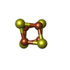 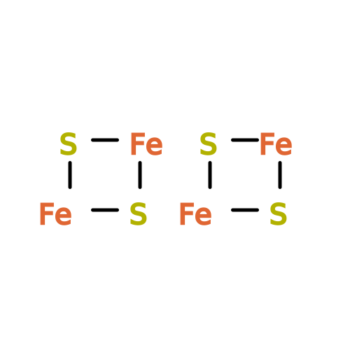 Mass: 351.640 Da / Num. of mol.: 3 / Source method: obtained synthetically / Formula: Fe4S4 / Feature type: SUBJECT OF INVESTIGATION Mass: 351.640 Da / Num. of mol.: 3 / Source method: obtained synthetically / Formula: Fe4S4 / Feature type: SUBJECT OF INVESTIGATIONHas ligand of interest | Y | Has protein modification | Y | |
|---|
-Experimental details
-Experiment
| Experiment | Method: ELECTRON MICROSCOPY |
|---|---|
| EM experiment | Aggregation state: PARTICLE / 3D reconstruction method: single particle reconstruction |
- Sample preparation
| Component | Name: Homodimer of carbon-monoxide dehydrogenase from Ruminococcus flavefaciens Type: COMPLEX / Entity ID: #1 / Source: RECOMBINANT |
|---|---|
| Molecular weight | Value: 0.158 MDa / Experimental value: NO |
| Source (natural) | Organism: Ruminococcus flavefaciens ATCC 19208 (bacteria) |
| Source (recombinant) | Organism: Escherichia coli (E. coli) |
| Buffer solution | pH: 8 / Details: 50 mM Tris-HCl pH 8, 20 mM NaCl, 5 % w/v glycerol, |
| Specimen | Conc.: 1 mg/ml / Embedding applied: NO / Shadowing applied: NO / Staining applied: NO / Vitrification applied: YES |
| Specimen support | Grid material: COPPER / Grid mesh size: 300 divisions/in. / Grid type: Quantifoil R1.2/1.3 |
| Vitrification | Cryogen name: ETHANE / Humidity: 0 % Details: Vetrification carried out by manually plunge-freezing in a nitrogen glove box |
- Electron microscopy imaging
| Experimental equipment |  Model: Titan Krios / Image courtesy: FEI Company |
|---|---|
| Microscopy | Model: TFS KRIOS |
| Electron gun | Electron source:  FIELD EMISSION GUN / Accelerating voltage: 300 kV / Illumination mode: FLOOD BEAM FIELD EMISSION GUN / Accelerating voltage: 300 kV / Illumination mode: FLOOD BEAM |
| Electron lens | Mode: BRIGHT FIELD / Nominal magnification: 130000 X / Nominal defocus max: 2000 nm / Nominal defocus min: 600 nm / Cs: 2.7 mm |
| Image recording | Electron dose: 41.65 e/Å2 / Film or detector model: GATAN K3 BIOQUANTUM (6k x 4k) |
- Processing
| EM software |
| ||||||||||||||||||||||||||||||||
|---|---|---|---|---|---|---|---|---|---|---|---|---|---|---|---|---|---|---|---|---|---|---|---|---|---|---|---|---|---|---|---|---|---|
| CTF correction | Type: PHASE FLIPPING AND AMPLITUDE CORRECTION | ||||||||||||||||||||||||||||||||
| 3D reconstruction | Resolution: 2.55 Å / Resolution method: FSC 0.143 CUT-OFF / Num. of particles: 123492 / Symmetry type: POINT | ||||||||||||||||||||||||||||||||
| Atomic model building | Protocol: RIGID BODY FIT | ||||||||||||||||||||||||||||||||
| Atomic model building | Source name: AlphaFold / Type: in silico model | ||||||||||||||||||||||||||||||||
| Refinement | Highest resolution: 2.55 Å Stereochemistry target values: REAL-SPACE (WEIGHTED MAP SUM AT ATOM CENTERS) |
