 Movie
Movie Controller


+ Open data

- Basic information
| Entry | Database: PDB / ID: 11mp | |||||||||||||||||||||||||||
|---|---|---|---|---|---|---|---|---|---|---|---|---|---|---|---|---|---|---|---|---|---|---|---|---|---|---|---|---|
| Title | E. coli SufE bound to SufBC2D | |||||||||||||||||||||||||||
 Components Components |
| |||||||||||||||||||||||||||
 Keywords Keywords | METAL BINDING PROTEIN / Fe-S / Suf pathway / persulfide / iron-sulfur / sulfur transfer | |||||||||||||||||||||||||||
| Function / homology |  Function and homology information Function and homology informationmetallo-sulfur cluster assembly / sulfurtransferase complex / sulfur compound metabolic process / sulfur carrier activity / iron-sulfur cluster assembly complex / iron-sulfur cluster assembly / response to radiation / 2 iron, 2 sulfur cluster binding / enzyme activator activity / 4 iron, 4 sulfur cluster binding ...metallo-sulfur cluster assembly / sulfurtransferase complex / sulfur compound metabolic process / sulfur carrier activity / iron-sulfur cluster assembly complex / iron-sulfur cluster assembly / response to radiation / 2 iron, 2 sulfur cluster binding / enzyme activator activity / 4 iron, 4 sulfur cluster binding / response to oxidative stress / ATP hydrolysis activity / ATP binding / cytosol Similarity search - Function | |||||||||||||||||||||||||||
| Biological species |   Escherichia coli (E. coli) Escherichia coli (E. coli) | |||||||||||||||||||||||||||
| Method | ELECTRON MICROSCOPY / single particle reconstruction / cryo EM / Resolution: 4.21 Å | |||||||||||||||||||||||||||
 Authors Authors | Chhikara, N. / Dunkle, J.A. / Frantom, P.A. | |||||||||||||||||||||||||||
| Funding support |  United States, 2items United States, 2items
| |||||||||||||||||||||||||||
 Citation Citation | Journal: bioRxiv / Year: 2026 Title: The structure of the SufBCD-SufE complex reveals the mechanism of sulfur transfer in bacterial Fe-S cluster assembly. Authors: Nidhi Chhikara / Nadia Mireku / Jack A Dunkle / Patrick A Frantom / Abstract: Iron-sulfur clusters are essential cofactors assembled in bacteria by the Suf pathway through a series of transient protein-protein interactions that transfer sulfur from L-cysteine to a scaffold ...Iron-sulfur clusters are essential cofactors assembled in bacteria by the Suf pathway through a series of transient protein-protein interactions that transfer sulfur from L-cysteine to a scaffold complex. While early steps in persulfide transfer are well characterized, the mechanism of sulfur delivery to the SufBCD scaffold has remained unresolved. Here, we report the first structure of the SufBCD-SufE complex, capturing the final step in persulfide transfer in the Suf pathway. The structure reveals coordinated conformational changes in both SufB and SufE that expose the otherwise buried C254 acceptor site and position the SufE C51 loop beneath the SufB-SufD axis. Biochemical analysis of SufB variants demonstrates that substitutions in the globally conserved 220s β-strand enhance SufE binding affinity and persulfide transfer rates, consistent with stabilization of a locally rearranged, transfer-competent conformation. Together, these results support a model in which conformational gating regulates persulfide transfer, providing a mechanism for controlling access to reactive sulfur intermediates. #1: Journal: Acta Crystallogr D Struct Biol / Year: 2019 Title: Macromolecular structure determination using X-rays, neutrons and electrons: recent developments in Phenix. Authors: Dorothee Liebschner / Pavel V Afonine / Matthew L Baker / Gábor Bunkóczi / Vincent B Chen / Tristan I Croll / Bradley Hintze / Li Wei Hung / Swati Jain / Airlie J McCoy / Nigel W Moriarty ...Authors: Dorothee Liebschner / Pavel V Afonine / Matthew L Baker / Gábor Bunkóczi / Vincent B Chen / Tristan I Croll / Bradley Hintze / Li Wei Hung / Swati Jain / Airlie J McCoy / Nigel W Moriarty / Robert D Oeffner / Billy K Poon / Michael G Prisant / Randy J Read / Jane S Richardson / David C Richardson / Massimo D Sammito / Oleg V Sobolev / Duncan H Stockwell / Thomas C Terwilliger / Alexandre G Urzhumtsev / Lizbeth L Videau / Christopher J Williams / Paul D Adams /   Abstract: Diffraction (X-ray, neutron and electron) and electron cryo-microscopy are powerful methods to determine three-dimensional macromolecular structures, which are required to understand biological ...Diffraction (X-ray, neutron and electron) and electron cryo-microscopy are powerful methods to determine three-dimensional macromolecular structures, which are required to understand biological processes and to develop new therapeutics against diseases. The overall structure-solution workflow is similar for these techniques, but nuances exist because the properties of the reduced experimental data are different. Software tools for structure determination should therefore be tailored for each method. Phenix is a comprehensive software package for macromolecular structure determination that handles data from any of these techniques. Tasks performed with Phenix include data-quality assessment, map improvement, model building, the validation/rebuilding/refinement cycle and deposition. Each tool caters to the type of experimental data. The design of Phenix emphasizes the automation of procedures, where possible, to minimize repetitive and time-consuming manual tasks, while default parameters are chosen to encourage best practice. A graphical user interface provides access to many command-line features of Phenix and streamlines the transition between programs, project tracking and re-running of previous tasks. | |||||||||||||||||||||||||||
| History |
|
- Structure visualization
| Structure viewer | Molecule: MolmilJmol/JSmol |
|---|
- Downloads & links
-Download
| PDBx/mmCIF format | 11mp.cif.gz | 313.8 KB | Display | PDBx/mmCIF format |
|---|---|---|---|---|
| PDB format | pdb11mp.ent.gz | 209.7 KB | Display | PDB format |
| PDBx/mmJSON format | 11mp.json.gz | Tree view | PDBx/mmJSON format | |
| Others |  Other downloads Other downloads |
-Validation report
| Arichive directory | https://data.pdbj.org/pub/pdb/validation_reports/1m/11mpftp://data.pdbj.org/pub/pdb/validation_reports/1m/11mp | HTTPS FTP |
|---|
-Related structure data
| Related structure data |  75839MC M: map data used to model this data C: citing same article ( |
|---|---|
| Similar structure data |
-Links
 PDBj
PDBj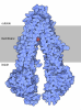
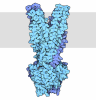
- Assembly
| Deposited unit | 
|
|---|---|
| 1 |
|
-Components
| #1: Protein | Mass: 56396.637 Da / Num. of mol.: 1 / Mutation: Y224A Source method: isolated from a genetically manipulated source Source: (gene. exp.) Escherichia coli (E. coli) / Gene: sufB, ynhE, b1683, JW5273 / Production host: Escherichia coli (E. coli) / References: UniProt: P77522 | ||||
|---|---|---|---|---|---|
| #2: Protein | Mass: 15817.281 Da / Num. of mol.: 1 Source method: isolated from a genetically manipulated source Source: (gene. exp.) Escherichia coli (E. coli) / Gene: sufE, ynhA, b1679, JW1669 / Production host: Escherichia coli (E. coli) / References: UniProt: P76194 | ||||
| #3: Protein | Mass: 27613.332 Da / Num. of mol.: 2 Source method: isolated from a genetically manipulated source Source: (gene. exp.) Escherichia coli (E. coli) / Gene: sufC, ynhD, b1682, JW1672 / Production host: Escherichia coli (E. coli) / References: UniProt: P77499#4: Protein | | Mass: 46884.641 Da / Num. of mol.: 1 Source method: isolated from a genetically manipulated source Source: (gene. exp.) Escherichia coli (E. coli) / Gene: sufD, ynhC, b1681, JW1671 / Production host: Escherichia coli (E. coli) / References: UniProt: P77689Has protein modification | N | |
-Experimental details
-Experiment
| Experiment | Method: ELECTRON MICROSCOPY |
|---|---|
| EM experiment | Aggregation state: PARTICLE / 3D reconstruction method: single particle reconstruction |
- Sample preparation
| Component | Name: SufBC2D-SufE complex / Type: COMPLEX / Entity ID: all / Source: RECOMBINANT |
|---|---|
| Molecular weight | Value: 0.174 MDa / Experimental value: NO |
| Source (natural) | Organism: Escherichia coli (E. coli) |
| Source (recombinant) | Organism: Escherichia coli (E. coli) |
| Buffer solution | pH: 8 |
| Specimen | Embedding applied: NO / Shadowing applied: NO / Staining applied: NO / Vitrification applied: YES |
| Vitrification | Cryogen name: ETHANE |
- Electron microscopy imaging
| Microscopy | Model: TFS GLACIOS |
|---|---|
| Electron gun | Electron source:  FIELD EMISSION GUN / Accelerating voltage: 200 kV / Illumination mode: FLOOD BEAM FIELD EMISSION GUN / Accelerating voltage: 200 kV / Illumination mode: FLOOD BEAM |
| Electron lens | Mode: BRIGHT FIELD / Nominal defocus max: 3341 nm / Nominal defocus min: 100 nm |
| Image recording | Electron dose: 60 e/Å2 / Film or detector model: FEI FALCON IV (4k x 4k) |
- Processing
| EM software |
| ||||||||||||||||||||||||
|---|---|---|---|---|---|---|---|---|---|---|---|---|---|---|---|---|---|---|---|---|---|---|---|---|---|
| CTF correction | Type: PHASE FLIPPING AND AMPLITUDE CORRECTION | ||||||||||||||||||||||||
| 3D reconstruction | Resolution: 4.21 Å / Resolution method: FSC 0.143 CUT-OFF / Num. of particles: 76000 / Symmetry type: POINT | ||||||||||||||||||||||||
| Refinement | Cross valid method: NONE Stereochemistry target values: GeoStd + Monomer Library + CDL v1.2 | ||||||||||||||||||||||||
| Displacement parameters | Biso mean: 313.39 Å2 | ||||||||||||||||||||||||
| Refine LS restraints |
|
