 Movie
Movie Controller

[English] 日本語
 Yorodumi
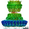
- EMDB-8398: Single particle cryo-EM reconstruction of the Salmonella SPI-1 ty... -
+ Open data

- Basic information
| Entry | Database: EMDB / ID: EMD-8398 | |||||||||
|---|---|---|---|---|---|---|---|---|---|---|
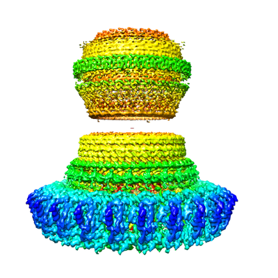
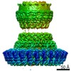
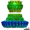
| Title | Single particle cryo-EM reconstruction of the Salmonella SPI-1 type III secretion injectisome basal body at 4.3 Angstrom resolution | |||||||||
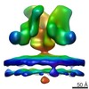
 Map data Map data | 24-fold averaged map of the basal body complex | |||||||||
 Sample Sample |
| |||||||||
 Keywords Keywords | Bacterial / secretion / injectisome / membrane protein | |||||||||
| Function / homology |  Function and homology information Function and homology information | |||||||||
| Biological species |  Salmonella enterica subsp. enterica serovar Typhimurium (bacteria) Salmonella enterica subsp. enterica serovar Typhimurium (bacteria) | |||||||||
| Method | single particle reconstruction / cryo EM / Resolution: 4.3 Å | |||||||||
 Authors Authors | Worrall LJ / Hong C | |||||||||
| Funding support |  Canada, 1 items Canada, 1 items
| |||||||||
 Citation Citation | Journal: Nature / Year: 2016 Title: Near-atomic-resolution cryo-EM analysis of the Salmonella T3S injectisome basal body. Authors: L J Worrall / C Hong / M Vuckovic / W Deng / J R C Bergeron / D D Majewski / R K Huang / T Spreter / B B Finlay / Z Yu / N C J Strynadka /  Abstract: The type III secretion (T3S) injectisome is a specialized protein nanomachine that is critical for the pathogenicity of many Gram-negative bacteria, including purveyors of plague, typhoid fever, ...The type III secretion (T3S) injectisome is a specialized protein nanomachine that is critical for the pathogenicity of many Gram-negative bacteria, including purveyors of plague, typhoid fever, whooping cough, sexually transmitted infections and major nosocomial infections. This syringe-shaped 3.5-MDa macromolecular assembly spans both bacterial membranes and that of the infected host cell. The internal channel formed by the injectisome allows for the direct delivery of partially unfolded virulence effectors into the host cytoplasm. The structural foundation of the injectisome is the basal body, a molecular lock-nut structure composed predominantly of three proteins that form highly oligomerized concentric rings spanning the inner and outer membranes. Here we present the structure of the prototypical Salmonella enterica serovar Typhimurium pathogenicity island 1 basal body, determined using single-particle cryo-electron microscopy, with the inner-membrane-ring and outer-membrane-ring oligomers defined at 4.3 Å and 3.6 Å resolution, respectively. This work presents the first, to our knowledge, high-resolution structural characterization of the major components of the basal body in the assembled state, including that of the widespread class of outer-membrane portals known as secretins. | |||||||||
| History |
|
- Structure visualization
| Movie |
Movie viewer |
|---|---|
| Structure viewer | EM map: SurfViewMolmilJmol/JSmol |
| Supplemental images |
- Downloads & links
-EMDB archive
| Map data | emd_8398.map.gz | 86.7 MB | EMDB map data format | |
|---|---|---|---|---|
| Header (meta data) | emd-8398-v30.xmlemd-8398.xml | 16.3 KB 16.3 KB | Display Display | EMDB header |
| Images |  emd_8398.png emd_8398.png | 149.9 KB | ||
| Filedesc metadata | emd-8398.cif.gz | 5.9 KB | ||
| Archive directory |  http://ftp.pdbj.org/pub/emdb/structures/EMD-8398ftp://ftp.pdbj.org/pub/emdb/structures/EMD-8398 http://ftp.pdbj.org/pub/emdb/structures/EMD-8398ftp://ftp.pdbj.org/pub/emdb/structures/EMD-8398 | HTTPS FTP |
-Related structure data
| Related structure data |  5tcpMC  8399C 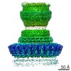 8400C  8401C 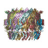 5tcqC 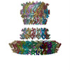 5tcrC C: citing same article ( M: atomic model generated by this map |
|---|---|
| Similar structure data |

-Links
| EMDB pages | EMDB (EBI/PDBe) / EMDataResource |
|---|---|
| Related items in Molecule of the Month |


-Map
| File | Download / File: emd_8398.map.gz / Format: CCP4 / Size: 103 MB / Type: IMAGE STORED AS FLOATING POINT NUMBER (4 BYTES) | ||||||||||||||||||||||||||||||||||||||||||||||||||||||||||||||||||||
|---|---|---|---|---|---|---|---|---|---|---|---|---|---|---|---|---|---|---|---|---|---|---|---|---|---|---|---|---|---|---|---|---|---|---|---|---|---|---|---|---|---|---|---|---|---|---|---|---|---|---|---|---|---|---|---|---|---|---|---|---|---|---|---|---|---|---|---|---|---|
| Annotation | 24-fold averaged map of the basal body complex | ||||||||||||||||||||||||||||||||||||||||||||||||||||||||||||||||||||
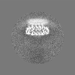
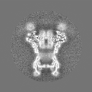
| Projections & slices | Image control
Images are generated by Spider. | ||||||||||||||||||||||||||||||||||||||||||||||||||||||||||||||||||||
| Voxel size | X=Y=Z: 1.71 Å | ||||||||||||||||||||||||||||||||||||||||||||||||||||||||||||||||||||
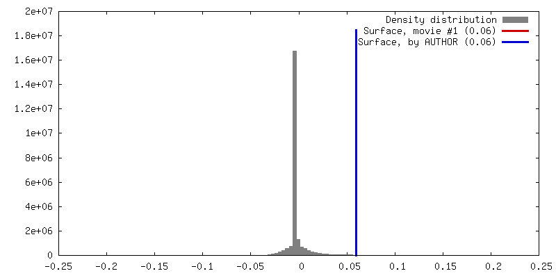
| Density |
| ||||||||||||||||||||||||||||||||||||||||||||||||||||||||||||||||||||
| Symmetry | Space group: 1 | ||||||||||||||||||||||||||||||||||||||||||||||||||||||||||||||||||||
| Details | EMDB XML:
CCP4 map header:
| ||||||||||||||||||||||||||||||||||||||||||||||||||||||||||||||||||||
 Z (Sec.)
 Y (Row.)
 X (Col.)


-Supplemental data
- Sample components
-Entire : Type III secretion injectisome basal body
| Entire | Name: Type III secretion injectisome basal body |
|---|---|
| Components |
|
-Supramolecule #1: Type III secretion injectisome basal body
| Supramolecule | Name: Type III secretion injectisome basal body / type: complex / ID: 1 / Parent: 0 / Macromolecule list: all / Details: PrgH130-392 mutant |
|---|---|
| Source (natural) | Organism: Salmonella enterica subsp. enterica serovar Typhimurium (bacteria) |
| Molecular weight | Theoretical: 2 MDa |
-Macromolecule #1: Lipoprotein PrgK
| Macromolecule | Name: Lipoprotein PrgK / type: protein_or_peptide / ID: 1 / Number of copies: 24 / Enantiomer: LEVO |
|---|---|
| Source (natural) | Organism: Salmonella enterica subsp. enterica serovar Typhimurium (bacteria) |
| Molecular weight | Theoretical: 26.199723 KDa |
| Sequence | String: CKDKDLLKGL DQEQANEVIA VLQMHNIEAN KIDSGKLGYS ITVAEPDFTA AVYWIKTYQL PPRPRVEIAQ MFPADSLVSS PRAEKARLY SAIEQRLEQS LQTMEGVLSA RVHISYDIDA GENGRPPKPV HLSALAVYER GSPLAHQISD IKRFLKNSFA D VDYDNISV ...String: CKDKDLLKGL DQEQANEVIA VLQMHNIEAN KIDSGKLGYS ITVAEPDFTA AVYWIKTYQL PPRPRVEIAQ MFPADSLVSS PRAEKARLY SAIEQRLEQS LQTMEGVLSA RVHISYDIDA GENGRPPKPV HLSALAVYER GSPLAHQISD IKRFLKNSFA D VDYDNISV VLSERSDAQL QAPGTPVKRN SFATSWIVLI ILLSVMSAGF GVWYYKNHYA RNKKGITADD KAKSSNE UniProtKB: Lipoprotein PrgK |
-Macromolecule #2: Protein PrgH
| Macromolecule | Name: Protein PrgH / type: protein_or_peptide / ID: 2 / Number of copies: 24 / Enantiomer: LEVO |
|---|---|
| Source (natural) | Organism: Salmonella enterica subsp. enterica serovar Typhimurium (bacteria) |
| Molecular weight | Theoretical: 30.360537 KDa |
| Sequence | String: SAKKNEPRFK NGIVAALAGF FILGIGTVGT LWILNSPQRQ AAELDSLLGQ EKERFQVLPG RDKMLYVAAQ NERDTLWARQ VLARGDYDK NARVINENEE NKRISIWLDT YYPQLAYYRI HFDEPRKPVF WLSRQRNTMS KKELEVLSQK LRALMPYADS V NITLMDDV ...String: SAKKNEPRFK NGIVAALAGF FILGIGTVGT LWILNSPQRQ AAELDSLLGQ EKERFQVLPG RDKMLYVAAQ NERDTLWARQ VLARGDYDK NARVINENEE NKRISIWLDT YYPQLAYYRI HFDEPRKPVF WLSRQRNTMS KKELEVLSQK LRALMPYADS V NITLMDDV TAAGQAEAGL KQQALPYSRR NHKGGVTFVI QGALDDVEIL RARQFVDSYY RTWGGRYVQF AIELKDDWLK GR SFQYGAE GYIKMSPGHW YFPSPL UniProtKB: Protein PrgH |
-Experimental details
-Structure determination
| Method | cryo EM |
|---|---|
 Processing Processing | single particle reconstruction |
| Aggregation state | particle |
-Sample preparation
| Concentration | 10 mg/mL | ||||||||||||
|---|---|---|---|---|---|---|---|---|---|---|---|---|---|
| Buffer | pH: 8 Component:
| ||||||||||||
| Grid | Model: Quantifoil R1.2/1.3 / Material: GOLD / Mesh: 400 / Pretreatment - Type: GLOW DISCHARGE / Pretreatment - Time: 60 sec. / Pretreatment - Atmosphere: AIR / Pretreatment - Pressure: 0.038 kPa | ||||||||||||
| Vitrification | Cryogen name: ETHANE / Chamber humidity: 90 % / Chamber temperature: 298 K / Instrument: FEI VITROBOT MARK IV |
- Electron microscopy
| Microscope | FEI TITAN KRIOS |
|---|---|
| Temperature | Min: 80.0 K / Max: 80.0 K |
| Specialist optics | Energy filter - Name: Gatan GIF / Energy filter - Lower energy threshold: 0 eV / Energy filter - Upper energy threshold: 20 eV |
| Image recording | Film or detector model: GATAN K2 QUANTUM (4k x 4k) / Detector mode: SUPER-RESOLUTION / Digitization - Dimensions - Width: 7676 pixel / Digitization - Dimensions - Height: 7420 pixel / Digitization - Frames/image: 1-48 / Number grids imaged: 1 / Number real images: 2515 / Average exposure time: 0.375 sec. / Average electron dose: 1.3 e/Å2 |
| Electron beam | Acceleration voltage: 300 kV / Electron source:  FIELD EMISSION GUN FIELD EMISSION GUN |
| Electron optics | C2 aperture diameter: 70.0 µm / Calibrated defocus max: 3.2 µm / Calibrated defocus min: 1.3 µm / Calibrated magnification: 29240 / Illumination mode: FLOOD BEAM / Imaging mode: BRIGHT FIELD / Cs: 0.01 mm / Nominal defocus max: 3.0 µm / Nominal defocus min: 1.5 µm / Nominal magnification: 64000 |
| Sample stage | Specimen holder model: FEI TITAN KRIOS AUTOGRID HOLDER / Cooling holder cryogen: NITROGEN |
| Experimental equipment |  Model: Titan Krios / Image courtesy: FEI Company |
+Image processing
-Atomic model buiding 1
| Details | Initial fitting was carried out with Chimera, followed by rebuilding and refinement in Rosetta, Phenix, and Coot. |
|---|---|
| Refinement | Space: REAL / Protocol: FLEXIBLE FIT |
| Output model | PDB-5tcp: |
