 Movie
Movie Controller

[English] 日本語
 Yorodumi
- EMDB-77350: yeast 26S proteasome base assembly intermediate, base-Rpn14-Hsm3-... -
+ Open data

- Basic information
| Entry |  | |||||||||
|---|---|---|---|---|---|---|---|---|---|---|
| Title | yeast 26S proteasome base assembly intermediate, base-Rpn14-Hsm3-Nas6 with Rpt4-EQ | |||||||||
 Map data Map data | ||||||||||
 Sample Sample |
| |||||||||
 Keywords Keywords | proteasome / chaperone / base / 26S / AAA / motor / assembly / MOTOR PROTEIN | |||||||||
| Function / homology |  Function and homology information Function and homology informationRegulation of PTEN stability and activity / proteasome regulatory particle binding / proteasome regulatory particle, base subcomplex / proteasome regulatory particle assembly / protein-containing complex localization / proteasome-activating activity / proteasome regulatory particle, lid subcomplex / regulation of protein catabolic process / Cross-presentation of soluble exogenous antigens (endosomes) / TNFR2 non-canonical NF-kB pathway ...Regulation of PTEN stability and activity / proteasome regulatory particle binding / proteasome regulatory particle, base subcomplex / proteasome regulatory particle assembly / protein-containing complex localization / proteasome-activating activity / proteasome regulatory particle, lid subcomplex / regulation of protein catabolic process / Cross-presentation of soluble exogenous antigens (endosomes) / TNFR2 non-canonical NF-kB pathway / Proteasome assembly / nonfunctional rRNA decay / Ub-specific processing proteases / peptide catabolic process / positive regulation of RNA polymerase II transcription preinitiation complex assembly / mismatch repair / proteasome assembly / enzyme regulator activity / ERAD pathway / Neutrophil degranulation / proteasome storage granule / protein folding chaperone / proteasome complex / ubiquitin binding / positive regulation of transcription elongation by RNA polymerase II / nucleotide-excision repair / positive regulation of protein catabolic process / ubiquitin-dependent protein catabolic process / proteasome-mediated ubiquitin-dependent protein catabolic process / protein-macromolecule adaptor activity / chromatin remodeling / protein domain specific binding / ubiquitin protein ligase binding / endoplasmic reticulum / ATP hydrolysis activity / ATP binding / identical protein binding / nucleus / cytosol / cytoplasm Similarity search - Function | |||||||||
| Biological species |   Saccharomyces cerevisiae (brewer's yeast) Saccharomyces cerevisiae (brewer's yeast) | |||||||||
| Method | single particle reconstruction / cryo EM / Resolution: 3.93 Å | |||||||||
 Authors Authors | Hsieh HH / Martin A | |||||||||
| Funding support |  United States, 2 items United States, 2 items
| |||||||||
 Citation Citation | Journal: Acta Crystallogr D Struct Biol / Year: 2019 Title: Macromolecular structure determination using X-rays, neutrons and electrons: recent developments in Phenix. Authors: Dorothee Liebschner / Pavel V Afonine / Matthew L Baker / Gábor Bunkóczi / Vincent B Chen / Tristan I Croll / Bradley Hintze / Li Wei Hung / Swati Jain / Airlie J McCoy / Nigel W Moriarty ...Authors: Dorothee Liebschner / Pavel V Afonine / Matthew L Baker / Gábor Bunkóczi / Vincent B Chen / Tristan I Croll / Bradley Hintze / Li Wei Hung / Swati Jain / Airlie J McCoy / Nigel W Moriarty / Robert D Oeffner / Billy K Poon / Michael G Prisant / Randy J Read / Jane S Richardson / David C Richardson / Massimo D Sammito / Oleg V Sobolev / Duncan H Stockwell / Thomas C Terwilliger / Alexandre G Urzhumtsev / Lizbeth L Videau / Christopher J Williams / Paul D Adams /   Abstract: Diffraction (X-ray, neutron and electron) and electron cryo-microscopy are powerful methods to determine three-dimensional macromolecular structures, which are required to understand biological ...Diffraction (X-ray, neutron and electron) and electron cryo-microscopy are powerful methods to determine three-dimensional macromolecular structures, which are required to understand biological processes and to develop new therapeutics against diseases. The overall structure-solution workflow is similar for these techniques, but nuances exist because the properties of the reduced experimental data are different. Software tools for structure determination should therefore be tailored for each method. Phenix is a comprehensive software package for macromolecular structure determination that handles data from any of these techniques. Tasks performed with Phenix include data-quality assessment, map improvement, model building, the validation/rebuilding/refinement cycle and deposition. Each tool caters to the type of experimental data. The design of Phenix emphasizes the automation of procedures, where possible, to minimize repetitive and time-consuming manual tasks, while default parameters are chosen to encourage best practice. A graphical user interface provides access to many command-line features of Phenix and streamlines the transition between programs, project tracking and re-running of previous tasks. | |||||||||
| History |
|
- Structure visualization
| Supplemental images |
|---|

- Downloads & links
-EMDB archive
| Map data | emd_77350.map.gz | 254.8 MB | EMDB map data format | |
|---|---|---|---|---|
| Header (meta data) | emd-77350-v30.xmlemd-77350.xml | 38.2 KB 38.2 KB | Display Display | EMDB header |
| FSC (resolution estimation) | emd_77350_fsc.xml | 17 KB | Display | FSC data file |
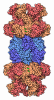
| Images |  emd_77350.png emd_77350.png | 106 KB | ||
| Filedesc metadata | emd-77350.cif.gz | 10.7 KB | ||
| Others | emd_77350_half_map_1.map.gzemd_77350_half_map_2.map.gz | 475 MB 475 MB | ||
| Archive directory |  https://data.pdbj.org/pub/emdb/structures/EMD-77350ftp://data.pdbj.org/pub/emdb/structures/EMD-77350 https://data.pdbj.org/pub/emdb/structures/EMD-77350ftp://data.pdbj.org/pub/emdb/structures/EMD-77350 | HTTPS FTP |
-Related structure data
| Related structure data |  36bdMC  35zrC  35zvC  35zwC  36axC  36blC  36bmC C: citing same article ( M: atomic model generated by this map |
|---|---|
| Similar structure data |


-Links
| EMDB pages | EMDB (EBI/PDBe) / EMDataResource |
|---|---|
| Related items in Molecule of the Month |



-Map
| File | Download / File: emd_77350.map.gz / Format: CCP4 / Size: 512 MB / Type: IMAGE STORED AS FLOATING POINT NUMBER (4 BYTES) | ||||||||||||||||||||||||||||||||||||
|---|---|---|---|---|---|---|---|---|---|---|---|---|---|---|---|---|---|---|---|---|---|---|---|---|---|---|---|---|---|---|---|---|---|---|---|---|---|
| Projections & slices | Image control
Images are generated by Spider. | ||||||||||||||||||||||||||||||||||||
| Voxel size | X=Y=Z: 1.048 Å | ||||||||||||||||||||||||||||||||||||
| Density |
| ||||||||||||||||||||||||||||||||||||
| Symmetry | Space group: 1 | ||||||||||||||||||||||||||||||||||||
| Details | EMDB XML:
|
 Z (Sec.)
 Y (Row.)
 X (Col.)


-Supplemental data
-Half map: #1
| File | emd_77350_half_map_1.map | ||||||||||||
|---|---|---|---|---|---|---|---|---|---|---|---|---|---|
| Projections & Slices |
| ||||||||||||
| Density Histograms |


-Half map: #2
| File | emd_77350_half_map_2.map | ||||||||||||
|---|---|---|---|---|---|---|---|---|---|---|---|---|---|
| Projections & Slices |
| ||||||||||||
| Density Histograms |


- Sample components
+Entire : yeast 26S proteasome base assembly intermediate, Hsm3-Rpt1-Rpt2 (...
+Supramolecule #1: yeast 26S proteasome base assembly intermediate, Hsm3-Rpt1-Rpt2 (...
+Macromolecule #1: 26S proteasome regulatory subunit 7 homolog

 Escherichia coli (E. coli)
+Macromolecule #2: 26S proteasome regulatory subunit 4 homolog
+Macromolecule #3: 26S proteasome regulatory subunit RPN1
+Macromolecule #4: DNA mismatch repair protein HSM3
+Macromolecule #5: 26S proteasome regulatory subunit 8 homolog
+Macromolecule #6: 26S proteasome regulatory subunit 6B homolog
+Macromolecule #7: 26S proteasome regulatory subunit RPN13
+Macromolecule #8: 26S proteasome regulatory subunit RPN14
+Macromolecule #9: Probable 26S proteasome regulatory subunit p28
+Macromolecule #10: 26S proteasome regulatory subunit RPN2
+Macromolecule #11: 26S proteasome subunit RPT4
+Macromolecule #12: 26S proteasome regulatory subunit 6A
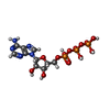
+Macromolecule #13: ADENOSINE-5'-TRIPHOSPHATE
-Experimental details
-Structure determination
| Method | cryo EM |
|---|---|
 Processing Processing | single particle reconstruction |
| Aggregation state | particle |
-Sample preparation
| Concentration | 6 mg/mL |
|---|---|
| Buffer | pH: 7.6 |
| Vitrification | Cryogen name: ETHANE |
- Electron microscopy
| Microscope | TFS KRIOS |
|---|---|
| Image recording | Film or detector model: GATAN K3 (6k x 4k) / Average electron dose: 50.0 e/Å2 |
| Electron beam | Acceleration voltage: 300 kV / Electron source:  FIELD EMISSION GUN FIELD EMISSION GUN |
| Electron optics | Illumination mode: FLOOD BEAM / Imaging mode: BRIGHT FIELD / Nominal defocus max: 2.0 µm / Nominal defocus min: 0.5 µm |
| Experimental equipment |  Model: Titan Krios / Image courtesy: FEI Company |

