 Movie
Movie Controller

[English] 日本語
 Yorodumi
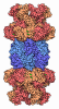
- EMDB-77307: yeast 26S proteasome base assembly intermediate, Rpn14-Rpt6 (base... -
+ Open data

- Basic information
| Entry |  | |||||||||
|---|---|---|---|---|---|---|---|---|---|---|
| Title | yeast 26S proteasome base assembly intermediate, Rpn14-Rpt6 (base-Rpn14-Hsm3-Nas6(2) with Rpt4-EQ) | |||||||||
 Map data Map data | ||||||||||
 Sample Sample |
| |||||||||
 Keywords Keywords | proteasome / chaperone / base / 26S / AAA / motor / assembly / MOTOR PROTEIN | |||||||||
| Function / homology |  Function and homology information Function and homology informationproteasome regulatory particle, base subcomplex / proteasome regulatory particle assembly / proteasome-activating activity / regulation of protein catabolic process / Cross-presentation of soluble exogenous antigens (endosomes) / TNFR2 non-canonical NF-kB pathway / Proteasome assembly / nonfunctional rRNA decay / Ub-specific processing proteases / positive regulation of RNA polymerase II transcription preinitiation complex assembly ...proteasome regulatory particle, base subcomplex / proteasome regulatory particle assembly / proteasome-activating activity / regulation of protein catabolic process / Cross-presentation of soluble exogenous antigens (endosomes) / TNFR2 non-canonical NF-kB pathway / Proteasome assembly / nonfunctional rRNA decay / Ub-specific processing proteases / positive regulation of RNA polymerase II transcription preinitiation complex assembly / enzyme regulator activity / Neutrophil degranulation / proteasome storage granule / protein folding chaperone / proteasome complex / positive regulation of transcription elongation by RNA polymerase II / nucleotide-excision repair / ubiquitin-dependent protein catabolic process / proteasome-mediated ubiquitin-dependent protein catabolic process / protein-macromolecule adaptor activity / chromatin remodeling / protein domain specific binding / ubiquitin protein ligase binding / endoplasmic reticulum / ATP hydrolysis activity / ATP binding / identical protein binding / nucleus / cytosol / cytoplasm Similarity search - Function | |||||||||
| Biological species |   Saccharomyces cerevisiae (brewer's yeast) Saccharomyces cerevisiae (brewer's yeast) | |||||||||
| Method | single particle reconstruction / cryo EM / Resolution: 3.11 Å | |||||||||
 Authors Authors | Hsieh HH / Martin A | |||||||||
| Funding support |  United States, 2 items United States, 2 items
| |||||||||
 Citation Citation | Journal: Acta Crystallogr D Struct Biol / Year: 2019 Title: Macromolecular structure determination using X-rays, neutrons and electrons: recent developments in Phenix. Authors: Dorothee Liebschner / Pavel V Afonine / Matthew L Baker / Gábor Bunkóczi / Vincent B Chen / Tristan I Croll / Bradley Hintze / Li Wei Hung / Swati Jain / Airlie J McCoy / Nigel W Moriarty ...Authors: Dorothee Liebschner / Pavel V Afonine / Matthew L Baker / Gábor Bunkóczi / Vincent B Chen / Tristan I Croll / Bradley Hintze / Li Wei Hung / Swati Jain / Airlie J McCoy / Nigel W Moriarty / Robert D Oeffner / Billy K Poon / Michael G Prisant / Randy J Read / Jane S Richardson / David C Richardson / Massimo D Sammito / Oleg V Sobolev / Duncan H Stockwell / Thomas C Terwilliger / Alexandre G Urzhumtsev / Lizbeth L Videau / Christopher J Williams / Paul D Adams /   Abstract: Diffraction (X-ray, neutron and electron) and electron cryo-microscopy are powerful methods to determine three-dimensional macromolecular structures, which are required to understand biological ...Diffraction (X-ray, neutron and electron) and electron cryo-microscopy are powerful methods to determine three-dimensional macromolecular structures, which are required to understand biological processes and to develop new therapeutics against diseases. The overall structure-solution workflow is similar for these techniques, but nuances exist because the properties of the reduced experimental data are different. Software tools for structure determination should therefore be tailored for each method. Phenix is a comprehensive software package for macromolecular structure determination that handles data from any of these techniques. Tasks performed with Phenix include data-quality assessment, map improvement, model building, the validation/rebuilding/refinement cycle and deposition. Each tool caters to the type of experimental data. The design of Phenix emphasizes the automation of procedures, where possible, to minimize repetitive and time-consuming manual tasks, while default parameters are chosen to encourage best practice. A graphical user interface provides access to many command-line features of Phenix and streamlines the transition between programs, project tracking and re-running of previous tasks. | |||||||||
| History |
|
- Structure visualization
| Supplemental images |
|---|

- Downloads & links
-EMDB archive
| Map data | emd_77307.map.gz | 256.8 MB | EMDB map data format | |
|---|---|---|---|---|
| Header (meta data) | emd-77307-v30.xmlemd-77307.xml | 23.6 KB 23.6 KB | Display Display | EMDB header |
| FSC (resolution estimation) | emd_77307_fsc.xml | 16.9 KB | Display | FSC data file |
| Images |  emd_77307.png emd_77307.png | 87.1 KB | ||
| Filedesc metadata | emd-77307.cif.gz | 7.8 KB | ||
| Others | emd_77307_half_map_1.map.gzemd_77307_half_map_2.map.gz | 474.3 MB 474.3 MB | ||
| Archive directory |  https://data.pdbj.org/pub/emdb/structures/EMD-77307ftp://data.pdbj.org/pub/emdb/structures/EMD-77307 https://data.pdbj.org/pub/emdb/structures/EMD-77307ftp://data.pdbj.org/pub/emdb/structures/EMD-77307 | HTTPS FTP |
-Related structure data
| Related structure data |  35zvMC  35zrC  35zwC  36axC  36bdC  36blC  36bmC M: atomic model generated by this map C: citing same article ( |
|---|---|
| Similar structure data |


-Links
| EMDB pages | EMDB (EBI/PDBe) / EMDataResource |
|---|---|
| Related items in Molecule of the Month |



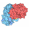
-Map
| File | Download / File: emd_77307.map.gz / Format: CCP4 / Size: 512 MB / Type: IMAGE STORED AS FLOATING POINT NUMBER (4 BYTES) | ||||||||||||||||||||||||||||||||||||
|---|---|---|---|---|---|---|---|---|---|---|---|---|---|---|---|---|---|---|---|---|---|---|---|---|---|---|---|---|---|---|---|---|---|---|---|---|---|
| Projections & slices | Image control
Images are generated by Spider. | ||||||||||||||||||||||||||||||||||||
| Voxel size | X=Y=Z: 1.048 Å | ||||||||||||||||||||||||||||||||||||
| Density |
| ||||||||||||||||||||||||||||||||||||
| Symmetry | Space group: 1 | ||||||||||||||||||||||||||||||||||||
| Details | EMDB XML:
|
 Z (Sec.)
 Y (Row.)
 X (Col.)


-Supplemental data
-Half map: #1
| File | emd_77307_half_map_1.map | ||||||||||||
|---|---|---|---|---|---|---|---|---|---|---|---|---|---|
| Projections & Slices |
| ||||||||||||
| Density Histograms |


-Half map: #2
| File | emd_77307_half_map_2.map | ||||||||||||
|---|---|---|---|---|---|---|---|---|---|---|---|---|---|
| Projections & Slices |
| ||||||||||||
| Density Histograms |


- Sample components
-Entire : yeast 26S proteasome base assembly intermediate, Hsm3-Rpt1-Rpt2 (...
| Entire | Name: yeast 26S proteasome base assembly intermediate, Hsm3-Rpt1-Rpt2 (base-Hsm3-Nas6) |
|---|---|
| Components |
|
-Supramolecule #1: yeast 26S proteasome base assembly intermediate, Hsm3-Rpt1-Rpt2 (...
| Supramolecule | Name: yeast 26S proteasome base assembly intermediate, Hsm3-Rpt1-Rpt2 (base-Hsm3-Nas6) type: complex / ID: 1 / Parent: 0 / Macromolecule list: #1-#4 |
|---|---|
| Source (natural) | Organism: Saccharomyces cerevisiae (brewer's yeast) |
-Macromolecule #1: 26S proteasome regulatory subunit RPN1
| Macromolecule | Name: 26S proteasome regulatory subunit RPN1 / type: protein_or_peptide / ID: 1 / Number of copies: 1 / Enantiomer: LEVO |
|---|---|
| Source (natural) | Organism: Saccharomyces cerevisiae (brewer's yeast) |
| Molecular weight | Theoretical: 109.601906 KDa |
| Recombinant expression | Organism:   Escherichia coli (E. coli) Escherichia coli (E. coli) |
| Sequence | String: MVDESDKKQQ TIDEQSQISP EKQTPNKKDK KKEEEEQLSE EDAKLKTDLE LLVERLKEDD SSLYEASLNA LKESIKNSTS SMTAVPKPL KFLRPTYPDL CSIYDKWTDP NLKSSLADVL SILAMTYSEN GKHDSLRYRL LSDVSDFEGW GHEYIRHLAL E IGEVYNDQ ...String: MVDESDKKQQ TIDEQSQISP EKQTPNKKDK KKEEEEQLSE EDAKLKTDLE LLVERLKEDD SSLYEASLNA LKESIKNSTS SMTAVPKPL KFLRPTYPDL CSIYDKWTDP NLKSSLADVL SILAMTYSEN GKHDSLRYRL LSDVSDFEGW GHEYIRHLAL E IGEVYNDQ VEKDAEDETS SDGSKSDGSA ATSGFEFSKE DTLRLCLDIV PYFLKHNGEE DAVDLLLEIE SIDKLPQFVD EN TFQRVCQ YMVACVPLLP PPEDVAFLKT AYSIYLSQNE LTDAIALAVR LGEEDMIRSV FDATSDPVMH KQLAYILAAQ KTS FEYEGV QDIIGNGKLS EHFLYLAKEL NLTGPKVPED IYKSHLDNSK SVFSSAGLDS AQQNLASSFV NGFLNLGYCN DKLI VDNDN WVYKTKGDGM TSAVASIGSI YQWNLDGLQQ LDKYLYVDEP EVKAGALLGI GISASGVHDG EVEPALLLLQ DYVTN PDTK ISSAAILGLG IAFAGSKNDE VLGLLLPIAA STDLPIETAA MASLALAHVF VGTCNGDITT SIMDNFLERT AIELKT DWV RFLALALGIL YMGQGEQVDD VLETISAIEH PMTSAIEVLV GSCAYTGTGD VLLIQDLLHR LTPKNVKGEE DADEEET AE GQTNSISDFL GEQVNEPTKN EEAEIEVDEM EVDAEGEEVE VKAEITEKKN GESLEGEEIK SEEKKGKSSD KDATTDGK N DDEEEEKEAG IVDELAYAVL GIALIALGED IGKEMSLRHF GHLMHYGNEH IRRMVPLAMG IVSVSDPQMK VFDTLTRFS HDADLEVSMN SIFAMGLCGA GTNNARLAQL LRQLASYYSR EQDALFITRL AQGLLHLGKG TMTMDVFNDA HVLNKVTLAS ILTTAVGLV SPSFMLKHHQ LFYMLNAGIR PKFILALNDE GEPIKVNVRV GQAVETVGQA GRPKKITGWI TQSTPVLLNH G ERAELETD EYISYTSHIE GVVILKKNPD YREEE UniProtKB: 26S proteasome regulatory subunit RPN1 |
-Macromolecule #2: 26S proteasome regulatory subunit 8 homolog
| Macromolecule | Name: 26S proteasome regulatory subunit 8 homolog / type: protein_or_peptide / ID: 2 / Number of copies: 1 / Enantiomer: LEVO |
|---|---|
| Source (natural) | Organism: Saccharomyces cerevisiae (brewer's yeast) |
| Molecular weight | Theoretical: 45.342742 KDa |
| Recombinant expression | Organism: Escherichia coli (E. coli) |
| Sequence | String: MTAAVTSSNI VLETHESGIK PYFEQKIQET ELKIRSKTEN VRRLEAQRNA LNDKVRFIKD ELRLLQEPGS YVGEVIKIVS DKKVLVKVQ PEGKYIVDVA KDINVKDLKA SQRVCLRSDS YMLHKVLENK ADPLVSLMMV EKVPDSTYDM VGGLTKQIKE I KEVIELPV ...String: MTAAVTSSNI VLETHESGIK PYFEQKIQET ELKIRSKTEN VRRLEAQRNA LNDKVRFIKD ELRLLQEPGS YVGEVIKIVS DKKVLVKVQ PEGKYIVDVA KDINVKDLKA SQRVCLRSDS YMLHKVLENK ADPLVSLMMV EKVPDSTYDM VGGLTKQIKE I KEVIELPV KHPELFESLG IAQPKGVILY GPPGTGKTLL ARAVAHHTDC KFIRVSGAEL VQKYIGEGSR MVRELFVMAR EH APSIIFM DEIDSIGSTR VEGSGGGDSE VQRTMLELLN QLDGFETSKN IKIIMATNRL DILDPALLRP GRIDRKIEFP PPS VAARAE ILRIHSRKMN LTRGINLRKV AEKMNGCSGA DVKGVCTEAG MYALRERRIH VTQEDFELAV GKVMNKNQET AISV AKLFK UniProtKB: 26S proteasome regulatory subunit 8 homolog |
-Macromolecule #3: 26S proteasome regulatory subunit 6B homolog
| Macromolecule | Name: 26S proteasome regulatory subunit 6B homolog / type: protein_or_peptide / ID: 3 / Number of copies: 1 / Enantiomer: LEVO |
|---|---|
| Source (natural) | Organism: Saccharomyces cerevisiae (brewer's yeast) |
| Molecular weight | Theoretical: 47.953676 KDa |
| Recombinant expression | Organism: Escherichia coli (E. coli) |
| Sequence | String: MEELGIVTPV EKAVEEKPAV KSYASLLAQL NGTVNNNSAL SNVNSDIYFK LKKLEKEYEL LTLQEDYIKD EQRHLKRELK RAQEEVKRI QSVPLVIGQF LEPIDQNTGI VSSTTGMSYV VRILSTLDRE LLKPSMSVAL HRHSNALVDI LPPDSDSSIS V MGENEKPD ...String: MEELGIVTPV EKAVEEKPAV KSYASLLAQL NGTVNNNSAL SNVNSDIYFK LKKLEKEYEL LTLQEDYIKD EQRHLKRELK RAQEEVKRI QSVPLVIGQF LEPIDQNTGI VSSTTGMSYV VRILSTLDRE LLKPSMSVAL HRHSNALVDI LPPDSDSSIS V MGENEKPD VTYADVGGLD MQKQEIREAV ELPLVQADLY EQIGIDPPRG VLLYGPPGTG KTMLVKAVAN STKAAFIRVN GS EFVHKYL GEGPRMVRDV FRLARENAPS IIFIDEVDSI ATKRFDAQTG SDREVQRILI ELLTQMDGFD QSTNVKVIMA TNR ADTLDP ALLRPGRLDR KIEFPSLRDR RERRLIFGTI ASKMSLAPEA DLDSLIIRND SLSGAVIAAI MQEAGLRAVR KNRY VILQS DLEEAYATQV KTDNTVDKFD FYK UniProtKB: 26S proteasome regulatory subunit 6B homolog |
-Macromolecule #4: 26S proteasome regulatory subunit RPN14
| Macromolecule | Name: 26S proteasome regulatory subunit RPN14 / type: protein_or_peptide / ID: 4 / Number of copies: 1 / Enantiomer: LEVO |
|---|---|
| Source (natural) | Organism: Saccharomyces cerevisiae (brewer's yeast) |
| Molecular weight | Theoretical: 46.433684 KDa |
| Recombinant expression | Organism: Escherichia coli (E. coli) |
| Sequence | String: MTKTITVAHI QYDFKAVLEE NDENDDEFYI NVDKNLNEIK EHKIVVLGNS RGVDAGKGNT FEKVGSHLYK ARLDGHDFLF NTIIRDGSK MLKRADYTAV DTAKLQMRRF ILGTTEGDIK VLDSNFNLQR EIDQAHVSEI TKLKFFPSGE ALISSSQDMQ L KIWSVKDG ...String: MTKTITVAHI QYDFKAVLEE NDENDDEFYI NVDKNLNEIK EHKIVVLGNS RGVDAGKGNT FEKVGSHLYK ARLDGHDFLF NTIIRDGSK MLKRADYTAV DTAKLQMRRF ILGTTEGDIK VLDSNFNLQR EIDQAHVSEI TKLKFFPSGE ALISSSQDMQ L KIWSVKDG SNPRTLIGHR ATVTDIAIID RGRNVLSASL DGTIRLWECG TGTTIHTFNR KENPHDGVNS IALFVGTDRQ LH EISTSKK NNLEFGTYGK YVIAGHVSGV ITVHNVFSKE QTIQLPSKFT CSCNSLTVDG NNANYIYAGY ENGMLAQWDL RSP ECPVGE FLINEGTPIN NVYFAAGALF VSSGFDTSIK LDIISDPESE RPAIEFETPT FLVSNDDEVS QFCYVSDDES NGEV LEVGK NNFCALYNLS NP UniProtKB: 26S proteasome regulatory subunit RPN14 |
-Macromolecule #5: ADENOSINE-5'-TRIPHOSPHATE
| Macromolecule | Name: ADENOSINE-5'-TRIPHOSPHATE / type: ligand / ID: 5 / Number of copies: 1 / Formula: ATP |
|---|---|
| Molecular weight | Theoretical: 507.181 Da |
| Chemical component information | 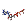 ChemComp-ATP: |
-Experimental details
-Structure determination
| Method | cryo EM |
|---|---|
 Processing Processing | single particle reconstruction |
| Aggregation state | particle |
-Sample preparation
| Concentration | 6 mg/mL |
|---|---|
| Buffer | pH: 7.6 |
| Vitrification | Cryogen name: ETHANE |
- Electron microscopy
| Microscope | TFS KRIOS |
|---|---|
| Image recording | Film or detector model: GATAN K3 (6k x 4k) / Average electron dose: 50.0 e/Å2 |
| Electron beam | Acceleration voltage: 300 kV / Electron source:  FIELD EMISSION GUN FIELD EMISSION GUN |
| Electron optics | Illumination mode: FLOOD BEAM / Imaging mode: BRIGHT FIELD / Nominal defocus max: 2.0 µm / Nominal defocus min: 0.5 µm |
| Experimental equipment |  Model: Titan Krios / Image courtesy: FEI Company |

