[English] 日本語
Yorodumi
- EMDB-77126: Human Single-ring Hsp10 -

+
Open data


ID or keywords:

Loading...

-
Basic information

Entry
Database: EMDB / ID: EMD-77126
TitleHuman Single-ring Hsp10
Map data
Sample
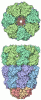
  • Complex: Single-Ring Human Hsp10
    • Protein or peptide: 10 kDa heat shock protein, mitochondrial
KeywordsHsp10 / Hsp60 / cochaperone / cryo-EM / CHAPERONE
Function / homology
Function and homology information


RHOG GTPase cycle / response to unfolded protein / Mitochondrial unfolded protein response (UPRmt) / protein folding chaperone / intrinsic apoptotic signaling pathway / osteoblast differentiation / : / protein-folding chaperone binding / protein folding / mitochondrial matrix ...RHOG GTPase cycle / response to unfolded protein / Mitochondrial unfolded protein response (UPRmt) / protein folding chaperone / intrinsic apoptotic signaling pathway / osteoblast differentiation / : / protein-folding chaperone binding / protein folding / mitochondrial matrix / mitochondrion / RNA binding / extracellular exosome / ATP binding / membrane / metal ion binding
Similarity search - Function
Chaperonin GroES, conserved site / Chaperonins cpn10 signature. / Chaperonin 10 Kd subunit / GroES chaperonin family / GroES chaperonin superfamily / Chaperonin 10 Kd subunit / GroES-like superfamily
Similarity search - Domain/homology
10 kDa heat shock protein, mitochondrial
Similarity search - Component
Biological speciesHomo sapiens (human)
Methodsingle particle reconstruction / cryo EM / Resolution: 4.0 Å
AuthorsPage AA / Walti MA
Funding support United States, 2 items
OrganizationGrant numberCountry
National Institutes of Health/National Institute of General Medical Sciences (NIH/NIGMS)R00GM132496 United States
National Institutes of Health/National Institute of General Medical Sciences (NIH/NIGMS)R35GM156786 United States
CitationJournal: bioRxiv / Year: 2026
Title: Hsp10: From Single to Double Rings-Structural Basis of Protein Homeostasis.
Authors: Abigail Page / Wyatt Hendricks / Aranea Dunckley / Marielle A Wälti
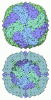
Abstract: The human chaperone complex Hsp60/Hsp10 is essential for maintaining cellular proteostasis by preventing protein misfolding and aggregation. Disruption of these processes contributes to ...The human chaperone complex Hsp60/Hsp10 is essential for maintaining cellular proteostasis by preventing protein misfolding and aggregation. Disruption of these processes contributes to neurodegenerative diseases, while overexpression of Hsp60 and Hsp10 is associated with various cancers. Understanding their molecular mechanisms is therefore of fundamental importance. Unlike its bacterial homolog GroEL, human Hsp60 adopts multiple oligomeric states, with both heptameric and tetradecameric forms binding Hsp10 to form a folding cavity for substrate refolding. Here, we determine the cryo-EM structure of apo Hsp10 and find that, in addition to its single-ring form, it also assembles into a compact double-ring state. This reveals that Hsp10, like Hsp60, exhibits structural behavior that differs markedly from its bacterial counterpart. Traditionally viewed as a passive cofactor assisting Hsp60, we show that Hsp10 alone possesses intrinsic chaperone activity: it supports folding of natural substrates such as malate dehydrogenase 1 and manganese superoxide dismutase. NMR analysis further shows that substrate binding occurs primarily at the core of Hsp10 rather than at the loops. Our findings suggest that Hsp10 exists in equilibrium between single- and double-ring complexes in the unbound state, and upon binding as a single-ring complex, it actively guides substrates into the folding chamber.
History
DepositionMay 12, 2026-
Header (metadata) releaseJul 15, 2026-
Map releaseJul 15, 2026-
UpdateJul 15, 2026-
Current statusJul 15, 2026Processing site: RCSB / Status: Released

-
Structure visualization

Supplemental images

Downloads & links

-
Map

FileDownload / File: emd_77126.map.gz / Format: CCP4 / Size: 163.6 MB / Type: IMAGE STORED AS FLOATING POINT NUMBER (4 BYTES)
Projections & slices

Image control

Size
Brightness
Contrast
Others
AxesZ (Sec.)Y (Row.)X (Col.)
0.41 Å/pix.
x 350 pix.
= 143.5 Å
0.41 Å/pix.
x 350 pix.
= 143.5 Å
0.41 Å/pix.
x 350 pix.
= 143.5 Å

Surface

Projections

Slices (1/3)

Slices (1/2)

Slices (2/3)

Images are generated by Spider.

Voxel sizeX=Y=Z: 0.41 Å
Density
Contour LevelBy AUTHOR: 0.03
Minimum - Maximum-0.09216604 - 0.19174923
Average (Standard dev.)0.00007423642 (±0.0072208354)
SymmetrySpace group: 1
Details

EMDB XML:

Map geometry
Axis orderXYZ
Origin000
Dimensions350350350
Spacing350350350
CellA=B=C: 143.5 Å
α=β=γ: 90.0 °

-
Supplemental data

-
Half map: #1

Fileemd_77126_half_map_1.map
Projections & Slices
AxesZYX

Projections

Slices (1/2)
Density Histograms

-
Half map: #2

Fileemd_77126_half_map_2.map
Projections & Slices
AxesZYX

Projections

Slices (1/2)
Density Histograms

-
Sample components

-
Entire : Single-Ring Human Hsp10

EntireName: Single-Ring Human Hsp10
Components
  • Complex: Single-Ring Human Hsp10
    • Protein or peptide: 10 kDa heat shock protein, mitochondrial

-
Supramolecule #1: Single-Ring Human Hsp10

SupramoleculeName: Single-Ring Human Hsp10 / type: complex / ID: 1 / Parent: 0 / Macromolecule list: all
Source (natural)Organism: Homo sapiens (human)
Molecular weightTheoretical: 76 kDa/nm

-
Macromolecule #1: 10 kDa heat shock protein, mitochondrial

MacromoleculeName: 10 kDa heat shock protein, mitochondrial / type: protein_or_peptide / ID: 1 / Number of copies: 7 / Enantiomer: LEVO
Source (natural)Organism: Homo sapiens (human)
Molecular weightTheoretical: 10.946674 KDa
Recombinant expressionOrganism: Escherichia coli (E. coli)
SequenceString:
MAGQAFRKFL PLFDRVLVER SAAETVTKGG IMLPEKSQGK VLQATVVAVG SGSKGKGGEI QPVSVKVGDK VLLPEYGGTK VVLDDKDYF LFRDGDILGK YVD

UniProtKB: 10 kDa heat shock protein, mitochondrial

-
Experimental details

-
Structure determination

Methodcryo EM
Processingsingle particle reconstruction
Aggregation stateparticle

-
Sample preparation

Concentration2 mg/mL
BufferpH: 7.4
Component:
ConcentrationFormulaName
300.0 mMNaClsodium chloride
10.0 mMMgCl2magnesium chloride
20.0 mMKClpotassium chloride
20.0 mMMg(CH3COO)2magnesium acetate
50.0 mMtris
VitrificationCryogen name: ETHANE / Chamber humidity: 80 % / Chamber temperature: 283 K / Instrument: LEICA EM GP

-
Electron microscopy

MicroscopeTFS KRIOS
Image recordingFilm or detector model: GATAN K2 BASE (4k x 4k) / Average electron dose: 50.0 e/Å2
Electron beamAcceleration voltage: 300 kV / Electron source: FIELD EMISSION GUN
Electron opticsIllumination mode: OTHER / Imaging mode: OTHER / Nominal defocus max: 5.0 µm / Nominal defocus min: 1.2 µm
Experimental equipment
Model: Titan Krios / Image courtesy: FEI Company

+
Image processing

CTF correctionType: PHASE FLIPPING ONLY
Startup modelType of model: NONE
Final reconstructionResolution.type: BY AUTHOR / Resolution: 4.0 Å / Resolution method: OTHER / Software - Name: cryoSPARC / Number images used: 46178
Initial angle assignmentType: NOT APPLICABLE
Final angle assignmentType: NOT APPLICABLE
FSC plot (resolution estimation)

+
About Yorodumi

-
News

-
Feb 9, 2022. New format data for meta-information of EMDB entries

New format data for meta-information of EMDB entries

  • Version 3 of the EMDB header file is now the official format.
  • The previous official version 1.9 will be removed from the archive.

Related info.:EMDB header

External links:wwPDB to switch to version 3 of the EMDB data model

-
Aug 12, 2020. Covid-19 info

Covid-19 info

URL: https://pdbj.org/emnavi/covid19.php

New page: Covid-19 featured information page in EM Navigator.

Related info.:Covid-19 info / Mar 5, 2020. Novel coronavirus structure data

+
Mar 5, 2020. Novel coronavirus structure data

Novel coronavirus structure data

Related info.:Yorodumi Speices / Aug 12, 2020. Covid-19 info

External links:COVID-19 featured content - PDBj / Molecule of the Month (242):Coronavirus Proteases

+
Jan 31, 2019. EMDB accession codes are about to change! (news from PDBe EMDB page)

EMDB accession codes are about to change! (news from PDBe EMDB page)

  • The allocation of 4 digits for EMDB accession codes will soon come to an end. Whilst these codes will remain in use, new EMDB accession codes will include an additional digit and will expand incrementally as the available range of codes is exhausted. The current 4-digit format prefixed with “EMD-” (i.e. EMD-XXXX) will advance to a 5-digit format (i.e. EMD-XXXXX), and so on. It is currently estimated that the 4-digit codes will be depleted around Spring 2019, at which point the 5-digit format will come into force.
  • The EM Navigator/Yorodumi systems omit the EMD- prefix.

Related info.:Q: What is EMD? / ID/Accession-code notation in Yorodumi/EM Navigator

External links:EMDB Accession Codes are Changing Soon! / Contact to PDBj

+
Jul 12, 2017. Major update of PDB

Major update of PDB

  • wwPDB released updated PDB data conforming to the new PDBx/mmCIF dictionary.
  • This is a major update changing the version number from 4 to 5, and with Remediation, in which all the entries are updated.
  • In this update, many items about electron microscopy experimental information are reorganized (e.g. em_software).
  • Now, EM Navigator and Yorodumi are based on the updated data.

External links:wwPDB Remediation / Enriched Model Files Conforming to OneDep Data Standards Now Available in the PDB FTP Archive

-
Yorodumi

Thousand views of thousand structures

  • Yorodumi is a browser for structure data from EMDB, PDB, SASBDB, etc.
  • This page is also the successor to EM Navigator detail page, and also detail information page/front-end page for Omokage search.
  • The word "yorodu" (or yorozu) is an old Japanese word meaning "ten thousand". "mi" (miru) is to see.

Related info.:EMDB / PDB / SASBDB / Comparison of 3 databanks / Yorodumi Search / Aug 31, 2016. New EM Navigator & Yorodumi / Yorodumi Papers / Jmol/JSmol / Function and homology information / Changes in new EM Navigator and Yorodumi

Read more