[English] 日本語
Yorodumi
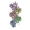
- EMDB-76301: Two gelsolin domains G2G3 bound to F-actin -

+
Open data


ID or keywords:

Loading...

-
Basic information

Entry
Database: EMDB / ID: EMD-76301
TitleTwo gelsolin domains G2G3 bound to F-actin
Map data
Sample
  • Complex: F-actin bound to gelsolin domains G2 and G3
    • Complex: gelsolin
      • Protein or peptide: Gelsolin
    • Complex: F-actin
      • Protein or peptide: Actin, alpha skeletal muscle
  • Ligand: ADENOSINE-5'-DIPHOSPHATE
  • Ligand: MAGNESIUM ION
  • Ligand: CALCIUM ION
Keywordsgelsolin / actin / STRUCTURAL PROTEIN
Function / homology
Function and homology information


striated muscle atrophy / regulation of establishment of T cell polarity / regulation of plasma membrane raft polarization / regulation of receptor clustering / positive regulation of keratinocyte apoptotic process / renal protein absorption / positive regulation of protein processing in phagocytic vesicle / phosphatidylinositol 3-kinase catalytic subunit binding / positive regulation of actin nucleation / actin cap ...striated muscle atrophy / regulation of establishment of T cell polarity / regulation of plasma membrane raft polarization / regulation of receptor clustering / positive regulation of keratinocyte apoptotic process / renal protein absorption / positive regulation of protein processing in phagocytic vesicle / phosphatidylinositol 3-kinase catalytic subunit binding / positive regulation of actin nucleation / actin cap / myosin II binding / host-mediated suppression of symbiont invasion / actin filament severing / barbed-end actin filament capping / cell projection assembly / actin polymerization or depolymerization / actin filament depolymerization / actin filament capping / relaxation of cardiac muscle / Sensory processing of sound by outer hair cells of the cochlea / phagocytosis, engulfment / cardiac muscle cell contraction / cytoskeletal motor activator activity / myosin heavy chain binding / hepatocyte apoptotic process / tropomyosin binding / actin filament bundle / troponin I binding / filamentous actin / mesenchyme migration / skeletal muscle myofibril / sarcoplasm / actin filament bundle assembly / striated muscle thin filament / skeletal muscle thin filament assembly / cilium assembly / actin monomer binding / Caspase-mediated cleavage of cytoskeletal proteins / skeletal muscle fiber development / phagocytic vesicle / response to muscle stretch / stress fiber / titin binding / actin filament polymerization / phosphatidylinositol-4,5-bisphosphate binding / actin filament organization / central nervous system development / filopodium / actin filament / protein destabilization / cellular response to type II interferon / Hydrolases; Acting on acid anhydrides; Acting on acid anhydrides to facilitate cellular and subcellular movement / calcium-dependent protein binding / actin filament binding / lamellipodium / actin cytoskeleton / actin binding / cell body / secretory granule lumen / blood microparticle / amyloid fibril formation / ficolin-1-rich granule lumen / Amyloid fiber formation / protein domain specific binding / focal adhesion / hydrolase activity / calcium ion binding / Neutrophil degranulation / positive regulation of gene expression / magnesium ion binding / : / extracellular exosome / extracellular region / ATP binding / identical protein binding / plasma membrane / cytoplasm / cytosol
Similarity search - Function
Villin/Gelsolin / Gelsolin homology domain / Gelsolin-like domain / Gelsolin repeat / ADF-H/Gelsolin-like domain superfamily / Actins signature 1. / Actin, conserved site / Actins signature 2. / Actin/actin-like conserved site / Actins and actin-related proteins signature. ...Villin/Gelsolin / Gelsolin homology domain / Gelsolin-like domain / Gelsolin repeat / ADF-H/Gelsolin-like domain superfamily / Actins signature 1. / Actin, conserved site / Actins signature 2. / Actin/actin-like conserved site / Actins and actin-related proteins signature. / Actin / Actin family / Actin / ATPase, nucleotide binding domain
Similarity search - Domain/homology
Gelsolin / Actin, alpha skeletal muscle
Similarity search - Component
Biological speciesHomo sapiens (human) / Oryctolagus cuniculus (rabbit)
Methodsingle particle reconstruction / cryo EM / Resolution: 3.44 Å
AuthorsSaks AJ / Dominguez R
Funding support United States, 3 items
OrganizationGrant numberCountry
National Institutes of Health/National Institute of General Medical Sciences (NIH/NIGMS)5R01GM152412-02 United States
National Institutes of Health/National Institute of General Medical Sciences (NIH/NIGMS)5R01GM073791-20 United States
National Institutes of Health/National Institute of General Medical Sciences (NIH/NIGMS)5T32GM132039-07 United States
CitationJournal: To Be Published
Title: Domains G2G3 Prime the Actin Filament for Severing by Gelsolin
Authors: Saks AJ / Boczkowska M / Barrie KR / Robinson RC / Dominguez R
History
DepositionMar 26, 2026-
Header (metadata) releaseJul 8, 2026-
Map releaseJul 8, 2026-
UpdateJul 8, 2026-
Current statusJul 8, 2026Processing site: RCSB / Status: Released

-
Structure visualization

Supplemental images

Downloads & links

-
Map

FileReleased
Projections & slices

Image control

Size
Brightness
Contrast
Others
AxesZ (Sec.)Y (Row.)X (Col.)
1.08 Å/pix.
x 320 pix.
= 345.6 Å
1.08 Å/pix.
x 320 pix.
= 345.6 Å
1.08 Å/pix.
x 320 pix.
= 345.6 Å

Surface

Projections

Slices (1/3)

Slices (1/2)

Slices (2/3)

Images are generated by Spider.

Voxel sizeX=Y=Z: 1.08 Å
Density
Contour LevelBy AUTHOR: 0.045
Minimum - Maximum-0.11636227 - 0.23962225
Average (Standard dev.)0.00010876341 (±0.008680216)
SymmetrySpace group: 1
Details

EMDB XML:

Map geometry
Axis orderXYZ
Origin000
Dimensions320320320
Spacing320320320
CellA=B=C: 345.6 Å
α=β=γ: 90.0 °

-
Supplemental data

-
Half map: #2

Fileemd_76301_half_map_1.map
Projections & Slices
AxesZYX

Projections

Slices (1/2)
Density Histograms

-
Half map: #1

Fileemd_76301_half_map_2.map
Projections & Slices
AxesZYX

Projections

Slices (1/2)
Density Histograms

-
Sample components

-
Entire : F-actin bound to gelsolin domains G2 and G3

EntireName: F-actin bound to gelsolin domains G2 and G3
Components
  • Complex: F-actin bound to gelsolin domains G2 and G3
    • Complex: gelsolin
      • Protein or peptide: Gelsolin
    • Complex: F-actin
      • Protein or peptide: Actin, alpha skeletal muscle
  • Ligand: ADENOSINE-5'-DIPHOSPHATE
  • Ligand: MAGNESIUM ION
  • Ligand: CALCIUM ION

-
Supramolecule #1: F-actin bound to gelsolin domains G2 and G3

SupramoleculeName: F-actin bound to gelsolin domains G2 and G3 / type: complex / ID: 1 / Parent: 0 / Macromolecule list: #1-#2
Molecular weightTheoretical: 1.5 kDa/nm

-
Supramolecule #2: gelsolin

SupramoleculeName: gelsolin / type: complex / ID: 2 / Parent: 1 / Macromolecule list: #2
Source (natural)Organism: Homo sapiens (human)

-
Supramolecule #3: F-actin

SupramoleculeName: F-actin / type: complex / ID: 3 / Parent: 1 / Macromolecule list: #1
Source (natural)Organism: Oryctolagus cuniculus (rabbit) / Tissue: Skeletal muscle

-
Macromolecule #1: Actin, alpha skeletal muscle

MacromoleculeName: Actin, alpha skeletal muscle / type: protein_or_peptide / ID: 1 / Number of copies: 8 / Enantiomer: LEVO
EC number: Hydrolases; Acting on acid anhydrides; Acting on acid anhydrides to facilitate cellular and subcellular movement
Source (natural)Organism: Oryctolagus cuniculus (rabbit)
Molecular weightTheoretical: 41.875633 KDa
SequenceString: DEDETTALVC DNGSGLVKAG FAGDDAPRAV FPSIVGRPRH QGVMVGMGQK DSYVGDEAQS KRGILTLKYP IE(HIC)GII TNW DDMEKIWHHT FYNELRVAPE EHPTLLTEAP LNPKANREKM TQIMFETFNV PAMYVAIQAV LSLYASGRTT GIVLDSG DG VTHNVPIYEG ...String:
DEDETTALVC DNGSGLVKAG FAGDDAPRAV FPSIVGRPRH QGVMVGMGQK DSYVGDEAQS KRGILTLKYP IE(HIC)GII TNW DDMEKIWHHT FYNELRVAPE EHPTLLTEAP LNPKANREKM TQIMFETFNV PAMYVAIQAV LSLYASGRTT GIVLDSG DG VTHNVPIYEG YALPHAIMRL DLAGRDLTDY LMKILTERGY SFVTTAEREI VRDIKEKLCY VALDFENEMA TAASSSSL E KSYELPDGQV ITIGNERFRC PETLFQPSFI GMESAGIHET TYNSIMKCDI DIRKDLYANN VMSGGTTMYP GIADRMQKE ITALAPSTMK IKIIAPPERK YSVWIGGSIL ASLSTFQQMW ITKQEYDEAG PSIVHRKCF

UniProtKB: Actin, alpha skeletal muscle

-
Macromolecule #2: Gelsolin

MacromoleculeName: Gelsolin / type: protein_or_peptide / ID: 2 / Number of copies: 2 / Enantiomer: LEVO
Source (natural)Organism: Homo sapiens (human)
Molecular weightTheoretical: 27.451873 KDa
Recombinant expressionOrganism: Escherichia coli (E. coli)
SequenceString: SGFKHVVPNE VVVQRLFQVK GRRVVRATEV PVSWESFNNG DCFILDLGNN IHQWCGSNSN RYERLKATQV SKGIRDNERS GRARVHVSE EGTEPEAMLQ VLGPKPALPA GTEDTAKEDA ANRKLAKLYK VSNGAGTMSV SLVADENPFA QGALKSEDCF I LDHGKDGK ...String:
SGFKHVVPNE VVVQRLFQVK GRRVVRATEV PVSWESFNNG DCFILDLGNN IHQWCGSNSN RYERLKATQV SKGIRDNERS GRARVHVSE EGTEPEAMLQ VLGPKPALPA GTEDTAKEDA ANRKLAKLYK VSNGAGTMSV SLVADENPFA QGALKSEDCF I LDHGKDGK IFVWKGKQAN TEERKAALKT ASDFITKMDY PKQTQVSVLP EGGETPLFKQ FFKNWRDPDQ TDGLGLSYLS WS HPQFEK

UniProtKB: Gelsolin

-
Macromolecule #3: ADENOSINE-5'-DIPHOSPHATE

MacromoleculeName: ADENOSINE-5'-DIPHOSPHATE / type: ligand / ID: 3 / Number of copies: 8 / Formula: ADP
Molecular weightTheoretical: 427.201 Da
Chemical component information

ChemComp-ADP:
ADENOSINE-5'-DIPHOSPHATE / ADP, energy-carrying molecule*YM

-
Macromolecule #4: MAGNESIUM ION

MacromoleculeName: MAGNESIUM ION / type: ligand / ID: 4 / Number of copies: 8 / Formula: MG
Molecular weightTheoretical: 24.305 Da

-
Macromolecule #5: CALCIUM ION

MacromoleculeName: CALCIUM ION / type: ligand / ID: 5 / Number of copies: 4 / Formula: CA
Molecular weightTheoretical: 40.078 Da

-
Experimental details

-
Structure determination

Methodcryo EM
Processingsingle particle reconstruction
Aggregation statefilament

-
Sample preparation

Concentration0.42 mg/mL
BufferpH: 7.5
Component:
ConcentrationFormulaName
20.0 mMC8H18N2O4SHEPES
50.0 mMNaClSodium chloride
2.0 mMCaCl2calcium chloride
2.0 mMMgCl2magnesium chloride
0.2 mMATPadenosine triphosphate
VitrificationCryogen name: ETHANE / Chamber humidity: 100 % / Chamber temperature: 277 K / Instrument: FEI VITROBOT MARK IV

-
Electron microscopy

MicroscopeTFS KRIOS
Image recordingFilm or detector model: GATAN K3 (6k x 4k) / Average electron dose: 50.0 e/Å2
Electron beamAcceleration voltage: 300 kV / Electron source: FIELD EMISSION GUN
Electron opticsIllumination mode: FLOOD BEAM / Imaging mode: BRIGHT FIELD / Nominal defocus max: 2.5 µm / Nominal defocus min: 0.5 µm
Experimental equipment
Model: Titan Krios / Image courtesy: FEI Company

+
Image processing

CTF correctionType: NONE
Startup modelType of model: NONE
Final reconstructionResolution.type: BY AUTHOR / Resolution: 3.44 Å / Resolution method: FSC 0.143 CUT-OFF / Software - Name: cryoSPARC / Number images used: 39272
Initial angle assignmentType: NOT APPLICABLE
Final angle assignmentType: NOT APPLICABLE
FSC plot (resolution estimation)

-
Atomic model buiding 1

Initial model
PDB IDChain

source_name: PDB, initial_model_type: experimental model

source_name: PDB, initial_model_type: experimental model
Output model

PDB-12bz:
Two gelsolin domains G2G3 bound to F-actin

+
About Yorodumi

-
News

-
Feb 9, 2022. New format data for meta-information of EMDB entries

New format data for meta-information of EMDB entries

  • Version 3 of the EMDB header file is now the official format.
  • The previous official version 1.9 will be removed from the archive.

Related info.:EMDB header

External links:wwPDB to switch to version 3 of the EMDB data model

-
Aug 12, 2020. Covid-19 info

Covid-19 info

URL: https://pdbj.org/emnavi/covid19.php

New page: Covid-19 featured information page in EM Navigator.

Related info.:Covid-19 info / Mar 5, 2020. Novel coronavirus structure data

+
Mar 5, 2020. Novel coronavirus structure data

Novel coronavirus structure data

Related info.:Yorodumi Speices / Aug 12, 2020. Covid-19 info

External links:COVID-19 featured content - PDBj / Molecule of the Month (242):Coronavirus Proteases

+
Jan 31, 2019. EMDB accession codes are about to change! (news from PDBe EMDB page)

EMDB accession codes are about to change! (news from PDBe EMDB page)

  • The allocation of 4 digits for EMDB accession codes will soon come to an end. Whilst these codes will remain in use, new EMDB accession codes will include an additional digit and will expand incrementally as the available range of codes is exhausted. The current 4-digit format prefixed with “EMD-” (i.e. EMD-XXXX) will advance to a 5-digit format (i.e. EMD-XXXXX), and so on. It is currently estimated that the 4-digit codes will be depleted around Spring 2019, at which point the 5-digit format will come into force.
  • The EM Navigator/Yorodumi systems omit the EMD- prefix.

Related info.:Q: What is EMD? / ID/Accession-code notation in Yorodumi/EM Navigator

External links:EMDB Accession Codes are Changing Soon! / Contact to PDBj

+
Jul 12, 2017. Major update of PDB

Major update of PDB

  • wwPDB released updated PDB data conforming to the new PDBx/mmCIF dictionary.
  • This is a major update changing the version number from 4 to 5, and with Remediation, in which all the entries are updated.
  • In this update, many items about electron microscopy experimental information are reorganized (e.g. em_software).
  • Now, EM Navigator and Yorodumi are based on the updated data.

External links:wwPDB Remediation / Enriched Model Files Conforming to OneDep Data Standards Now Available in the PDB FTP Archive

-
Yorodumi

Thousand views of thousand structures

  • Yorodumi is a browser for structure data from EMDB, PDB, SASBDB, etc.
  • This page is also the successor to EM Navigator detail page, and also detail information page/front-end page for Omokage search.
  • The word "yorodu" (or yorozu) is an old Japanese word meaning "ten thousand". "mi" (miru) is to see.

Related info.:EMDB / PDB / SASBDB / Comparison of 3 databanks / Yorodumi Search / Aug 31, 2016. New EM Navigator & Yorodumi / Yorodumi Papers / Jmol/JSmol / Function and homology information / Changes in new EM Navigator and Yorodumi

Read more