[English] 日本語
Yorodumi
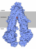
- EMDB-75839: E. coli SufE bound to SufBC2D -

+
Open data


ID or keywords:

Loading...

-
Basic information

Entry
Database: EMDB / ID: EMD-75839
TitleE. coli SufE bound to SufBC2D
Map data
Sample
  • Complex: SufBC2D-SufE complex
    • Protein or peptide: Iron-sulfur cluster assembly protein SufB
    • Protein or peptide: Cysteine desulfuration protein SufE
    • Protein or peptide: Probable ATP-dependent transporter SufC
    • Protein or peptide: Iron-sulfur cluster assembly protein SufD
KeywordsFe-S / Suf pathway / persulfide / iron-sulfur / sulfur transfer / METAL BINDING PROTEIN
Function / homology
Function and homology information


metallo-sulfur cluster assembly / sulfurtransferase complex / sulfur compound metabolic process / sulfur carrier activity / iron-sulfur cluster assembly complex / iron-sulfur cluster assembly / response to radiation / 2 iron, 2 sulfur cluster binding / enzyme activator activity / 4 iron, 4 sulfur cluster binding ...metallo-sulfur cluster assembly / sulfurtransferase complex / sulfur compound metabolic process / sulfur carrier activity / iron-sulfur cluster assembly complex / iron-sulfur cluster assembly / response to radiation / 2 iron, 2 sulfur cluster binding / enzyme activator activity / 4 iron, 4 sulfur cluster binding / response to oxidative stress / ATP hydrolysis activity / ATP binding / cytosol
Similarity search - Function
SUF system FeS cluster assembly, SufB / SUF system FeS cluster assembly, SufBD, N-terminal / SufBD protein N-terminal region / Cysteine desulfuration protein SufE / SUF system FeS cluster assembly, SufD / SUF system FeS cluster assembly, SufBD / Fe-S metabolism associated domain, SufE-like / SUF system FeS cluster assembly, SufBD superfamily / : / SUF system FeS cluster assembly, SufBD core domain ...SUF system FeS cluster assembly, SufB / SUF system FeS cluster assembly, SufBD, N-terminal / SufBD protein N-terminal region / Cysteine desulfuration protein SufE / SUF system FeS cluster assembly, SufD / SUF system FeS cluster assembly, SufBD / Fe-S metabolism associated domain, SufE-like / SUF system FeS cluster assembly, SufBD superfamily / : / SUF system FeS cluster assembly, SufBD core domain / Fe-S metabolism associated domain / FeS cluster assembly SUF system, ATPase SufC / ABC transporter-like, conserved site / ABC transporters family signature. / ABC transporter / ABC transporter-like, ATP-binding domain / ATP-binding cassette, ABC transporter-type domain profile. / ATPases associated with a variety of cellular activities / AAA+ ATPase domain / P-loop containing nucleoside triphosphate hydrolase
Similarity search - Domain/homology
Cysteine desulfuration protein SufE / Probable ATP-dependent transporter SufC / Iron-sulfur cluster assembly protein SufB / Iron-sulfur cluster assembly protein SufD
Similarity search - Component
Biological speciesEscherichia coli (E. coli)
Methodsingle particle reconstruction / cryo EM / Resolution: 4.21 Å
AuthorsChhikara N / Dunkle JA / Frantom PA
Funding support United States, 2 items
OrganizationGrant numberCountry
National Institutes of Health/National Institute of General Medical Sciences (NIH/NIGMS)R01GM112919 United States
National Institutes of Health/National Institute of General Medical Sciences (NIH/NIGMS)R35GM142966 United States
CitationJournal: Acta Crystallogr D Struct Biol / Year: 2019
Title: Macromolecular structure determination using X-rays, neutrons and electrons: recent developments in Phenix.
Authors: Dorothee Liebschner / Pavel V Afonine / Matthew L Baker / Gábor Bunkóczi / Vincent B Chen / Tristan I Croll / Bradley Hintze / Li Wei Hung / Swati Jain / Airlie J McCoy / Nigel W Moriarty ...Authors: Dorothee Liebschner / Pavel V Afonine / Matthew L Baker / Gábor Bunkóczi / Vincent B Chen / Tristan I Croll / Bradley Hintze / Li Wei Hung / Swati Jain / Airlie J McCoy / Nigel W Moriarty / Robert D Oeffner / Billy K Poon / Michael G Prisant / Randy J Read / Jane S Richardson / David C Richardson / Massimo D Sammito / Oleg V Sobolev / Duncan H Stockwell / Thomas C Terwilliger / Alexandre G Urzhumtsev / Lizbeth L Videau / Christopher J Williams / Paul D Adams /
Abstract: Diffraction (X-ray, neutron and electron) and electron cryo-microscopy are powerful methods to determine three-dimensional macromolecular structures, which are required to understand biological ...Diffraction (X-ray, neutron and electron) and electron cryo-microscopy are powerful methods to determine three-dimensional macromolecular structures, which are required to understand biological processes and to develop new therapeutics against diseases. The overall structure-solution workflow is similar for these techniques, but nuances exist because the properties of the reduced experimental data are different. Software tools for structure determination should therefore be tailored for each method. Phenix is a comprehensive software package for macromolecular structure determination that handles data from any of these techniques. Tasks performed with Phenix include data-quality assessment, map improvement, model building, the validation/rebuilding/refinement cycle and deposition. Each tool caters to the type of experimental data. The design of Phenix emphasizes the automation of procedures, where possible, to minimize repetitive and time-consuming manual tasks, while default parameters are chosen to encourage best practice. A graphical user interface provides access to many command-line features of Phenix and streamlines the transition between programs, project tracking and re-running of previous tasks.
History
DepositionMar 5, 2026-
Header (metadata) releaseJul 15, 2026-
Map releaseJul 15, 2026-
UpdateJul 15, 2026-
Current statusJul 15, 2026Processing site: RCSB / Status: Released

-
Structure visualization

Supplemental images

Downloads & links

-
Map

FileDownload / File: emd_75839.map.gz / Format: CCP4 / Size: 244.1 MB / Type: IMAGE STORED AS FLOATING POINT NUMBER (4 BYTES)
Projections & slices

Image control

Size
Brightness
Contrast
Others
AxesZ (Sec.)Y (Row.)X (Col.)
0.73 Å/pix.
x 400 pix.
= 292. Å
0.73 Å/pix.
x 400 pix.
= 292. Å
0.73 Å/pix.
x 400 pix.
= 292. Å

Surface

Projections

Slices (1/3)

Slices (1/2)

Slices (2/3)

Images are generated by Spider.

Voxel sizeX=Y=Z: 0.73 Å
Density
Contour LevelBy AUTHOR: 0.015
Minimum - Maximum-0.08761956 - 0.14602815
Average (Standard dev.)-0.00006992974 (±0.0044832416)
SymmetrySpace group: 1
Details

EMDB XML:

Map geometry
Axis orderXYZ
Origin000
Dimensions400400400
Spacing400400400
CellA=B=C: 292.0 Å
α=β=γ: 90.0 °

-
Supplemental data

-
Mask #1

Fileemd_75839_msk_1.map
Projections & Slices
AxesZYX

Projections

Slices (1/2)
Density Histograms

-
Half map: #2

Fileemd_75839_half_map_1.map
Projections & Slices
AxesZYX

Projections

Slices (1/2)
Density Histograms

-
Half map: #1

Fileemd_75839_half_map_2.map
Projections & Slices
AxesZYX

Projections

Slices (1/2)
Density Histograms

-
Sample components

-
Entire : SufBC2D-SufE complex

EntireName: SufBC2D-SufE complex
Components
  • Complex: SufBC2D-SufE complex
    • Protein or peptide: Iron-sulfur cluster assembly protein SufB
    • Protein or peptide: Cysteine desulfuration protein SufE
    • Protein or peptide: Probable ATP-dependent transporter SufC
    • Protein or peptide: Iron-sulfur cluster assembly protein SufD

-
Supramolecule #1: SufBC2D-SufE complex

SupramoleculeName: SufBC2D-SufE complex / type: complex / ID: 1 / Parent: 0 / Macromolecule list: all
Source (natural)Organism: Escherichia coli (E. coli)
Molecular weightTheoretical: 174 KDa

-
Macromolecule #1: Iron-sulfur cluster assembly protein SufB

MacromoleculeName: Iron-sulfur cluster assembly protein SufB / type: protein_or_peptide / ID: 1 / Number of copies: 1 / Enantiomer: LEVO
Source (natural)Organism: Escherichia coli (E. coli)
Molecular weightTheoretical: 56.396637 KDa
Recombinant expressionOrganism: Escherichia coli (E. coli)
SequenceString: MGSSHHHHHH SQDPNSSRNT EATDDVKTWT GGPLNYKEGF FTQLATDELA KGINEEVVRA ISAKRNEPEW MLEFRLNAYR AWLEMEEPH WLKAHYDKLN YQDYSYYSAP SCGNCDDTCA SEPGAVQQTG ANAFLSKEVE AAFEQLGVPV REGKEVAVDA I FDSVSVAT ...String:
MGSSHHHHHH SQDPNSSRNT EATDDVKTWT GGPLNYKEGF FTQLATDELA KGINEEVVRA ISAKRNEPEW MLEFRLNAYR AWLEMEEPH WLKAHYDKLN YQDYSYYSAP SCGNCDDTCA SEPGAVQQTG ANAFLSKEVE AAFEQLGVPV REGKEVAVDA I FDSVSVAT TYREKLAEQG IIFCSFGEAI HDHPELVRKY LGTVVPGNDN FFAALNAAVA SDGTFIYVPK GVRCPMELST AF RINAEKT GQFERTILVA DEDSYVSYIE GCSAPVRDSY QLHAAVVEVI IHKNAEVKYS TVQNWFPGDN NTGGILNFVT KRA LCEGEN SKMSWTQSET GSAITWKYPS CILRGDNSIG EFYSVALTSG HQQADTGTKM IHIGKNTKST IISKGISAGH SQNS YRGLV KIMPTATNAR NFTQCDSMLI GANCGAHTFP YVECRNNSAQ LEHEATTSRI GEDQLFYCLQ RGISEEDAIS MIVNG FCKD VFSELPLEFA VEAQKLLAIS LEHSVG

UniProtKB: Iron-sulfur cluster assembly protein SufB

-
Macromolecule #2: Cysteine desulfuration protein SufE

MacromoleculeName: Cysteine desulfuration protein SufE / type: protein_or_peptide / ID: 2 / Number of copies: 1 / Enantiomer: LEVO
Source (natural)Organism: Escherichia coli (E. coli)
Molecular weightTheoretical: 15.817281 KDa
Recombinant expressionOrganism: Escherichia coli (E. coli)
SequenceString:
MALLPDKEKL LRNFLRCANW EEKYLYIIEL GQRLPELRDE DRSPQNSIQG CQSQVWIVMR QNAQGIIELQ GDSDAAIVKG LIAVVFILY DQMTPQDIVN FDVRPWFEKM ALTQHLTPSR SQGLEAMIRA IRAKAAALS

UniProtKB: Cysteine desulfuration protein SufE

-
Macromolecule #3: Probable ATP-dependent transporter SufC

MacromoleculeName: Probable ATP-dependent transporter SufC / type: protein_or_peptide / ID: 3 / Number of copies: 2 / Enantiomer: LEVO
Source (natural)Organism: Escherichia coli (E. coli)
Molecular weightTheoretical: 27.613332 KDa
Recombinant expressionOrganism: Escherichia coli (E. coli)
SequenceString: MLSIKDLHVS VEDKAILRGL SLDVHPGEVH AIMGPNGSGK STLSATLAGR EDYEVTGGTV EFKGKDLLAL SPEDRAGEGI FMAFQYPVE IPGVSNQFFL QTALNAVRSY RGQETLDRFD FQDLMEEKIA LLKMPEDLLT RSVNVGFSGG EKKRNDILQM A VLEPELCI ...String:
MLSIKDLHVS VEDKAILRGL SLDVHPGEVH AIMGPNGSGK STLSATLAGR EDYEVTGGTV EFKGKDLLAL SPEDRAGEGI FMAFQYPVE IPGVSNQFFL QTALNAVRSY RGQETLDRFD FQDLMEEKIA LLKMPEDLLT RSVNVGFSGG EKKRNDILQM A VLEPELCI LDESDSGLDI DALKVVADGV NSLRDGKRSF IIVTHYQRIL DYIKPDYVHV LYQGRIVKSG DFTLVKQLEE QG YGWLTEQ Q

UniProtKB: Probable ATP-dependent transporter SufC

-
Macromolecule #4: Iron-sulfur cluster assembly protein SufD

MacromoleculeName: Iron-sulfur cluster assembly protein SufD / type: protein_or_peptide / ID: 4 / Number of copies: 1 / Enantiomer: LEVO
Source (natural)Organism: Escherichia coli (E. coli)
Molecular weightTheoretical: 46.884641 KDa
Recombinant expressionOrganism: Escherichia coli (E. coli)
SequenceString: MAGLPNSSNA LQQWHHLFEA EGTKRSPQAQ QHLQQLLRTG LPTRKHENWK YTPLEGLINS QFVSIAGEIS PQQRDALALT LDSVRLVFV DGRYVPALSD ATEGSGYEVS INDDRQGLPD AIQAEVFLHL TESLAQSVTH IAVKRGQRPA KPLLLMHITQ G VAGEEVNT ...String:
MAGLPNSSNA LQQWHHLFEA EGTKRSPQAQ QHLQQLLRTG LPTRKHENWK YTPLEGLINS QFVSIAGEIS PQQRDALALT LDSVRLVFV DGRYVPALSD ATEGSGYEVS INDDRQGLPD AIQAEVFLHL TESLAQSVTH IAVKRGQRPA KPLLLMHITQ G VAGEEVNT AHYRHHLDLA EGAEATVIEH FVSLNDARHF TGARFTINVA ANAHLQHIKL AFENPLSHHF AHNDLLLAED AT AFSHSFL LGGAVLRHNT STQLNGENST LRINSLAMPV KNEVCDTRTW LEHNKGFCNS RQLHKTIVSD KGRAVFNGLI NVA QHAIKT DGQMTNNNLL MGKLAEVDTK PQLEIYADDV KCSHGATVGR IDDEQIFYLR SRGINQQDAQ QMIIYAFAAE LTEA LRDEG LKQQVLARIG QRLPGGAR

UniProtKB: Iron-sulfur cluster assembly protein SufD

-
Experimental details

-
Structure determination

Methodcryo EM
Processingsingle particle reconstruction
Aggregation stateparticle

-
Sample preparation

BufferpH: 8
VitrificationCryogen name: ETHANE

-
Electron microscopy

MicroscopeTFS GLACIOS
Image recordingFilm or detector model: FEI FALCON IV (4k x 4k) / Average electron dose: 60.0 e/Å2
Electron beamAcceleration voltage: 200 kV / Electron source: FIELD EMISSION GUN
Electron opticsIllumination mode: FLOOD BEAM / Imaging mode: BRIGHT FIELD / Nominal defocus max: 3.341 µm / Nominal defocus min: 0.1 µm

+
Image processing

CTF correctionType: PHASE FLIPPING AND AMPLITUDE CORRECTION
Startup modelType of model: INSILICO MODEL
Final reconstructionResolution.type: BY AUTHOR / Resolution: 4.21 Å / Resolution method: FSC 0.143 CUT-OFF / Software - Name: cryoSPARC (ver. 4.6) / Number images used: 76000
Initial angle assignmentType: MAXIMUM LIKELIHOOD
Final angle assignmentType: MAXIMUM LIKELIHOOD
FSC plot (resolution estimation)

+
About Yorodumi

-
News

-
Feb 9, 2022. New format data for meta-information of EMDB entries

New format data for meta-information of EMDB entries

  • Version 3 of the EMDB header file is now the official format.
  • The previous official version 1.9 will be removed from the archive.

Related info.:EMDB header

External links:wwPDB to switch to version 3 of the EMDB data model

-
Aug 12, 2020. Covid-19 info

Covid-19 info

URL: https://pdbj.org/emnavi/covid19.php

New page: Covid-19 featured information page in EM Navigator.

Related info.:Covid-19 info / Mar 5, 2020. Novel coronavirus structure data

+
Mar 5, 2020. Novel coronavirus structure data

Novel coronavirus structure data

Related info.:Yorodumi Speices / Aug 12, 2020. Covid-19 info

External links:COVID-19 featured content - PDBj / Molecule of the Month (242):Coronavirus Proteases

+
Jan 31, 2019. EMDB accession codes are about to change! (news from PDBe EMDB page)

EMDB accession codes are about to change! (news from PDBe EMDB page)

  • The allocation of 4 digits for EMDB accession codes will soon come to an end. Whilst these codes will remain in use, new EMDB accession codes will include an additional digit and will expand incrementally as the available range of codes is exhausted. The current 4-digit format prefixed with “EMD-” (i.e. EMD-XXXX) will advance to a 5-digit format (i.e. EMD-XXXXX), and so on. It is currently estimated that the 4-digit codes will be depleted around Spring 2019, at which point the 5-digit format will come into force.
  • The EM Navigator/Yorodumi systems omit the EMD- prefix.

Related info.:Q: What is EMD? / ID/Accession-code notation in Yorodumi/EM Navigator

External links:EMDB Accession Codes are Changing Soon! / Contact to PDBj

+
Jul 12, 2017. Major update of PDB

Major update of PDB

  • wwPDB released updated PDB data conforming to the new PDBx/mmCIF dictionary.
  • This is a major update changing the version number from 4 to 5, and with Remediation, in which all the entries are updated.
  • In this update, many items about electron microscopy experimental information are reorganized (e.g. em_software).
  • Now, EM Navigator and Yorodumi are based on the updated data.

External links:wwPDB Remediation / Enriched Model Files Conforming to OneDep Data Standards Now Available in the PDB FTP Archive

-
Yorodumi

Thousand views of thousand structures

  • Yorodumi is a browser for structure data from EMDB, PDB, SASBDB, etc.
  • This page is also the successor to EM Navigator detail page, and also detail information page/front-end page for Omokage search.
  • The word "yorodu" (or yorozu) is an old Japanese word meaning "ten thousand". "mi" (miru) is to see.

Related info.:EMDB / PDB / SASBDB / Comparison of 3 databanks / Yorodumi Search / Aug 31, 2016. New EM Navigator & Yorodumi / Yorodumi Papers / Jmol/JSmol / Function and homology information / Changes in new EM Navigator and Yorodumi

Read more