Movie
Movie Controller

[English] 日本語
 Yorodumi
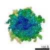
- EMDB-73311: GTPBP1*GDP*Phe-tRNA*ribosome in the post-GTP hydrolysis state, St... -
+ Open data

- Basic information
| Entry |  | |||||||||
|---|---|---|---|---|---|---|---|---|---|---|
| Title | GTPBP1*GDP*Phe-tRNA*ribosome in the post-GTP hydrolysis state, Structure IV | |||||||||
 Map data Map data | ||||||||||
 Sample Sample |
| |||||||||
 Keywords Keywords | GTPBP1 / tRNA / GCP / complex / RIBOSOME | |||||||||
| Function / homology |  Function and homology information Function and homology informationalpha-aminoacyl-tRNA binding / RNA surveillance / cytoplasmic exosome (RNase complex) / positive regulation of mRNA catabolic process / GTP metabolic process / protein-synthesizing GTPase / laminin receptor activity / translational elongation / 90S preribosome / ubiquitin ligase inhibitor activity ...alpha-aminoacyl-tRNA binding / RNA surveillance / cytoplasmic exosome (RNase complex) / positive regulation of mRNA catabolic process / GTP metabolic process / protein-synthesizing GTPase / laminin receptor activity / translational elongation / 90S preribosome / ubiquitin ligase inhibitor activity / translation elongation factor activity / positive regulation of signal transduction by p53 class mediator / phagocytic cup / protein-RNA complex assembly / translation regulator activity / rough endoplasmic reticulum / ribosomal small subunit export from nucleus / laminin binding / gastrulation / MDM2/MDM4 family protein binding / class I DNA-(apurinic or apyrimidinic site) endonuclease activity / cytosolic ribosome / DNA-(apurinic or apyrimidinic site) lyase / ribosomal large subunit biogenesis / maturation of LSU-rRNA from tricistronic rRNA transcript (SSU-rRNA, 5.8S rRNA, LSU-rRNA) / maturation of SSU-rRNA from tricistronic rRNA transcript (SSU-rRNA, 5.8S rRNA, LSU-rRNA) / positive regulation of apoptotic signaling pathway / maturation of SSU-rRNA / small-subunit processome / spindle / rRNA processing / rhythmic process / regulation of translation / positive regulation of canonical Wnt signaling pathway / antimicrobial humoral immune response mediated by antimicrobial peptide / heparin binding / large ribosomal subunit / ribosomal small subunit assembly / virus receptor activity / ribosome binding / ribosomal small subunit biogenesis / 5S rRNA binding / ribosomal large subunit assembly / small ribosomal subunit / small ribosomal subunit rRNA binding / cytosolic small ribosomal subunit / large ribosomal subunit rRNA binding / killing of cells of another organism / cytosolic large ribosomal subunit / defense response to Gram-negative bacterium / perikaryon / cell differentiation / cytoplasmic translation / tRNA binding / mitochondrial inner membrane / negative regulation of translation / postsynaptic density / rRNA binding / immune response / structural constituent of ribosome / ribosome / translation / ribonucleoprotein complex / cell division / DNA repair / mRNA binding / GTPase activity / apoptotic process / centrosome / synapse / dendrite / GTP binding / nucleolus / perinuclear region of cytoplasm / endoplasmic reticulum / Golgi apparatus / signal transduction / DNA binding / RNA binding / zinc ion binding / membrane / nucleus / plasma membrane / cytoplasm / cytosol Similarity search - Function | |||||||||
| Biological species |    Homo sapiens (human) Homo sapiens (human) | |||||||||
| Method | single particle reconstruction / cryo EM / Resolution: 2.9 Å | |||||||||
 Authors Authors | Susorov D / Korostelev AA | |||||||||
| Funding support |  United States, 2 items United States, 2 items
| |||||||||
 Citation Citation | Journal: Acta Crystallogr D Struct Biol / Year: 2019 Title: Macromolecular structure determination using X-rays, neutrons and electrons: recent developments in Phenix. Authors: Dorothee Liebschner / Pavel V Afonine / Matthew L Baker / Gábor Bunkóczi / Vincent B Chen / Tristan I Croll / Bradley Hintze / Li Wei Hung / Swati Jain / Airlie J McCoy / Nigel W Moriarty ...Authors: Dorothee Liebschner / Pavel V Afonine / Matthew L Baker / Gábor Bunkóczi / Vincent B Chen / Tristan I Croll / Bradley Hintze / Li Wei Hung / Swati Jain / Airlie J McCoy / Nigel W Moriarty / Robert D Oeffner / Billy K Poon / Michael G Prisant / Randy J Read / Jane S Richardson / David C Richardson / Massimo D Sammito / Oleg V Sobolev / Duncan H Stockwell / Thomas C Terwilliger / Alexandre G Urzhumtsev / Lizbeth L Videau / Christopher J Williams / Paul D Adams /   Abstract: Diffraction (X-ray, neutron and electron) and electron cryo-microscopy are powerful methods to determine three-dimensional macromolecular structures, which are required to understand biological ...Diffraction (X-ray, neutron and electron) and electron cryo-microscopy are powerful methods to determine three-dimensional macromolecular structures, which are required to understand biological processes and to develop new therapeutics against diseases. The overall structure-solution workflow is similar for these techniques, but nuances exist because the properties of the reduced experimental data are different. Software tools for structure determination should therefore be tailored for each method. Phenix is a comprehensive software package for macromolecular structure determination that handles data from any of these techniques. Tasks performed with Phenix include data-quality assessment, map improvement, model building, the validation/rebuilding/refinement cycle and deposition. Each tool caters to the type of experimental data. The design of Phenix emphasizes the automation of procedures, where possible, to minimize repetitive and time-consuming manual tasks, while default parameters are chosen to encourage best practice. A graphical user interface provides access to many command-line features of Phenix and streamlines the transition between programs, project tracking and re-running of previous tasks. | |||||||||
| History |
|
- Structure visualization
| Supplemental images |
|---|

- Downloads & links
-EMDB archive
| Map data | emd_73311.map.gz | 193.8 MB | EMDB map data format | |
|---|---|---|---|---|
| Header (meta data) | emd-73311-v30.xmlemd-73311.xml | 115.6 KB 115.6 KB | Display Display | EMDB header |
| FSC (resolution estimation) | emd_73311_fsc.xml | 14.1 KB | Display | FSC data file |
| Images |  emd_73311.png emd_73311.png | 146.8 KB | ||
| Filedesc metadata | emd-73311.cif.gz | 22 KB | ||
| Others | emd_73311_additional_1.map.gzemd_73311_additional_2.map.gzemd_73311_half_map_1.map.gzemd_73311_half_map_2.map.gz | 220.6 MB 193.7 MB 193.9 MB 193.8 MB | ||
| Archive directory |  http://ftp.pdbj.org/pub/emdb/structures/EMD-73311ftp://ftp.pdbj.org/pub/emdb/structures/EMD-73311 http://ftp.pdbj.org/pub/emdb/structures/EMD-73311ftp://ftp.pdbj.org/pub/emdb/structures/EMD-73311 | HTTPS FTP |
-Related structure data
| Related structure data |  9ypwMC  9ypgC  9ypoC  9ypsC  9yptC  9ypvC  9ypyC  9ypzC  9yq0C  9yq1C M: atomic model generated by this map C: citing same article ( |
|---|---|
| Similar structure data |


-Links
| EMDB pages | EMDB (EBI/PDBe) / EMDataResource |
|---|---|
| Related items in Molecule of the Month |


-Map
| File | Download / File: emd_73311.map.gz / Format: CCP4 / Size: 244.1 MB / Type: IMAGE STORED AS FLOATING POINT NUMBER (4 BYTES) | ||||||||||||||||||||||||||||||||||||
|---|---|---|---|---|---|---|---|---|---|---|---|---|---|---|---|---|---|---|---|---|---|---|---|---|---|---|---|---|---|---|---|---|---|---|---|---|---|
| Projections & slices | Image control
Images are generated by Spider. | ||||||||||||||||||||||||||||||||||||
| Voxel size | X=Y=Z: 1.167 Å | ||||||||||||||||||||||||||||||||||||
| Density |
| ||||||||||||||||||||||||||||||||||||
| Symmetry | Space group: 1 | ||||||||||||||||||||||||||||||||||||
| Details | EMDB XML:
|
 Z (Sec.)
 Y (Row.)
 X (Col.)


-Supplemental data
-Additional map: #1
| File | emd_73311_additional_1.map | ||||||||||||
|---|---|---|---|---|---|---|---|---|---|---|---|---|---|
| Projections & Slices |
| ||||||||||||
| Density Histograms |


-Additional map: #2
| File | emd_73311_additional_2.map | ||||||||||||
|---|---|---|---|---|---|---|---|---|---|---|---|---|---|
| Projections & Slices |
| ||||||||||||
| Density Histograms |


-Half map: #1
| File | emd_73311_half_map_1.map | ||||||||||||
|---|---|---|---|---|---|---|---|---|---|---|---|---|---|
| Projections & Slices |
| ||||||||||||
| Density Histograms |


-Half map: #2
| File | emd_73311_half_map_2.map | ||||||||||||
|---|---|---|---|---|---|---|---|---|---|---|---|---|---|
| Projections & Slices |
| ||||||||||||
| Density Histograms |


- Sample components
+Entire : GTPBP1*GDP*Phe-tRNA complex with 80S ribosome
+Supramolecule #1: GTPBP1*GDP*Phe-tRNA complex with 80S ribosome
+Macromolecule #1: 28S ribosomal RNA
+Macromolecule #2: 5S ribosomal RNA
+Macromolecule #3: 5.8S ribosomal RNA
+Macromolecule #4: 18S ribosomal RNA
+Macromolecule #79: MF mRNA
+Macromolecule #80: Phe-tRNA
+Macromolecule #81: Met-tRNA
+Macromolecule #5: Ribosomal protein L8
+Macromolecule #6: Ribosomal protein L3
+Macromolecule #7: 60S ribosomal protein L4
+Macromolecule #8: Large ribosomal subunit protein uL18
+Macromolecule #9: 60S ribosomal protein L6
+Macromolecule #10: 60S ribosomal protein L7a
+Macromolecule #11: 60S ribosomal protein L9
+Macromolecule #12: 60S ribosomal protein L10
+Macromolecule #13: Ribosomal protein L11
+Macromolecule #14: 60S ribosomal protein L7
+Macromolecule #15: 60S ribosomal protein L13
+Macromolecule #16: 60S ribosomal protein L14
+Macromolecule #17: Ribosomal protein L15
+Macromolecule #18: Large ribosomal subunit protein uL13
+Macromolecule #19: Large ribosomal subunit protein uL22
+Macromolecule #20: Ribosomal protein L18
+Macromolecule #21: Ribosomal protein L19
+Macromolecule #22: 60S ribosomal protein L18a
+Macromolecule #23: eL21
+Macromolecule #24: eL22
+Macromolecule #25: Ribosomal protein L23
+Macromolecule #26: eL24
+Macromolecule #27: eL23
+Macromolecule #28: uL24
+Macromolecule #29: 60S ribosomal protein L27
+Macromolecule #30: 60S ribosomal protein L27a
+Macromolecule #31: Large ribosomal subunit protein eL29
+Macromolecule #32: eL30
+Macromolecule #33: eL31
+Macromolecule #34: Ribosomal protein L32
+Macromolecule #35: eL33
+Macromolecule #36: Large ribosomal subunit protein eL34
+Macromolecule #37: eL35
+Macromolecule #38: 60S ribosomal protein L36
+Macromolecule #39: eL38
+Macromolecule #40: eL39
+Macromolecule #41: Ubiquitin-ribosomal protein eL40 fusion protein
+Macromolecule #42: eL41
+Macromolecule #43: Large ribosomal subunit protein eL42
+Macromolecule #44: eL43
+Macromolecule #45: eL28
+Macromolecule #46: uS2 (SA)
+Macromolecule #47: 40S ribosomal protein S3a
+Macromolecule #48: Small ribosomal subunit protein uS5
+Macromolecule #49: Ribosomal protein S3
+Macromolecule #50: eS4 (S4 X isoform)
+Macromolecule #51: Ribosomal protein S5
+Macromolecule #52: 40S ribosomal protein S6
+Macromolecule #53: 40S ribosomal protein S7
+Macromolecule #54: 40S ribosomal protein S8
+Macromolecule #55: Ribosomal protein S9 (Predicted)
+Macromolecule #56: Small ribosomal subunit protein eS10
+Macromolecule #57: Ribosomal protein S11
+Macromolecule #58: 40S ribosomal protein S12
+Macromolecule #59: Ribosomal protein S13
+Macromolecule #60: Small ribosomal subunit protein uS11
+Macromolecule #61: uS19
+Macromolecule #62: uS9
+Macromolecule #63: eS17
+Macromolecule #64: uS13
+Macromolecule #65: eS19
+Macromolecule #66: uS10
+Macromolecule #67: eS21
+Macromolecule #68: Ribosomal protein S15a
+Macromolecule #69: uS12
+Macromolecule #70: Small ribosomal subunit protein eS24
+Macromolecule #71: eS25
+Macromolecule #72: 40S ribosomal protein S26
+Macromolecule #73: 40S ribosomal protein S27
+Macromolecule #74: Ribosomal protein S28
+Macromolecule #75: eS29
+Macromolecule #76: 40S ribosomal protein S30
+Macromolecule #77: Ribosomal protein S27a
+Macromolecule #78: RACK1
+Macromolecule #82: Ribosomal protein L37
+Macromolecule #83: GTP-binding protein 1

+Macromolecule #84: SPERMIDINE

+Macromolecule #85: ZINC ION
+Macromolecule #86: GUANOSINE-5'-TRIPHOSPHATE

+Macromolecule #87: PHENYLALANINE

+Macromolecule #88: ADENOSINE-5'-TRIPHOSPHATE

+Macromolecule #89: METHIONINE

+Macromolecule #90: POTASSIUM ION
+Macromolecule #91: GUANOSINE-5'-DIPHOSPHATE

+Macromolecule #92: MAGNESIUM ION
-Experimental details
-Structure determination
| Method | cryo EM |
|---|---|
 Processing Processing | single particle reconstruction |
| Aggregation state | particle |
-Sample preparation
| Buffer | pH: 7.5 |
|---|---|
| Vitrification | Cryogen name: ETHANE |
- Electron microscopy
| Microscope | TFS KRIOS |
|---|---|
| Image recording | Film or detector model: GATAN K3 BIOQUANTUM (6k x 4k) / Average electron dose: 29.7531 e/Å2 |
| Electron beam | Acceleration voltage: 300 kV / Electron source:  FIELD EMISSION GUN FIELD EMISSION GUN |
| Electron optics | Illumination mode: FLOOD BEAM / Imaging mode: BRIGHT FIELD / Nominal defocus max: 1.5 µm / Nominal defocus min: 0.5 µm |
| Experimental equipment |  Model: Titan Krios / Image courtesy: FEI Company |