 Movie
Movie Controller


+ Open data

- Basic information
| Entry |  | |||||||||
|---|---|---|---|---|---|---|---|---|---|---|
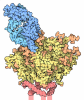
| Title | RSV-F SC-DM bound to a designed protein minibinder, cb13 | |||||||||
 Map data Map data | Unsharpened | |||||||||
 Sample Sample |
| |||||||||
 Keywords Keywords | RSV / SC-DM / RSV-F / DE NOVO PROTEIN / minibinder / glycoprotein / respiratory syncytial virus / cryoEM / cryo-EM / PROTEIN BINDING | |||||||||
| Function / homology |  Function and homology information Function and homology informationsymbiont-mediated induction of syncytium formation / host cell Golgi membrane / entry receptor-mediated virion attachment to host cell / fusion of virus membrane with host plasma membrane / viral envelope / symbiont entry into host cell / host cell plasma membrane / virion membrane Similarity search - Function | |||||||||
| Biological species | unidentified (others) / synthetic construct (others) /  Homo sapiens (human) Homo sapiens (human) | |||||||||
| Method | single particle reconstruction / cryo EM / Resolution: 4.63 Å | |||||||||
 Authors Authors | Borst AJ / Skotheim R | |||||||||
| Funding support |  United States, 1 items United States, 1 items
| |||||||||
 Citation Citation | Journal: To Be Published Title: Accelerating protein design by scaling experimental characterization Authors: Qian J / Milles LF / Wicky BIM / Ragotte RJ / Borst AJ | |||||||||
| History |
|
- Structure visualization
| Supplemental images |
|---|

- Downloads & links
-EMDB archive
| Map data | emd_72769.map.gz | 120.7 MB | EMDB map data format | |
|---|---|---|---|---|
| Header (meta data) | emd-72769-v30.xmlemd-72769.xml | 18.3 KB 18.3 KB | Display Display | EMDB header |
| FSC (resolution estimation) | emd_72769_fsc.xml | 13.3 KB | Display | FSC data file |
| Images |  emd_72769.png emd_72769.png | 47.8 KB | ||
| Filedesc metadata | emd-72769.cif.gz | 5.8 KB | ||
| Others | emd_72769_additional_1.map.gzemd_72769_half_map_1.map.gzemd_72769_half_map_2.map.gz | 229.9 MB 226.8 MB 226.8 MB | ||
| Archive directory |  https://data.pdbj.org/pub/emdb/structures/EMD-72769ftp://data.pdbj.org/pub/emdb/structures/EMD-72769 https://data.pdbj.org/pub/emdb/structures/EMD-72769ftp://data.pdbj.org/pub/emdb/structures/EMD-72769 | HTTPS FTP |
-Related structure data
| Related structure data |  9ycfMC M: atomic model generated by this map C: citing same article ( |
|---|---|
| Similar structure data |
-Links
| EMDB pages | EMDB (EBI/PDBe) / EMDataResource |
|---|---|
| Related items in Molecule of the Month |
-Map
| File | Download / File: emd_72769.map.gz / Format: CCP4 / Size: 244.1 MB / Type: IMAGE STORED AS FLOATING POINT NUMBER (4 BYTES) | ||||||||||||||||||||||||||||||||||||
|---|---|---|---|---|---|---|---|---|---|---|---|---|---|---|---|---|---|---|---|---|---|---|---|---|---|---|---|---|---|---|---|---|---|---|---|---|---|
| Annotation | Unsharpened | ||||||||||||||||||||||||||||||||||||
| Projections & slices | Image control
Images are generated by Spider. | ||||||||||||||||||||||||||||||||||||
| Voxel size | X=Y=Z: 0.885 Å | ||||||||||||||||||||||||||||||||||||
| Density |
| ||||||||||||||||||||||||||||||||||||
| Symmetry | Space group: 1 | ||||||||||||||||||||||||||||||||||||
| Details | EMDB XML:
|
 Z (Sec.)
 Y (Row.)
 X (Col.)


-Supplemental data
-Additional map: Sharpened
| File | emd_72769_additional_1.map | ||||||||||||
|---|---|---|---|---|---|---|---|---|---|---|---|---|---|
| Annotation | Sharpened | ||||||||||||
| Projections & Slices |
| ||||||||||||
| Density Histograms |


-Half map: Half B
| File | emd_72769_half_map_1.map | ||||||||||||
|---|---|---|---|---|---|---|---|---|---|---|---|---|---|
| Annotation | Half B | ||||||||||||
| Projections & Slices |
| ||||||||||||
| Density Histograms |


-Half map: Half A
| File | emd_72769_half_map_2.map | ||||||||||||
|---|---|---|---|---|---|---|---|---|---|---|---|---|---|
| Annotation | Half A | ||||||||||||
| Projections & Slices |
| ||||||||||||
| Density Histograms |


- Sample components
-Entire : RSV-F SC-DM bound to de novo minibinder, cb13
| Entire | Name: RSV-F SC-DM bound to de novo minibinder, cb13 |
|---|---|
| Components |
|
-Supramolecule #1: RSV-F SC-DM bound to de novo minibinder, cb13
| Supramolecule | Name: RSV-F SC-DM bound to de novo minibinder, cb13 / type: complex / ID: 1 / Parent: 0 / Macromolecule list: all |
|---|---|
| Source (natural) | Organism: unidentified (others) |
-Macromolecule #1: cb13
| Macromolecule | Name: cb13 / type: protein_or_peptide / ID: 1 / Number of copies: 1 / Enantiomer: LEVO |
|---|---|
| Source (natural) | Organism: synthetic construct (others) |
| Molecular weight | Theoretical: 8.798856 KDa |
| Recombinant expression | Organism:   Escherichia coli (E. coli) Escherichia coli (E. coli) |
| Sequence | String: MSHHHHHHHH SENLYFQSGL EKQLENLVRT AEVLVRHNDR LNAFFILEQA KDLAERLNDP ETLREVKELH KEL |
-Macromolecule #2: Fusion glycoprotein F0
| Macromolecule | Name: Fusion glycoprotein F0 / type: protein_or_peptide / ID: 2 / Number of copies: 3 / Enantiomer: LEVO |
|---|---|
| Source (natural) | Organism: Homo sapiens (human) |
| Molecular weight | Theoretical: 56.201781 KDa |
| Recombinant expression | Organism: Homo sapiens (human) |
| Sequence | String: QNITEEFYQS TCSAVSKGYL SALRTGWYTS VITIELSNIK EIKCNGTDAK VKLINQELDK YKNAVTELQL LMQSTTAANN RARGSGSGR FLGFLLGVGS AIASGIAVSK VLHLEGEVNK IKSALLSTNK AVVSLSNGVS VLTSKVLDLK NYIDKQLLPI V NKQSCRIP ...String: QNITEEFYQS TCSAVSKGYL SALRTGWYTS VITIELSNIK EIKCNGTDAK VKLINQELDK YKNAVTELQL LMQSTTAANN RARGSGSGR FLGFLLGVGS AIASGIAVSK VLHLEGEVNK IKSALLSTNK AVVSLSNGVS VLTSKVLDLK NYIDKQLLPI V NKQSCRIP NIETVIEFQQ KNNRLLEITR EFSVNAGVTT PVSTYMLTNS ELLSLINDMP ITNDQKKLMS NNVQIVRQQS YS IMSIIKE EVLAYVVQLP LYGVIDTPCW KLHTSPLCTT NTKEGSNICL TRTDRGWYCD NAGSVSFFPQ AETCKVQSNR VFC DTMNSL TLPSEVNLCN VDIFNPKYDC KIMTSKTDVS SSVITSLGAI VSCYGKTKCT ASNKNRGIIK TFSNGCDYVS NKGV DTVSV GNTLYYVNKQ EGKSLYVKGE PIINFYDPLV FPSDEFDASI SQVNEKINQS LAFIRKSDEL LSAIGGYIPE APRDG OAYV RKDGEWVLLS TFLGSGSGSH HHHHH UniProtKB: Fusion glycoprotein F0 |
-Experimental details
-Structure determination
| Method | cryo EM |
|---|---|
 Processing Processing | single particle reconstruction |
| Aggregation state | particle |
-Sample preparation
| Buffer | pH: 7.5 |
|---|---|
| Vitrification | Cryogen name: ETHANE |
- Electron microscopy
| Microscope | TFS GLACIOS |
|---|---|
| Image recording | Film or detector model: GATAN K3 (6k x 4k) / Average electron dose: 50.0 e/Å2 |
| Electron beam | Acceleration voltage: 200 kV / Electron source:  FIELD EMISSION GUN FIELD EMISSION GUN |
| Electron optics | Illumination mode: FLOOD BEAM / Imaging mode: BRIGHT FIELD / Nominal defocus max: 2.0 µm / Nominal defocus min: 1.0 µm |

