Movie
Movie Controller

[English] 日本語
 Yorodumi
- EMDB-72225: Cryo EM structure of alpha-glucosidase (yicI) from Klebsiella aer... -
+ Open data

- Basic information
| Entry |  | |||||||||
|---|---|---|---|---|---|---|---|---|---|---|
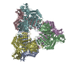
| Title | Cryo EM structure of alpha-glucosidase (yicI) from Klebsiella aerogenes | |||||||||
 Map data Map data | Main map | |||||||||
 Sample Sample |
| |||||||||
 Keywords Keywords | SSGCID / STRUCTURAL GENOMICS / SEATTLE STRUCTURAL GENOMICS CENTER FOR INFECTIOUS DISEASE / alpha-glucosidase / Klebsiella aerogenes / HYDROLASE | |||||||||
| Function / homology |  Function and homology information Function and homology informationalpha-D-xyloside xylohydrolase / alpha-D-xyloside xylohydrolase activity / carbohydrate binding / carbohydrate metabolic process Similarity search - Function | |||||||||
| Biological species |  Klebsiella aerogenes KCTC 2190 (bacteria) Klebsiella aerogenes KCTC 2190 (bacteria) | |||||||||
| Method | single particle reconstruction / cryo EM / Resolution: 3.19 Å | |||||||||
 Authors Authors | Lovell S / Liu L / Ingham DJ / Seattle Structural Genomics Center for Infectious Disease (SSGCID) | |||||||||
| Funding support |  United States, 1 items United States, 1 items
| |||||||||
 Citation Citation | Journal: To be published Title: Cryo EM structure of alpha-glucosidase (yicI) from Klebsiella aerogenes Authors: Liu L / Lovell S / Ingham DJ | |||||||||
| History |
|
- Structure visualization
| Supplemental images |
|---|

- Downloads & links
-EMDB archive
| Map data | emd_72225.map.gz | 137.7 MB | EMDB map data format | |
|---|---|---|---|---|
| Header (meta data) | emd-72225-v30.xmlemd-72225.xml | 20 KB 20 KB | Display Display | EMDB header |
| FSC (resolution estimation) | emd_72225_fsc.xml | 19.3 KB | Display | FSC data file |
| Images |  emd_72225.png emd_72225.png | 50 KB | ||
| Filedesc metadata | emd-72225.cif.gz | 6.8 KB | ||
| Others | emd_72225_half_map_1.map.gzemd_72225_half_map_2.map.gz | 262.3 MB 262.3 MB | ||
| Archive directory |  http://ftp.pdbj.org/pub/emdb/structures/EMD-72225ftp://ftp.pdbj.org/pub/emdb/structures/EMD-72225 http://ftp.pdbj.org/pub/emdb/structures/EMD-72225ftp://ftp.pdbj.org/pub/emdb/structures/EMD-72225 | HTTPS FTP |
-Related structure data
| Related structure data |  9q5cMC M: atomic model generated by this map C: citing same article ( |
|---|---|
| Similar structure data |
-Links
| EMDB pages | EMDB (EBI/PDBe) / EMDataResource |
|---|---|
| Related items in Molecule of the Month |


-Map
| File | Download / File: emd_72225.map.gz / Format: CCP4 / Size: 282.6 MB / Type: IMAGE STORED AS FLOATING POINT NUMBER (4 BYTES) | ||||||||||||||||||||||||||||||||||||
|---|---|---|---|---|---|---|---|---|---|---|---|---|---|---|---|---|---|---|---|---|---|---|---|---|---|---|---|---|---|---|---|---|---|---|---|---|---|
| Annotation | Main map | ||||||||||||||||||||||||||||||||||||
| Projections & slices | Image control
Images are generated by Spider. | ||||||||||||||||||||||||||||||||||||
| Voxel size | X=Y=Z: 0.54 Å | ||||||||||||||||||||||||||||||||||||
| Density |
| ||||||||||||||||||||||||||||||||||||
| Symmetry | Space group: 1 | ||||||||||||||||||||||||||||||||||||
| Details | EMDB XML:
|
 Z (Sec.)
 Y (Row.)
 X (Col.)


-Supplemental data
-Half map: #2
| File | emd_72225_half_map_1.map | ||||||||||||
|---|---|---|---|---|---|---|---|---|---|---|---|---|---|
| Projections & Slices |
| ||||||||||||
| Density Histograms |


-Half map: #1
| File | emd_72225_half_map_2.map | ||||||||||||
|---|---|---|---|---|---|---|---|---|---|---|---|---|---|
| Projections & Slices |
| ||||||||||||
| Density Histograms |


- Sample components
-Entire : alpha-glucosidase
| Entire | Name: alpha-glucosidase |
|---|---|
| Components |
|
-Supramolecule #1: alpha-glucosidase
| Supramolecule | Name: alpha-glucosidase / type: organelle_or_cellular_component / ID: 1 / Parent: 0 / Macromolecule list: all |
|---|---|
| Source (natural) | Organism: Klebsiella aerogenes KCTC 2190 (bacteria) |
| Molecular weight | Theoretical: 531.87 KDa |
-Macromolecule #1: Alpha-glucosidase yicI
| Macromolecule | Name: Alpha-glucosidase yicI / type: protein_or_peptide / ID: 1 / Number of copies: 6 / Enantiomer: LEVO |
|---|---|
| Source (natural) | Organism: Klebsiella aerogenes KCTC 2190 (bacteria) |
| Molecular weight | Theoretical: 88.650727 KDa |
| Recombinant expression | Organism: |
| Sequence | String: MAHHHHHHMK ISDGNWLIQP GLNLIQPVQV YEVEQQGNEM VVYAAPRDVR ERAWQLDTPL FTLRFFSPQE GIIGVRMEHF QGALDNSPH YPLNVQKDVH VEIENTAEFA ELKSGSLSVR VTKGEFWALD FLRDGVRITG SQLKNNGYVQ DSKTQRNYMF E RLDLGVGE ...String: MAHHHHHHMK ISDGNWLIQP GLNLIQPVQV YEVEQQGNEM VVYAAPRDVR ERAWQLDTPL FTLRFFSPQE GIIGVRMEHF QGALDNSPH YPLNVQKDVH VEIENTAEFA ELKSGSLSVR VTKGEFWALD FLRDGVRITG SQLKNNGYVQ DSKTQRNYMF E RLDLGVGE TVYGLGERFT ALVRNGQTVE TWNEDGGTST EQSYKNIPFY LTNRGYGVLV NHPQRVSFEV GSEKVSKVQF SV EGEYLEY FVIDGPTPKA VLNRYTQFTG RPALPPAWSF GLWLTTSFTT NYDEATVNSF IDGMAERHLP LHVFHFDCFW MKA FQWCDF EWDPLTFPDP EGMIKRLKAK GLKVCVWINP YIGQRSPVFK ELKEKGYLLK RPDGSLWQWD KWQPGLAIYD FTNP EACQW YASKLKGLVA MGVDCFKTDF GERIPTDVQW FDGSDPQKMH NHYAFIYNEL VWKVLKETVG EQEAVLFARS ASVGA QQFP VHWGGDCYAN YESMAESLRG GLSIGMSGFG FWSHDIGGFE NTAPAHVYKR WCAFGLLSSH SRLHGSKSYR VPWAYD EES CDVVRHFTQL KCRMMPYLYR QAALANEFGT PMLRAMMLEF PDDPACDYLD RQYMLGDSVL VAPVFSEAGE VQFYLPE GR WTHLWHNDEL PGSRWHKQRH DALSLPVYVR DNTLLALGNN DQKPDYAWHE GTAFQLFQLG DGNETVCQVP AADGSAIF T LKAKRQGNTI TVSGEGEARG WTLCLRNIPQ IAGVEGGTQA GSELGVVVSA QGNALTISL UniProtKB: alpha-D-xyloside xylohydrolase |
-Experimental details
-Structure determination
| Method | cryo EM |
|---|---|
 Processing Processing | single particle reconstruction |
| Aggregation state | 2D array |
-Sample preparation
| Concentration | 2.09 mg/mL |
|---|---|
| Buffer | pH: 7 Details: 25 mM HEPES pH 7.0, 500 mM NaCl, 5% Glycerol, 2 mM DTT, 0.025% Azide |
| Grid | Model: Quantifoil R1.2/1.3 / Material: COPPER / Mesh: 300 / Support film - Material: CARBON / Support film - topology: HOLEY / Pretreatment - Type: GLOW DISCHARGE / Pretreatment - Time: 1 sec. / Pretreatment - Pressure: 0.005 kPa |
| Vitrification | Cryogen name: ETHANE / Chamber humidity: 95 % / Chamber temperature: 277 K / Instrument: FEI VITROBOT MARK IV |
- Electron microscopy
| Microscope | TFS KRIOS |
|---|---|
| Image recording | Film or detector model: GATAN K3 (6k x 4k) / Detector mode: COUNTING / Number grids imaged: 1 / Average electron dose: 60.39 e/Å2 |
| Electron beam | Acceleration voltage: 300 kV / Electron source:  FIELD EMISSION GUN FIELD EMISSION GUN |
| Electron optics | Illumination mode: FLOOD BEAM / Imaging mode: 4D-STEM / Cs: 2.7 mm / Nominal defocus max: 0.6 µm / Nominal defocus min: 0.2 µm / Nominal magnification: 81000 |
| Sample stage | Specimen holder model: FEI TITAN KRIOS AUTOGRID HOLDER / Cooling holder cryogen: NITROGEN |
| Experimental equipment |  Model: Titan Krios / Image courtesy: FEI Company |