 Movie
Movie Controller

[English] 日本語
 Yorodumi
- EMDB-70678: Herpes simplex virus type 1 (HSV-1) A-capsid pUL6 portal protein,... -
+ Open data

- Basic information
| Entry |  | |||||||||||||||||||||||||||
|---|---|---|---|---|---|---|---|---|---|---|---|---|---|---|---|---|---|---|---|---|---|---|---|---|---|---|---|---|
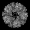
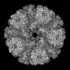
| Title | Herpes simplex virus type 1 (HSV-1) A-capsid pUL6 portal protein, dodecameric complex | |||||||||||||||||||||||||||
 Map data Map data | ||||||||||||||||||||||||||||
 Sample Sample |
| |||||||||||||||||||||||||||
 Keywords Keywords | Complex / VIRAL PROTEIN | |||||||||||||||||||||||||||
| Function / homology |  Function and homology information Function and homology informationassemblin / nuclear capsid assembly / viral release from host cell / chromosome organization / virion component / host cell cytoplasm / serine-type endopeptidase activity / host cell nucleus / proteolysis / identical protein binding Similarity search - Function | |||||||||||||||||||||||||||
| Biological species |  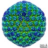 Human alphaherpesvirus 1 strain KOS Human alphaherpesvirus 1 strain KOS | |||||||||||||||||||||||||||
| Method | single particle reconstruction / cryo EM / Resolution: 3.6 Å | |||||||||||||||||||||||||||
 Authors Authors | Crofut EH / Kashyap S / Stevens A / Jih J / Liu Y-T / Zhou ZH | |||||||||||||||||||||||||||
| Funding support |  United States, 8 items United States, 8 items
| |||||||||||||||||||||||||||
 Citation Citation | Journal: J Virol / Year: 2025 Title: Structure of a new capsid form and comparison with A-, B-, and C-capsids clarify herpesvirus assembly. Authors: Alexander Stevens / Saarang Kashyap / Ethan Crofut / Ana Lucia Alvarez-Cabrera / Jonathan Jih / Yun-Tao Liu / Z Hong Zhou / Abstract: Three capsid types have been recognized from the nuclei of herpesvirus-infected cells: empty A-capsids, scaffolding-containing B-capsids, and DNA-filled C-capsids. Despite progress in determining ...Three capsid types have been recognized from the nuclei of herpesvirus-infected cells: empty A-capsids, scaffolding-containing B-capsids, and DNA-filled C-capsids. Despite progress in determining atomic structures of these capsids and extracellular virions in recent years, debate persists concerning the origins and temporal relationships among these capsids during capsid assembly and genome packaging. Here, we have imaged over 300,000 capsids of herpes simplex virus type 1 by cryogenic electron microscopy (cryoEM) and exhaustively classified them to characterize the structural heterogeneity of the DNA-translocating portal complex and their functional states. The resultant atomic structures reveal not only the expected A-, B-, and C-capsids but also capsids with portal vertices similar to C-capsids but no resolvable genome in the capsid lumen, which we named D-capsids. The dodecameric dsDNA-translocating portal complex varies across these capsid types in their radial positions in icosahedral capsids and exhibits structural dynamics within each capsid type. In D-capsids, terminal DNA density exists in multiple conformations including one reminiscent of that in C-capsids, suggesting D-capsids are products of failed DNA retention. This interpretation is supported by varying amounts of DNA outside individual D-capsids and by the correlation of capsid counts observed of infected cell nuclei and those after purification. Additionally, an "anchoring" segment of the scaffold protein is resolved interacting with the portal baskets of A- and B-capsids but not D- and C-capsids. Taken together, our data indicate that A-capsids arise from failed DNA packaging and D-capsids from failed genome retention, clarifying the origins of empty capsids in herpesvirus assembly.IMPORTANCEAs the prototypical herpesvirus, herpes simplex virus 1 (HSV-1) exhibits a global seroprevalence of 67% and approaching 90% in some localities. Herpesvirus infections can cause devastating cancers and birth defects, with HSV-1 infections leading to cold sores among the general population worldwide and blindness in developing nations. Here, we present atomic structures of the capsids sorted out from the nuclear isolates of HSV-1 infected cells, including the previously recognized A-, B-, and C-capsids, as well as the newly identified D-capsid. The structures show the details of protein-protein and protein-DNA interactions within each capsid type and the positional and interactional variability of the viral DNA-translocating portal vertex among these capsids. Importantly, our findings suggest that A-capsids are products of failed dsDNA packaging and D-capsids of failed genome retention. Together, the high-resolution 3D structures clarify the processes of genome packaging, maintenance, and ejection during capsid assembly, which are conserved across all herpesviruses. | |||||||||||||||||||||||||||
| History |
|
- Structure visualization
| Supplemental images |
|---|

- Downloads & links
-EMDB archive
| Map data | emd_70678.map.gz | 397.5 MB | EMDB map data format | |
|---|---|---|---|---|
| Header (meta data) | emd-70678-v30.xmlemd-70678.xml | 22.8 KB 22.8 KB | Display Display | EMDB header |
| FSC (resolution estimation) | emd_70678_fsc.xml | 17 KB | Display | FSC data file |
| Images |  emd_70678.png emd_70678.png | 114.3 KB | ||
| Masks | emd_70678_msk_1.map | 421.9 MB | Mask map | |
| Filedesc metadata | emd-70678.cif.gz | 7 KB | ||
| Others | emd_70678_half_map_1.map.gzemd_70678_half_map_2.map.gz | 389.6 MB 389.6 MB | ||
| Archive directory |  http://ftp.pdbj.org/pub/emdb/structures/EMD-70678ftp://ftp.pdbj.org/pub/emdb/structures/EMD-70678 http://ftp.pdbj.org/pub/emdb/structures/EMD-70678ftp://ftp.pdbj.org/pub/emdb/structures/EMD-70678 | HTTPS FTP |
-Related structure data
| Related structure data |  9op4MC  9op5C  9op8C  9opbC  9opcC  9opvC M: atomic model generated by this map C: citing same article ( |
|---|---|
| Similar structure data |


-Links
| EMDB pages | EMDB (EBI/PDBe) / EMDataResource |
|---|
-Map
| File | Download / File: emd_70678.map.gz / Format: CCP4 / Size: 421.9 MB / Type: IMAGE STORED AS FLOATING POINT NUMBER (4 BYTES) | ||||||||||||||||||||||||||||||||||||
|---|---|---|---|---|---|---|---|---|---|---|---|---|---|---|---|---|---|---|---|---|---|---|---|---|---|---|---|---|---|---|---|---|---|---|---|---|---|
| Projections & slices | Image control
Images are generated by Spider. | ||||||||||||||||||||||||||||||||||||
| Voxel size | X=Y=Z: 1.1 Å | ||||||||||||||||||||||||||||||||||||
| Density |
| ||||||||||||||||||||||||||||||||||||
| Symmetry | Space group: 1 | ||||||||||||||||||||||||||||||||||||
| Details | EMDB XML:
|
 Z (Sec.)
 Y (Row.)
 X (Col.)


-Supplemental data
-Mask #1
| File | emd_70678_msk_1.map | ||||||||||||
|---|---|---|---|---|---|---|---|---|---|---|---|---|---|
| Projections & Slices |
| ||||||||||||
| Density Histograms |


-Half map: #2
| File | emd_70678_half_map_1.map | ||||||||||||
|---|---|---|---|---|---|---|---|---|---|---|---|---|---|
| Projections & Slices |
| ||||||||||||
| Density Histograms |


-Half map: #1
| File | emd_70678_half_map_2.map | ||||||||||||
|---|---|---|---|---|---|---|---|---|---|---|---|---|---|
| Projections & Slices |
| ||||||||||||
| Density Histograms |


- Sample components
-Entire : Dodecameric complex of pUL6 with bound loops from scaffolding protein
| Entire | Name: Dodecameric complex of pUL6 with bound loops from scaffolding protein |
|---|---|
| Components |
|
-Supramolecule #1: Dodecameric complex of pUL6 with bound loops from scaffolding protein
| Supramolecule | Name: Dodecameric complex of pUL6 with bound loops from scaffolding protein type: complex / ID: 1 / Parent: 0 / Macromolecule list: all |
|---|---|
| Source (natural) | Organism: Human alphaherpesvirus 1 strain KOS |
-Macromolecule #1: Capsid scaffolding protein
| Macromolecule | Name: Capsid scaffolding protein / type: protein_or_peptide / ID: 1 / Number of copies: 12 / Enantiomer: LEVO |
|---|---|
| Source (natural) | Organism: Human alphaherpesvirus 1 strain KOS |
| Molecular weight | Theoretical: 66.501609 KDa |
| Sequence | String: MAADAPGDRM EEPLPDRAVP IYVAGFLALY DSGDSGELAL DPDTVRAALP PDNPLPINVD HRAGCEVGRV LAVVDDPRGP FFVGLIACV QLERVLETAA SAAIFERRGP PLSREERLLY LITNYLPSVS LATKRLGGEA HPDRTLFAHV ALCAIGRRLG T IVTYDTGL ...String: MAADAPGDRM EEPLPDRAVP IYVAGFLALY DSGDSGELAL DPDTVRAALP PDNPLPINVD HRAGCEVGRV LAVVDDPRGP FFVGLIACV QLERVLETAA SAAIFERRGP PLSREERLLY LITNYLPSVS LATKRLGGEA HPDRTLFAHV ALCAIGRRLG T IVTYDTGL DAAIAPFRHL SPASREGARR LAAEAEIALS GRTWAPGVEA LTHTLLSTAV NNMMLRDRWS LVAERRRQAG IA GHTYLQA SEKFKMWGAE PVSAPARGYK NGAPESTDIP PGSIAAAPQG DRCPIVRQRG VALSPVLPPM NPVPTSGTPA PAP PGDGSY LWIPASHYNQ LVAGHAAPQP QPHSAFGFPA AAGAVAYGPH GAGLSQHYPP HVAHQYPGVL FSGPSPLEAQ IAAL VGAIA ADRQAGGQPA AGDPGVRGSG KRRRYEAGPS ESYCDQDEPD ADYPYYPGEA RGGPRGVDSR RAARQSPGTN ETITA LMGA VTSLQQELAH MRARTSAPYG MYTPVAHYRP QVGEPEPTTT HPALCPPEAV YRPPPHSAPY GPPQGPASHA PTPPYA PAA CPPGPPPPPC PSTQTRAPLP TEPAFPPAAT GSQPEASNAE AGALVNASSA AHVDVDTARA ADLFVSQMMG AR UniProtKB: Capsid scaffolding protein |
-Macromolecule #2: Capsid portal protein
| Macromolecule | Name: Capsid portal protein / type: protein_or_peptide / ID: 2 / Number of copies: 12 / Enantiomer: LEVO |
|---|---|
| Source (natural) | Organism: Human alphaherpesvirus 1 strain KOS |
| Molecular weight | Theoretical: 74.178523 KDa |
| Sequence | String: MTAPRSWAPT TRARGDTEAL CSPEDGWVKV HPTPGTMLFR EILHGQLGYT EGQGVYNVVR SSEATTRQLQ AAIFHALLNA TTYRDLEAD WLGHVAARGL QPQRLVRRYR NAREADIAGV AERVFDTWRN TLRTTLLDFA HGLVACFAPG GPSGPSSFPK Y IDWLTCLG ...String: MTAPRSWAPT TRARGDTEAL CSPEDGWVKV HPTPGTMLFR EILHGQLGYT EGQGVYNVVR SSEATTRQLQ AAIFHALLNA TTYRDLEAD WLGHVAARGL QPQRLVRRYR NAREADIAGV AERVFDTWRN TLRTTLLDFA HGLVACFAPG GPSGPSSFPK Y IDWLTCLG LVPILRKRQE GGVTQGLRAF LKQHPLTRQL ATVAEAAERA GPGFFELALA FDSTRVADYD RVYIYYNHRR GD WLVRDPI SGQRGECLVL WPPLWTGDRL VFDSPVQRLF PEIVACHSLR EHAHVCRLRN TASVKVLLGR KSDSERGVAG AAR VVNKVL GEDDETKAGS AASRLVRLII NMKGMRHVGD INDTVRSYLD EAGGHLIDAP AVDGTLPGFG KGGNSRGSAG QDQG GRAPQ LRQAFRTAVV NNINGVLEGY INNLFGTIER LRETNAGLAT QLQERDRELR RATAGALERQ QRAADLAAES VTGGC GSRP AGADLLRADY DIIDVSKSMD DDTYVANSFQ HPYIPSYAQD LERLSRLWEH ELVRCFKILC HRNNQGQETS ISYSSG AIA AFVAPYFESV LRAPRVGAPI TGSDVILGEE ELWDAVFKKT RLQTYLTDIA ALFVADVQHA ALPPPPSPVG ADFRPGA SP RGRSRSRSPG RTAPGAPDQG GGIGHRDGRR DGRR UniProtKB: Capsid portal protein |
-Experimental details
-Structure determination
| Method | cryo EM |
|---|---|
 Processing Processing | single particle reconstruction |
| Aggregation state | particle |
-Sample preparation
| Buffer | pH: 7.4 |
|---|---|
| Grid | Model: Quantifoil / Material: COPPER / Mesh: 200 / Support film - Material: CARBON / Support film - topology: HOLEY / Pretreatment - Type: GLOW DISCHARGE / Pretreatment - Time: 30 sec. |
| Vitrification | Cryogen name: ETHANE |
- Electron microscopy
| Microscope | TFS KRIOS |
|---|---|
| Image recording | Film or detector model: GATAN K3 (6k x 4k) / Average electron dose: 45.0 e/Å2 |
| Electron beam | Acceleration voltage: 300 kV / Electron source:  FIELD EMISSION GUN FIELD EMISSION GUN |
| Electron optics | Illumination mode: FLOOD BEAM / Imaging mode: BRIGHT FIELD / Cs: 2.7 mm / Nominal defocus max: 2.5 µm / Nominal defocus min: 1.5 µm / Nominal magnification: 81000 |
| Sample stage | Specimen holder model: FEI TITAN KRIOS AUTOGRID HOLDER / Cooling holder cryogen: NITROGEN |
| Experimental equipment |  Model: Titan Krios / Image courtesy: FEI Company |

