 Movie
Movie Controller

[English] 日本語
 Yorodumi
- EMDB-70204: Cryo-EM Structure of YfdQ Reveals a Widespread Novel Family of Ba... -
+ Open data

- Basic information
| Entry |  | |||||||||||||||
|---|---|---|---|---|---|---|---|---|---|---|---|---|---|---|---|---|
| Title | Cryo-EM Structure of YfdQ Reveals a Widespread Novel Family of Bacteriophage-Associated Proteins with Shell-Like Assemblies | |||||||||||||||
 Map data Map data | Raw map | |||||||||||||||
 Sample Sample |
| |||||||||||||||
 Keywords Keywords | D18 symmetry / shell-like structure / DUF2303 / YfdQ / VIRAL PROTEIN | |||||||||||||||
| Biological species |  Caudoviricetes (bacterial and archaeal viruses with head-tail morphology) Caudoviricetes (bacterial and archaeal viruses with head-tail morphology) | |||||||||||||||
| Method | single particle reconstruction / cryo EM / Resolution: 3.9 Å | |||||||||||||||
 Authors Authors | Guzzo CR / Araujo GG / Merighi DGS | |||||||||||||||
| Funding support |  Brazil, 4 items Brazil, 4 items
| |||||||||||||||
 Citation Citation | Journal: Structure / Year: 2026 Title: Cryo-EM structure of YfdQ reveals a widespread family of bacteriophage-associated proteins with shell-like assemblies. Authors: Davi Gabriel Salustiano Merighi / Guilherme Camargo Pompeu / Gabriel Guarany de Araujo / Anacleto Silva de Souza / Germán Gustavo Sgro / Chuck Shaker Farah / Cristiane Rodrigues Guzzo / Abstract: Bacteriophages are key players in microbial ecosystems with strong biotechnological potential, yet many phage-encoded proteins remain uncharacterized. We present the structure of the YfdQ complex ...Bacteriophages are key players in microbial ecosystems with strong biotechnological potential, yet many phage-encoded proteins remain uncharacterized. We present the structure of the YfdQ complex from the Xanthomonas citri ΦXacm4-11 phage, a conserved DUF2303 protein encoded by double-stranded DNA bacteriophages of the Caudoviricetes class. Using cryo-electron microscopy, we report that native YfdQ assembles into a shell-like structure with D18 symmetry forming a ∼1.1 MegaDalton complex, a hollow interior and a unique equatorial β strand stitching oligomerization interface that holds together the two hemispheres. YfdQ homologs are widely distributed among phages infecting diverse bacterial hosts, suggesting a conserved biological role. YfdQ are found in prophages and cryptic phages. Our results suggest that heterologous expression of YfdQ in X. citri and in Escherichia coli allows the obtention of structurally heterogeneous YfdQ complexes, suggesting that its proper assembly into the shell-like structure may depend on still unidentified viral factors. | |||||||||||||||
| History |
|
- Structure visualization
| Supplemental images |
|---|

- Downloads & links
-EMDB archive
| Map data | emd_70204.map.gz | 178 MB |  EMDB map data format EMDB map data format | |
|---|---|---|---|---|
| Header (meta data) | emd-70204-v30.xmlemd-70204.xml | 20.6 KB 20.6 KB | Display Display | EMDB header |
| FSC (resolution estimation) | emd_70204_fsc.xml | 15.2 KB | Display | FSC data file |
| Images |  emd_70204.png emd_70204.png | 94.8 KB | ||
| Masks | emd_70204_msk_1.map | 371.3 MB | Mask map | |
| Filedesc metadata | emd-70204.cif.gz | 6.8 KB | ||
| Others | emd_70204_half_map_1.map.gzemd_70204_half_map_2.map.gz | 343.4 MB 343.4 MB | ||
| Archive directory |  https://data.pdbj.org/pub/emdb/structures/EMD-70204ftp://data.pdbj.org/pub/emdb/structures/EMD-70204 https://data.pdbj.org/pub/emdb/structures/EMD-70204ftp://data.pdbj.org/pub/emdb/structures/EMD-70204 | HTTPS FTP |
-Related structure data
-Links
| EMDB pages | EMDB (EBI/PDBe) / EMDataResource |
|---|
-Map
| File | Download / File: emd_70204.map.gz / Format: CCP4 / Size: 371.3 MB / Type: IMAGE STORED AS FLOATING POINT NUMBER (4 BYTES) | ||||||||||||||||||||||||||||||||||||
|---|---|---|---|---|---|---|---|---|---|---|---|---|---|---|---|---|---|---|---|---|---|---|---|---|---|---|---|---|---|---|---|---|---|---|---|---|---|
| Annotation | Raw map | ||||||||||||||||||||||||||||||||||||
| Projections & slices | Image control
Images are generated by Spider. | ||||||||||||||||||||||||||||||||||||
| Voxel size | X=Y=Z: 0.86 Å | ||||||||||||||||||||||||||||||||||||
| Density |
| ||||||||||||||||||||||||||||||||||||
| Symmetry | Space group: 1 | ||||||||||||||||||||||||||||||||||||
| Details | EMDB XML:
|
 Z (Sec.)
 Y (Row.)
 X (Col.)


-Supplemental data
-Mask #1
| File | emd_70204_msk_1.map | ||||||||||||
|---|---|---|---|---|---|---|---|---|---|---|---|---|---|
| Projections & Slices |
| ||||||||||||
| Density Histograms |


-Half map: Half map
| File | emd_70204_half_map_1.map | ||||||||||||
|---|---|---|---|---|---|---|---|---|---|---|---|---|---|
| Annotation | Half map | ||||||||||||
| Projections & Slices |
| ||||||||||||
| Density Histograms |


-Half map: Half map
| File | emd_70204_half_map_2.map | ||||||||||||
|---|---|---|---|---|---|---|---|---|---|---|---|---|---|
| Annotation | Half map | ||||||||||||
| Projections & Slices |
| ||||||||||||
| Density Histograms |


- Sample components
-Entire : YfdQ pprotein complex
| Entire | Name: YfdQ pprotein complex |
|---|---|
| Components |
|
-Supramolecule #1: YfdQ pprotein complex
| Supramolecule | Name: YfdQ pprotein complex / type: complex / ID: 1 / Parent: 0 / Macromolecule list: all / Details: YfdQ |
|---|---|
| Source (natural) | Organism: Caudoviricetes (bacterial and archaeal viruses with head-tail morphology) Strain: phiXacm4-11 |
| Molecular weight | Theoretical: 1.059 MDa |
-Macromolecule #1: YfdQ Protein
| Macromolecule | Name: YfdQ Protein / type: protein_or_peptide / ID: 1 / Number of copies: 36 / Enantiomer: LEVO |
|---|---|
| Source (natural) | Organism: Caudoviricetes (bacterial and archaeal viruses with head-tail morphology) Strain: phiXacm4-11 |
| Molecular weight | Theoretical: 29.457115 KDa |
| Sequence | String: MDKSAIEAIQ DSSAAAAKAT QEQIPAGITH LVAVPNGVTL QNVEISLAGR TRFRGKLVTS SIPDFVTYVK NREGGHGFID TDKLGATVF FNLGDADKPG HADHTARLTL QATPAYLAML AAGGKAFSQR DALDFIEDWS HLIGAHQVSD DGAFSPIPLA R AIAAIRKV ...String: MDKSAIEAIQ DSSAAAAKAT QEQIPAGITH LVAVPNGVTL QNVEISLAGR TRFRGKLVTS SIPDFVTYVK NREGGHGFID TDKLGATVF FNLGDADKPG HADHTARLTL QATPAYLAML AAGGKAFSQR DALDFIEDWS HLIGAHQVSD DGAFSPIPLA R AIAAIRKV KIKAESTTEQ TEGNFQQSRS VLESVAASSD EGLPDVLSFR TEPYLGLPER TFHMRVSVST TGREPSLRFR VI ALEQEQD EIAKQFKALL LGEVGEAASM TIGTFTP |
-Experimental details
-Structure determination
| Method | cryo EM |
|---|---|
 Processing Processing | single particle reconstruction |
| Aggregation state | particle |
-Sample preparation
| Buffer | pH: 7.5 Details: 50 mM Tris-HCl, 100 mM NaCl, 10 mM MgSO47(H2O), pH 7.5 |
|---|---|
| Vitrification | Cryogen name: ETHANE / Chamber humidity: 100 % / Chamber temperature: 295 K Details: A purified YfdQ proteins were pipetted over Glow-Discharged Quantifoil CU-R2/2 400 Mesh grids with a 2 nm ultrathin carbon layer and vitrified by plunge-freezing using a Vitrobot (Thermo ...Details: A purified YfdQ proteins were pipetted over Glow-Discharged Quantifoil CU-R2/2 400 Mesh grids with a 2 nm ultrathin carbon layer and vitrified by plunge-freezing using a Vitrobot (Thermo Fisher Scientific). The sample was drained by 3.5 s blot and 0 force, and 4.5 s and 0 force programs.. |
| Details | native particles produced during phiXacm4-11 infection |
- Electron microscopy
| Microscope | TFS KRIOS |
|---|---|
| Image recording | Film or detector model: FEI FALCON III (4k x 4k) / Detector mode: COUNTING / Average electron dose: 44.89 e/Å2 Details: Data were acquired using a Titan Krios G3i cryo-electron microscope, 300 kV (Thermo Fisher Scientific), at the LNNano facility, CNPEM, Campinas, Brazil. The microscope was equipped with a 4k- ...Details: Data were acquired using a Titan Krios G3i cryo-electron microscope, 300 kV (Thermo Fisher Scientific), at the LNNano facility, CNPEM, Campinas, Brazil. The microscope was equipped with a 4k-4k CMOS camera, phase plate, spherical aberration corrector, and Falcon 3 detector without energy filter or a K3 detector with energy filter. For the native YfdQ dataset, data collection utilized the Falcon 3 detector in counting mode. |
| Electron beam | Acceleration voltage: 300 kV / Electron source:  FIELD EMISSION GUN FIELD EMISSION GUN |
| Electron optics | C2 aperture diameter: 70.0 µm / Illumination mode: FLOOD BEAM / Imaging mode: BRIGHT FIELD / Cs: 0.001 mm / Nominal defocus max: 2.0 µm / Nominal defocus min: 0.5 µm |
| Experimental equipment |  Model: Titan Krios / Image courtesy: FEI Company |
+Image processing

-Atomic model buiding 1
| Refinement | Space: REAL / Protocol: RIGID BODY FIT |
|---|---|
| Output model | 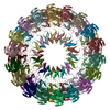 PDB-9o7m: |
