 Movie
Movie Controller


+ Open data

- Basic information
| Entry |  | |||||||||
|---|---|---|---|---|---|---|---|---|---|---|
| Title | Perinereis linea erythrocruorin | |||||||||
 Map data Map data | ||||||||||
 Sample Sample |
| |||||||||
 Keywords Keywords | invertebrate / annelid / hemoglobin / erythrocruorin / OXYGEN TRANSPORT | |||||||||
| Biological species |  Perinereis linea (invertebrata) Perinereis linea (invertebrata) | |||||||||
| Method | single particle reconstruction / cryo EM / Resolution: 2.84 Å | |||||||||
 Authors Authors | Deng JX / Jiang YL / Zhou CZ | |||||||||
| Funding support |  China, 1 items China, 1 items
| |||||||||
 Citation Citation | Journal: Structure / Year: 2026 Title: Structure of Perinereis linea erythrocruorin reveals a compact extracellular globin megacomplex. Authors: Jia-Xin Deng / Wen-Bin Cheng / Kang Xu / Pu Hou / Yuxing Chen / Yong-Liang Jiang / Cong-Zhao Zhou / Abstract: Many invertebrates lack erythrocytes and instead rely on extracellular hemoglobin assemblies, termed erythrocruorins, for oxygen transport. Here we report a 2.84 Å cryo-electron microscopy (cryo-EM) ...Many invertebrates lack erythrocytes and instead rely on extracellular hemoglobin assemblies, termed erythrocruorins, for oxygen transport. Here we report a 2.84 Å cryo-electron microscopy (cryo-EM) structure of Perinereis linea erythrocruorin (PlEc). PlEc is a ∼3.3 MDa megacomplex composed of 180 polypeptide chains organized into 12 protomers, forming a hexagonal bilayer with D6 symmetry. Each protomer consists of 12 globin subunits and three linker subunits, adopting a mushroom-like architecture. The cap of the mushroom is formed by a globin dodecamer associated with a heterotrimeric linker head, and the stem consists of a triple-stranded coiled coil derived from the N-terminal helices of three linker subunits. Biochemical assays show that PlEc has thermal stability and auto-oxidation rate comparable to those of other erythrocruorins, but displays relatively lower oxygen-binding affinity. These findings provide mechanistic insights into the quaternary assembly of invertebrate erythrocruorins and lay the groundwork for the potential biomedical applications. | |||||||||
| History |
|
- Structure visualization
| Supplemental images |
|---|

- Downloads & links
-EMDB archive
| Map data | emd_68387.map.gz | 483.3 MB |  EMDB map data format EMDB map data format | |
|---|---|---|---|---|
| Header (meta data) | emd-68387-v30.xmlemd-68387.xml | 20.1 KB 20.1 KB | Display Display | EMDB header |
| Images |  emd_68387.png emd_68387.png | 61.7 KB | ||
| Masks | emd_68387_msk_1.map | 512 MB | Mask map | |
| Filedesc metadata | emd-68387.cif.gz | 6.1 KB | ||
| Others | emd_68387_half_map_1.map.gzemd_68387_half_map_2.map.gz | 473.2 MB 473.2 MB | ||
| Archive directory |  http://ftp.pdbj.org/pub/emdb/structures/EMD-68387ftp://ftp.pdbj.org/pub/emdb/structures/EMD-68387 http://ftp.pdbj.org/pub/emdb/structures/EMD-68387ftp://ftp.pdbj.org/pub/emdb/structures/EMD-68387 | HTTPS FTP |
-Related structure data

-Links
| EMDB pages | EMDB (EBI/PDBe) / EMDataResource |
|---|
-Map
| File | Download / File: emd_68387.map.gz / Format: CCP4 / Size: 512 MB / Type: IMAGE STORED AS FLOATING POINT NUMBER (4 BYTES) | ||||||||||||||||||||||||||||||||||||
|---|---|---|---|---|---|---|---|---|---|---|---|---|---|---|---|---|---|---|---|---|---|---|---|---|---|---|---|---|---|---|---|---|---|---|---|---|---|
| Projections & slices | Image control
Images are generated by Spider. | ||||||||||||||||||||||||||||||||||||
| Voxel size | X=Y=Z: 1.07 Å | ||||||||||||||||||||||||||||||||||||
| Density |
| ||||||||||||||||||||||||||||||||||||
| Symmetry | Space group: 1 | ||||||||||||||||||||||||||||||||||||
| Details | EMDB XML:
|
 Z (Sec.)
 Y (Row.)
 X (Col.)


-Supplemental data
-Mask #1
| File | emd_68387_msk_1.map | ||||||||||||
|---|---|---|---|---|---|---|---|---|---|---|---|---|---|
| Projections & Slices |
| ||||||||||||
| Density Histograms |


-Half map: #2
| File | emd_68387_half_map_1.map | ||||||||||||
|---|---|---|---|---|---|---|---|---|---|---|---|---|---|
| Projections & Slices |
| ||||||||||||
| Density Histograms |


-Half map: #1
| File | emd_68387_half_map_2.map | ||||||||||||
|---|---|---|---|---|---|---|---|---|---|---|---|---|---|
| Projections & Slices |
| ||||||||||||
| Density Histograms |


- Sample components
-Entire : Perinereis linea erythrocruorin
| Entire | Name: Perinereis linea erythrocruorin |
|---|---|
| Components |
|
-Supramolecule #1: Perinereis linea erythrocruorin
| Supramolecule | Name: Perinereis linea erythrocruorin / type: complex / ID: 1 / Parent: 0 / Macromolecule list: #1-#6 |
|---|---|
| Source (natural) | Organism: Perinereis linea (invertebrata) |
-Macromolecule #1: Extracellular globin A
| Macromolecule | Name: Extracellular globin A / type: protein_or_peptide / ID: 1 / Number of copies: 3 / Enantiomer: LEVO |
|---|---|
| Source (natural) | Organism: Perinereis linea (invertebrata) |
| Molecular weight | Theoretical: 16.43865 KDa |
| Sequence | String: DQCCSIEDRH EVQALWQSIW SAENTGKRTL IGRRIFEELF DINPGTKALF GRVNVDDMGS PEFKAHVLRV MNGLDTLIGV LDDPATGTS LIQHLAEQHK ARDGFKAAYF KDIGVALRRV LPQVASCFNP EAWNHCFDGF VASITAAMA |
-Macromolecule #2: Extracellular globin B
| Macromolecule | Name: Extracellular globin B / type: protein_or_peptide / ID: 2 / Number of copies: 3 / Enantiomer: LEVO |
|---|---|
| Source (natural) | Organism: Perinereis linea (invertebrata) |
| Molecular weight | Theoretical: 16.476941 KDa |
| Sequence | String: DTHCGPLQRL KVKQQWAKAY GVGHERVELG IALWKSMFSQ DPEARKLFDR VHGEDVRSPA FEAHIARVFN GFDRIISSLT DEDVLNAQL AHLKEQHIKL GITAHHFKLM RTGLGYVLPA QLGRCFDKAA WSACWDEVIY PGIKSL |
-Macromolecule #3: Extracellular globin C
| Macromolecule | Name: Extracellular globin C / type: protein_or_peptide / ID: 3 / Number of copies: 3 / Enantiomer: LEVO |
|---|---|
| Source (natural) | Organism: Perinereis linea (invertebrata) |
| Molecular weight | Theoretical: 16.570662 KDa |
| Sequence | String: DDCCSAADRH AVLSDWQNVW SAEFTGRRVA IGKAIFEELF AIDASAKGVF GRVHVDDQSS PEWAAHVIRV INGLDLAINL LEDPRALNE ELHHLARQHR ERDGVKAVYF DEIEKALLKV LPQVSSNFNA GAWDRCFTRI ASVIKAELP |
-Macromolecule #4: Extracellular globin D
| Macromolecule | Name: Extracellular globin D / type: protein_or_peptide / ID: 4 / Number of copies: 3 / Enantiomer: LEVO |
|---|---|
| Source (natural) | Organism: Perinereis linea (invertebrata) |
| Molecular weight | Theoretical: 16.101162 KDa |
| Sequence | String: DCNNLRRLKV KYQWSMVYDT THDRSQFATA VWRQFFKTYP DRSLFSNVRG ENIYSPEFRA HMVRVFAGFD ILISVLDSEP VLNAALAHY NTFHKQFDSI PFKQFGEVLL DTLSQFIPNE FDQDAWKECY AVIVAGIGA |
-Macromolecule #5: Hemoglobin linker 1
| Macromolecule | Name: Hemoglobin linker 1 / type: protein_or_peptide / ID: 5 / Number of copies: 2 / Enantiomer: LEVO |
|---|---|
| Source (natural) | Organism: Perinereis linea (invertebrata) |
| Molecular weight | Theoretical: 24.84693 KDa |
| Sequence | String: WAGIASKADA QEARINRLAG RVESVAEKLR TGGERVKNFM GEFREMEFRV NELEGNGCDK RHFQCGGNSR ECISDLLTCD GSPDCQNGA DEADDICHIP IPAGTVLVGH LNTDHDFCTK RKPNEMDLYI TSVTRSKYMQ SRLKVKANLN IKYTAEGSEV E DVLPVSGY ...String: WAGIASKADA QEARINRLAG RVESVAEKLR TGGERVKNFM GEFREMEFRV NELEGNGCDK RHFQCGGNSR ECISDLLTCD GSPDCQNGA DEADDICHIP IPAGTVLVGH LNTDHDFCTK RKPNEMDLYI TSVTRSKYMQ SRLKVKANLN IKYTAEGSEV E DVLPVSGY YNFGTHQLVI LPPEDDRLGI ICRFRAGNDN RCMASIVHEA SLTHCGDDFV FVAQH |
-Macromolecule #6: Hemoglobin linker 2
| Macromolecule | Name: Hemoglobin linker 2 / type: protein_or_peptide / ID: 6 / Number of copies: 1 / Enantiomer: LEVO |
|---|---|
| Source (natural) | Organism: Perinereis linea (invertebrata) |
| Molecular weight | Theoretical: 25.39807 KDa |
| Sequence | String: SAFSDPGVAE GRLANQDARL EQLENYLEGV ISKYEKFTQG REARVRRWNA LQDRVWGLEA HHCDDEHFSC RDDVYNCVGH QLVCDGTKD CLNGRDEDEE TCRVVPGVGS AFEGTLVKQD PCTSRKPRTF RFVVTSVDTS PNFPQEPKVK AAVIMQSDQT G ETVESSMT ...String: SAFSDPGVAE GRLANQDARL EQLENYLEGV ISKYEKFTQG REARVRRWNA LQDRVWGLEA HHCDDEHFSC RDDVYNCVGH QLVCDGTKD CLNGRDEDEE TCRVVPGVGS AFEGTLVKQD PCTSRKPRTF RFVVTSVDTS PNFPQEPKVK AAVIMQSDQT G ETVESSMT ANGVYDYTHR RLILYSPDND SLVFTCTFDR YNDDLCRGEI RRESGTSCVE FGLARLG |
-Macromolecule #7: PROTOPORPHYRIN IX CONTAINING FE
| Macromolecule | Name: PROTOPORPHYRIN IX CONTAINING FE / type: ligand / ID: 7 / Number of copies: 12 / Formula: HEM |
|---|---|
| Molecular weight | Theoretical: 616.487 Da |
| Chemical component information | 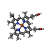 ChemComp-HEM: |
-Macromolecule #8: CALCIUM ION
| Macromolecule | Name: CALCIUM ION / type: ligand / ID: 8 / Number of copies: 3 / Formula: CA |
|---|---|
| Molecular weight | Theoretical: 40.078 Da |
-Experimental details
-Structure determination
| Method | cryo EM |
|---|---|
 Processing Processing | single particle reconstruction |
| Aggregation state | particle |
-Sample preparation
| Buffer | pH: 7.4 |
|---|---|
| Vitrification | Cryogen name: ETHANE |
- Electron microscopy
| Microscope | FEI TECNAI F30 |
|---|---|
| Image recording | Film or detector model: GATAN K2 QUANTUM (4k x 4k) / Average electron dose: 50.0 e/Å2 |
| Electron beam | Acceleration voltage: 300 kV / Electron source:  FIELD EMISSION GUN FIELD EMISSION GUN |
| Electron optics | Illumination mode: SPOT SCAN / Imaging mode: BRIGHT FIELD / Nominal defocus max: 2.0 µm / Nominal defocus min: 1.0 µm |
| Experimental equipment |  Model: Tecnai F30 / Image courtesy: FEI Company |
