 Movie
Movie Controller


+ Open data

- Basic information
| Entry |  | |||||||||
|---|---|---|---|---|---|---|---|---|---|---|
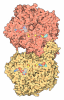
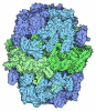
| Title | DPR epimerase complex, PMD-bound state | |||||||||
 Map data Map data | ||||||||||
 Sample Sample |
| |||||||||
 Keywords Keywords | Epimerase complex / OXIDOREDUCTASE / ISOMERASE | |||||||||
| Function / homology |  Function and homology information Function and homology informationdecaprenylphospho-beta-D-erythro-pentofuranosid-2-ulose 2-reductase / arabinan biosynthetic process / cell wall polysaccharide biosynthetic process / decaprenylphospho-beta-D-ribofuranose 2-dehydrogenase / D-arabinono-1,4-lactone oxidase activity / capsule polysaccharide biosynthetic process / FAD binding / cell wall organization / periplasmic space / oxidoreductase activity ...decaprenylphospho-beta-D-erythro-pentofuranosid-2-ulose 2-reductase / arabinan biosynthetic process / cell wall polysaccharide biosynthetic process / decaprenylphospho-beta-D-ribofuranose 2-dehydrogenase / D-arabinono-1,4-lactone oxidase activity / capsule polysaccharide biosynthetic process / FAD binding / cell wall organization / periplasmic space / oxidoreductase activity / response to antibiotic / plasma membrane Similarity search - Function | |||||||||
| Biological species |  Mycobacterium tuberculosis H37Rv (bacteria) Mycobacterium tuberculosis H37Rv (bacteria) | |||||||||
| Method | single particle reconstruction / cryo EM / Resolution: 2.73 Å | |||||||||
 Authors Authors | Wu F / Gao S / Rao Z / Zhang L | |||||||||
| Funding support |  China, 1 items China, 1 items
| |||||||||
 Citation Citation | Journal: To Be Published Title: DPR epimerase complex, PMD-bound state Authors: Wu F / Gao S / Rao Z / Zhang L | |||||||||
| History |
|
- Structure visualization
| Supplemental images |
|---|

- Downloads & links
-EMDB archive
| Map data | emd_67659.map.gz | 140.9 MB | EMDB map data format | |
|---|---|---|---|---|
| Header (meta data) | emd-67659-v30.xmlemd-67659.xml | 18.7 KB 18.7 KB | Display Display | EMDB header |
| Images |  emd_67659.png emd_67659.png | 57.6 KB | ||
| Filedesc metadata | emd-67659.cif.gz | 6.6 KB | ||
| Others | emd_67659_half_map_1.map.gzemd_67659_half_map_2.map.gz | 138.3 MB 138.3 MB | ||
| Archive directory |  https://data.pdbj.org/pub/emdb/structures/EMD-67659ftp://data.pdbj.org/pub/emdb/structures/EMD-67659 https://data.pdbj.org/pub/emdb/structures/EMD-67659ftp://data.pdbj.org/pub/emdb/structures/EMD-67659 | HTTPS FTP |
-Related structure data
| Related structure data |  21grMC M: atomic model generated by this map C: citing same article ( |
|---|---|
| Similar structure data |
-Links
| EMDB pages | EMDB (EBI/PDBe) / EMDataResource |
|---|---|
| Related items in Molecule of the Month |
-Map
| File | Download / File: emd_67659.map.gz / Format: CCP4 / Size: 149.9 MB / Type: IMAGE STORED AS FLOATING POINT NUMBER (4 BYTES) | ||||||||||||||||||||||||||||||||||||
|---|---|---|---|---|---|---|---|---|---|---|---|---|---|---|---|---|---|---|---|---|---|---|---|---|---|---|---|---|---|---|---|---|---|---|---|---|---|
| Projections & slices | Image control
Images are generated by Spider. | ||||||||||||||||||||||||||||||||||||
| Voxel size | X=Y=Z: 0.832 Å | ||||||||||||||||||||||||||||||||||||
| Density |
| ||||||||||||||||||||||||||||||||||||
| Symmetry | Space group: 1 | ||||||||||||||||||||||||||||||||||||
| Details | EMDB XML:
|
 Z (Sec.)
 Y (Row.)
 X (Col.)


-Supplemental data
-Half map: #2
| File | emd_67659_half_map_1.map | ||||||||||||
|---|---|---|---|---|---|---|---|---|---|---|---|---|---|
| Projections & Slices |
| ||||||||||||
| Density Histograms |


-Half map: #1
| File | emd_67659_half_map_2.map | ||||||||||||
|---|---|---|---|---|---|---|---|---|---|---|---|---|---|
| Projections & Slices |
| ||||||||||||
| Density Histograms |


- Sample components
-Entire : DPR epimerise complex
| Entire | Name: DPR epimerise complex |
|---|---|
| Components |
|
-Supramolecule #1: DPR epimerise complex
| Supramolecule | Name: DPR epimerise complex / type: complex / ID: 1 / Parent: 0 / Macromolecule list: #1-#2 |
|---|---|
| Source (natural) | Organism: Mycobacterium tuberculosis H37Rv (bacteria) |
| Molecular weight | Theoretical: 155 KDa |
-Macromolecule #1: Decaprenylphosphoryl-2-keto-beta-D-erythro-pentose reductase
| Macromolecule | Name: Decaprenylphosphoryl-2-keto-beta-D-erythro-pentose reductase type: protein_or_peptide / ID: 1 / Number of copies: 2 / Enantiomer: LEVO EC number: decaprenylphospho-beta-D-erythro-pentofuranosid-2-ulose 2-reductase |
|---|---|
| Source (natural) | Organism: Mycobacterium tuberculosis H37Rv (bacteria) / Strain: ATCC 25618 / H37Rv |
| Molecular weight | Theoretical: 27.501812 KDa |
| Recombinant expression | Organism: Mycolicibacterium smegmatis MC2 155 (bacteria) |
| Sequence | String: MVLDAVGNPQ TVLLLGGTSE IGLAICERYL HNSAARIVLA CLPDDPRRED AAAAMKQAGA RSVELIDFDA LDTDSHPKMI EAAFSGGDV DVAIVAFGLL GDAEELWQNQ RKAVQIAEIN YTAAVSVGVL LAEKMRAQGF GQIIAMSSAA GERVRRANFV Y GSTKAGLD ...String: MVLDAVGNPQ TVLLLGGTSE IGLAICERYL HNSAARIVLA CLPDDPRRED AAAAMKQAGA RSVELIDFDA LDTDSHPKMI EAAFSGGDV DVAIVAFGLL GDAEELWQNQ RKAVQIAEIN YTAAVSVGVL LAEKMRAQGF GQIIAMSSAA GERVRRANFV Y GSTKAGLD GFYLGLSEAL REYGVRVLVI RPGQVRTRMS AHLKEAPLTV DKEYVANLAV TASAKGKELV WAPAAFRYVM MV LRHIPRS IFRKLPI UniProtKB: Decaprenylphosphoryl-2-keto-beta-D-erythro-pentose reductase |
-Macromolecule #2: Decaprenylphosphoryl-beta-D-ribose oxidase
| Macromolecule | Name: Decaprenylphosphoryl-beta-D-ribose oxidase / type: protein_or_peptide / ID: 2 / Number of copies: 2 / Enantiomer: LEVO EC number: decaprenylphospho-beta-D-ribofuranose 2-dehydrogenase |
|---|---|
| Source (natural) | Organism: Mycobacterium tuberculosis H37Rv (bacteria) / Strain: ATCC 25618 / H37Rv |
| Molecular weight | Theoretical: 50.219977 KDa |
| Recombinant expression | Organism: Mycolicibacterium smegmatis MC2 155 (bacteria) |
| Sequence | String: MLSVGATTTA TRLTGWGRTA PSVANVLRTP DAEMIVKAVA RVAESGGGRG AIARGLGRSY GDNAQNGGGL VIDMTPLNTI HSIDADTKL VDIDAGVNLD QLMKAALPFG LWVPVLPGTR QVTVGGAIAC DIHGKNHHSA GSFGNHVRSM DLLTADGEIR H LTPTGEDA ...String: MLSVGATTTA TRLTGWGRTA PSVANVLRTP DAEMIVKAVA RVAESGGGRG AIARGLGRSY GDNAQNGGGL VIDMTPLNTI HSIDADTKL VDIDAGVNLD QLMKAALPFG LWVPVLPGTR QVTVGGAIAC DIHGKNHHSA GSFGNHVRSM DLLTADGEIR H LTPTGEDA ELFWATVGGN GLTGIIMRAT IEMTPTSTAY FIADGDVTAS LDETIALHSD GSEARYTYSS AWFDAISAPP KL GRAAVSR GRLATVEQLP AKLRSEPLKF DAPQLLTLPD VFPNGLANKY TFGPIGELWY RKSGTYRGKV QNLTQFYHPL DMF GEWNRA YGPAGFLQYQ FVIPTEAVDE FKKIIGVIQA SGHYSFLNVF KLFGPRNQAP LSFPIPGWNI CVDFPIKDGL GKFV SELDR RVLEFGGRLY TAKDSRTTAE TFHAMYPRVD EWISVRRKVD PLRVFASDMA RRLELL UniProtKB: Decaprenylphosphoryl-beta-D-ribose oxidase |
-Macromolecule #3: pretomanid
| Macromolecule | Name: pretomanid / type: ligand / ID: 3 / Number of copies: 2 / Formula: A1L5K |
|---|---|
| Molecular weight | Theoretical: 360.265 Da |
-Macromolecule #4: 1,4-DIHYDRONICOTINAMIDE ADENINE DINUCLEOTIDE
| Macromolecule | Name: 1,4-DIHYDRONICOTINAMIDE ADENINE DINUCLEOTIDE / type: ligand / ID: 4 / Number of copies: 2 / Formula: NAI |
|---|---|
| Molecular weight | Theoretical: 665.441 Da |
| Chemical component information | 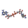 ChemComp-NAI: |
-Macromolecule #5: FLAVIN-ADENINE DINUCLEOTIDE
| Macromolecule | Name: FLAVIN-ADENINE DINUCLEOTIDE / type: ligand / ID: 5 / Number of copies: 2 / Formula: FAD |
|---|---|
| Molecular weight | Theoretical: 785.55 Da |
| Chemical component information | 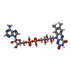 ChemComp-FAD: |
-Experimental details
-Structure determination
| Method | cryo EM |
|---|---|
 Processing Processing | single particle reconstruction |
| Aggregation state | particle |
-Sample preparation
| Concentration | 0.5 mg/mL |
|---|---|
| Buffer | pH: 7.5 / Details: 150mM NaaCl, 20mM Hepes |
| Grid | Model: Quantifoil R1.2/1.3 / Material: COPPER / Mesh: 300 / Pretreatment - Type: PLASMA CLEANING / Pretreatment - Time: 30 sec. |
| Vitrification | Cryogen name: ETHANE / Chamber humidity: 100 % / Chamber temperature: 281 K / Instrument: FEI VITROBOT MARK II |
- Electron microscopy
| Microscope | FEI TALOS ARCTICA |
|---|---|
| Specialist optics | Energy filter - Name: GIF Bioquantum |
| Image recording | Film or detector model: GATAN K3 BIOQUANTUM (6k x 4k) / Average electron dose: 60.0 e/Å2 |
| Electron beam | Acceleration voltage: 300 kV / Electron source:  FIELD EMISSION GUN FIELD EMISSION GUN |
| Electron optics | Illumination mode: SPOT SCAN / Imaging mode: BRIGHT FIELD / Nominal defocus max: 2.0 µm / Nominal defocus min: 1.2 µm / Nominal magnification: 105000 |
| Sample stage | Specimen holder model: FEI TITAN KRIOS AUTOGRID HOLDER / Cooling holder cryogen: NITROGEN |
| Experimental equipment | 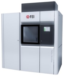 Model: Talos Arctica / Image courtesy: FEI Company |
