 Movie
Movie Controller


+ Open data

- Basic information
| Entry |  | |||||||||
|---|---|---|---|---|---|---|---|---|---|---|
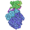
| Title | Cryo-EM structure of the human UBR1 in complex with tryptophan | |||||||||
 Map data Map data | ||||||||||
 Sample Sample |
| |||||||||
 Keywords Keywords | Ubiquitination / LIGASE | |||||||||
| Function / homology |  Function and homology information Function and homology informationL-leucine binding / ubiquitin-dependent protein catabolic process via the N-end rule pathway / cellular response to L-leucine / negative regulation of TOR signaling / ubiquitin ligase complex / proteasome complex / RING-type E3 ubiquitin transferase / ubiquitin protein ligase activity / Antigen processing: Ubiquitination & Proteasome degradation / proteasome-mediated ubiquitin-dependent protein catabolic process ...L-leucine binding / ubiquitin-dependent protein catabolic process via the N-end rule pathway / cellular response to L-leucine / negative regulation of TOR signaling / ubiquitin ligase complex / proteasome complex / RING-type E3 ubiquitin transferase / ubiquitin protein ligase activity / Antigen processing: Ubiquitination & Proteasome degradation / proteasome-mediated ubiquitin-dependent protein catabolic process / protein ubiquitination / zinc ion binding / cytosol / cytoplasm Similarity search - Function | |||||||||
| Biological species |  Homo sapiens (human) Homo sapiens (human) | |||||||||
| Method | single particle reconstruction / cryo EM / Resolution: 3.44 Å | |||||||||
 Authors Authors | Yan R / Hu Z | |||||||||
| Funding support |  China, 1 items China, 1 items
| |||||||||
 Citation Citation | Journal: To Be Published Title: Cryo-EM structure of the human UBR1 in complex with tryptophan Authors: Yan R / Hu Z | |||||||||
| History |
|
- Structure visualization
| Supplemental images |
|---|

- Downloads & links
-EMDB archive
| Map data | emd_65850.map.gz | 327.9 MB | EMDB map data format | |
|---|---|---|---|---|
| Header (meta data) | emd-65850-v30.xmlemd-65850.xml | 15.3 KB 15.3 KB | Display Display | EMDB header |
| Images |  emd_65850.png emd_65850.png | 39.2 KB | ||
| Filedesc metadata | emd-65850.cif.gz | 6.2 KB | ||
| Others | emd_65850_half_map_1.map.gzemd_65850_half_map_2.map.gz | 322.5 MB 322.5 MB | ||
| Archive directory |  http://ftp.pdbj.org/pub/emdb/structures/EMD-65850ftp://ftp.pdbj.org/pub/emdb/structures/EMD-65850 http://ftp.pdbj.org/pub/emdb/structures/EMD-65850ftp://ftp.pdbj.org/pub/emdb/structures/EMD-65850 | HTTPS FTP |
-Related structure data
| Related structure data |  9wbwMC M: atomic model generated by this map C: citing same article ( |
|---|---|
| Similar structure data |
-Links
| EMDB pages | EMDB (EBI/PDBe) / EMDataResource |
|---|---|
| Related items in Molecule of the Month |
-Map
| File | Download / File: emd_65850.map.gz / Format: CCP4 / Size: 347.6 MB / Type: IMAGE STORED AS FLOATING POINT NUMBER (4 BYTES) | ||||||||||||||||||||||||||||||||||||
|---|---|---|---|---|---|---|---|---|---|---|---|---|---|---|---|---|---|---|---|---|---|---|---|---|---|---|---|---|---|---|---|---|---|---|---|---|---|
| Projections & slices | Image control
Images are generated by Spider. | ||||||||||||||||||||||||||||||||||||
| Voxel size | X=Y=Z: 0.67 Å | ||||||||||||||||||||||||||||||||||||
| Density |
| ||||||||||||||||||||||||||||||||||||
| Symmetry | Space group: 1 | ||||||||||||||||||||||||||||||||||||
| Details | EMDB XML:
|
 Z (Sec.)
 Y (Row.)
 X (Col.)


-Supplemental data
-Half map: #2
| File | emd_65850_half_map_1.map | ||||||||||||
|---|---|---|---|---|---|---|---|---|---|---|---|---|---|
| Projections & Slices |
| ||||||||||||
| Density Histograms |


-Half map: #1
| File | emd_65850_half_map_2.map | ||||||||||||
|---|---|---|---|---|---|---|---|---|---|---|---|---|---|
| Projections & Slices |
| ||||||||||||
| Density Histograms |


- Sample components
-Entire : Cryo-EM structure of the human UBR1 in complex with tryptophan
| Entire | Name: Cryo-EM structure of the human UBR1 in complex with tryptophan |
|---|---|
| Components |
|
-Supramolecule #1: Cryo-EM structure of the human UBR1 in complex with tryptophan
| Supramolecule | Name: Cryo-EM structure of the human UBR1 in complex with tryptophan type: complex / ID: 1 / Parent: 0 / Macromolecule list: #1 |
|---|---|
| Source (natural) | Organism: Homo sapiens (human) |
-Macromolecule #1: E3 ubiquitin-protein ligase UBR1
| Macromolecule | Name: E3 ubiquitin-protein ligase UBR1 / type: protein_or_peptide / ID: 1 / Number of copies: 1 / Enantiomer: LEVO / EC number: RING-type E3 ubiquitin transferase |
|---|---|
| Source (natural) | Organism: Homo sapiens (human) |
| Molecular weight | Theoretical: 200.446344 KDa |
| Recombinant expression | Organism: Eukaryota (eukaryotes) |
| Sequence | String: MADEEAGGTE RMEISAELPQ TPQRLASWWD QQVDFYTAFL HHLAQLVPEI YFAEMDPDLE KQEESVQMSI FTPLEWYLFG EDPDICLEK LKHSGAFQLC GRVFKSGETT YSCRDCAIDP TCVLCMDCFQ DSVHKNHRYK MHTSTGGGFC DCGDTEAWKT G PFCVNHEP ...String: MADEEAGGTE RMEISAELPQ TPQRLASWWD QQVDFYTAFL HHLAQLVPEI YFAEMDPDLE KQEESVQMSI FTPLEWYLFG EDPDICLEK LKHSGAFQLC GRVFKSGETT YSCRDCAIDP TCVLCMDCFQ DSVHKNHRYK MHTSTGGGFC DCGDTEAWKT G PFCVNHEP GRAGTIKENS RCPLNEEVIV QARKIFPSVI KYVVEMTIWE EEKELPPELQ IREKNERYYC VLFNDEHHSY DH VIYSLQR ALDCELAEAQ LHTTAIDKEG RRAVKAGAYA ACQEAKEDIK SHSENVSQHP LHVEVLHSEI MAHQKFALRL GSW MNKIMS YSSDFRQIFC QACLREEPDS ENPCLISRLM LWDAKLYKGA RKILHELIFS SFFMEMEYKK LFAMEFVKYY KQLQ KEYIS DDHDRSISIT ALSVQMFTVP TLARHLIEEQ NVISVITETL LEVLPEYLDR NNKFNFQGYS QDKLGRVYAV ICDLK YILI SKPTIWTERL RMQFLEGFRS FLKILTCMQG MEEIRRQVGQ HIEVDPDWEA AIAIQMQLKN ILLMFQEWCA CDEELL LVA YKECHKAVMR CSTSFISSSK TVVQSCGHSL ETKSYRVSED LVSIHLPLSR TLAGLHVRLS RLGAVSRLHE FVSFEDF QV EVLVEYPLRC LVLVAQVVAE MWRRNGLSLI SQVFYYQDVK CREEMYDKDI IMLQIGASLM DPNKFLLLVL QRYELAEA F NKTISTKDQD LIKQYNTLIE EMLQVLIYIV GERYVPGVGN VTKEEVTMRE IIHLLCIEPM PHSAIAKNLP ENENNETGL ENVINKVATF KKPGVSGHGV YELKDESLKD FNMYFYHYSK TQHSKAEHMQ KKRRKQENKD EALPPPPPPE FCPAFSKVIN LLNCDIMMY ILRTVFERAI DTDSNLWTEG MLQMAFHILA LGLLEEKQQL QKAPEEEVTF DFYHKASRLG SSAMNIQMLL E KLKGIPQL EGQKDMITWI LQMFDTVKRL REKSCLIVAT TSGSESIKND EITHDKEKAE RKRKAEAARL HRQKIMAQMS AL QKNFIET HKLMYDNTSE MPGKEDSIME EESTPAVSDY SRIALGPKRG PSVTEKEVLT CILCQEEQEV KIENNAMVLS ACV QKSTAL TQHRGKPIEL SGEALDPLFM DPDLAYGTYT GSCGHVMHAV CWQKYFEAVQ LSSQQRIHVD LFDLESGEYL CPLC KSLCN TVIPIIPLQP QKINSENADA LAQLLTLARW IQTVLARISG YNIRHAKGEN PIPIFFNQGM GDSTLEFHSI LSFGV ESSI KYSNSIKEMV ILFATTIYRI GLKVPPDERD PRVPMLTWST CAFTIQAIEN LLGDEGKPLF GALQNRQHNG LKALMQ FAV AQRITCPQVL IQKHLVRLLS VVLPNIKSED TPCLLSIDLF HVLVGAVLAF PSLYWDDPVD LQPSSVSSSY NHLYLFH LI TMAHMLQILL TVDTGLPLAQ VQEDSEEAHS ASSFFAEISQ YTSGSIGCDI PGWYLWVSLK NGITPYLRCA ALFFHYLL G VTPPEELHTN SAEGEYSALC SYLSLPTNLF LLFQEYWDTV RPLLQRWCAD PALLNCLKQK NTVVRYPRKR NSLIELPDD YSCLLNQASH FRCPRSADDE RKHPVLCLFC GAILCSQNIC CQEIVNGEEV GACIFHALHC GAGVCIFLKI RECRVVLVEG KARGCAYPA PYLDEYGETD PGLKRGNPLH LSRERYRKLH LVWQQHCIIE EIARSQETNQ MLFGFNWQLL UniProtKB: E3 ubiquitin-protein ligase UBR1 |
-Macromolecule #2: TRYPTOPHAN
| Macromolecule | Name: TRYPTOPHAN / type: ligand / ID: 2 / Number of copies: 1 / Formula: TRP |
|---|---|
| Molecular weight | Theoretical: 204.225 Da |
| Chemical component information | 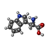 ChemComp-TRP: |
-Experimental details
-Structure determination
| Method | cryo EM |
|---|---|
 Processing Processing | single particle reconstruction |
| Aggregation state | particle |
-Sample preparation
| Buffer | pH: 7.5 |
|---|---|
| Vitrification | Cryogen name: NITROGEN |
- Electron microscopy
| Microscope | FEI POLARA 300 |
|---|---|
| Image recording | Film or detector model: DIRECT ELECTRON APOLLO (4k x 4k) / Average electron dose: 1.5625 e/Å2 |
| Electron beam | Acceleration voltage: 300 kV / Electron source:  FIELD EMISSION GUN FIELD EMISSION GUN |
| Electron optics | Illumination mode: FLOOD BEAM / Imaging mode: BRIGHT FIELD / Nominal defocus max: 1.8 µm / Nominal defocus min: 1.4000000000000001 µm |
| Experimental equipment | 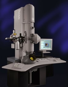 Model: Tecnai Polara / Image courtesy: FEI Company |
