 Movie
Movie Controller

[English] 日本語
 Yorodumi
- EMDB-65849: Cryo-EM structure of the human UBR2 N-domain in complex with tryp... -
+ Open data

- Basic information
| Entry |  | |||||||||
|---|---|---|---|---|---|---|---|---|---|---|
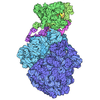
| Title | Cryo-EM structure of the human UBR2 N-domain in complex with tryptophan | |||||||||
 Map data Map data | ||||||||||
 Sample Sample |
| |||||||||
 Keywords Keywords | Ubiquitinalytion / METAL BINDING PROTEIN | |||||||||
| Function / homology |  Function and homology information Function and homology informationhistone H2A ubiquitin ligase activity / L-leucine binding / ubiquitin-dependent protein catabolic process via the N-end rule pathway / male meiotic nuclear division / transposable element silencing / cellular response to L-leucine / positive regulation of T cell receptor signaling pathway / negative regulation of TOR signaling / reciprocal meiotic recombination / ubiquitin ligase complex ...histone H2A ubiquitin ligase activity / L-leucine binding / ubiquitin-dependent protein catabolic process via the N-end rule pathway / male meiotic nuclear division / transposable element silencing / cellular response to L-leucine / positive regulation of T cell receptor signaling pathway / negative regulation of TOR signaling / reciprocal meiotic recombination / ubiquitin ligase complex / protein K63-linked ubiquitination / RING-type E3 ubiquitin transferase / ubiquitin protein ligase activity / Antigen processing: Ubiquitination & Proteasome degradation / heterochromatin formation / spermatogenesis / proteasome-mediated ubiquitin-dependent protein catabolic process / protein ubiquitination / chromatin / zinc ion binding / nucleus / cytosol / cytoplasm Similarity search - Function | |||||||||
| Biological species |  Homo sapiens (human) Homo sapiens (human) | |||||||||
| Method | single particle reconstruction / cryo EM / Resolution: 3.13 Å | |||||||||
 Authors Authors | Yan R / Hu Z | |||||||||
| Funding support |  China, 1 items China, 1 items
| |||||||||
 Citation Citation | Journal: To Be Published Title: Cryo-EM structure of the human UBR2 N-domain in complex with tryptophan Authors: Yan R / Hu Z | |||||||||
| History |
|
- Structure visualization
| Supplemental images |
|---|

- Downloads & links
-EMDB archive
| Map data | emd_65849.map.gz | 229.9 MB | EMDB map data format | |
|---|---|---|---|---|
| Header (meta data) | emd-65849-v30.xmlemd-65849.xml | 15.4 KB 15.4 KB | Display Display | EMDB header |
| Images |  emd_65849.png emd_65849.png | 47.3 KB | ||
| Filedesc metadata | emd-65849.cif.gz | 6.2 KB | ||
| Others | emd_65849_half_map_1.map.gzemd_65849_half_map_2.map.gz | 226.6 MB 226.6 MB | ||
| Archive directory |  http://ftp.pdbj.org/pub/emdb/structures/EMD-65849ftp://ftp.pdbj.org/pub/emdb/structures/EMD-65849 http://ftp.pdbj.org/pub/emdb/structures/EMD-65849ftp://ftp.pdbj.org/pub/emdb/structures/EMD-65849 | HTTPS FTP |
-Related structure data
| Related structure data |  9wbvMC M: atomic model generated by this map C: citing same article ( |
|---|---|
| Similar structure data |
-Links
| EMDB pages | EMDB (EBI/PDBe) / EMDataResource |
|---|---|
| Related items in Molecule of the Month |
-Map
| File | Download / File: emd_65849.map.gz / Format: CCP4 / Size: 244.1 MB / Type: IMAGE STORED AS FLOATING POINT NUMBER (4 BYTES) | ||||||||||||||||||||||||||||||||||||
|---|---|---|---|---|---|---|---|---|---|---|---|---|---|---|---|---|---|---|---|---|---|---|---|---|---|---|---|---|---|---|---|---|---|---|---|---|---|
| Projections & slices | Image control
Images are generated by Spider. | ||||||||||||||||||||||||||||||||||||
| Voxel size | X=Y=Z: 0.668 Å | ||||||||||||||||||||||||||||||||||||
| Density |
| ||||||||||||||||||||||||||||||||||||
| Symmetry | Space group: 1 | ||||||||||||||||||||||||||||||||||||
| Details | EMDB XML:
|
 Z (Sec.)
 Y (Row.)
 X (Col.)


-Supplemental data
-Half map: #2
| File | emd_65849_half_map_1.map | ||||||||||||
|---|---|---|---|---|---|---|---|---|---|---|---|---|---|
| Projections & Slices |
| ||||||||||||
| Density Histograms |


-Half map: #1
| File | emd_65849_half_map_2.map | ||||||||||||
|---|---|---|---|---|---|---|---|---|---|---|---|---|---|
| Projections & Slices |
| ||||||||||||
| Density Histograms |


- Sample components
-Entire : Cryo-EM structure of the human UBR2 N-domain in complex with tryp...
| Entire | Name: Cryo-EM structure of the human UBR2 N-domain in complex with tryptophan |
|---|---|
| Components |
|
-Supramolecule #1: Cryo-EM structure of the human UBR2 N-domain in complex with tryp...
| Supramolecule | Name: Cryo-EM structure of the human UBR2 N-domain in complex with tryptophan type: complex / ID: 1 / Parent: 0 / Macromolecule list: #1 |
|---|---|
| Source (natural) | Organism: Homo sapiens (human) |
-Macromolecule #1: E3 ubiquitin-protein ligase UBR2
| Macromolecule | Name: E3 ubiquitin-protein ligase UBR2 / type: protein_or_peptide / ID: 1 / Number of copies: 1 / Enantiomer: LEVO / EC number: RING-type E3 ubiquitin transferase |
|---|---|
| Source (natural) | Organism: Homo sapiens (human) |
| Molecular weight | Theoretical: 200.781016 KDa |
| Recombinant expression | Organism: Eukaryota (eukaryotes) |
| Sequence | String: MASELEPEVQ AIDRSLLECS AEEIAGKWLQ ATDLTREVYQ HLAHYVPKIY CRGPNPFPQK EDMLAQHVLL GPMEWYLCGE DPAFGFPKL EQANKPSHLC GRVFKVGEPT YSCRDCAVDP TCVLCMECFL GSIHRDHRYR MTTSGGGGFC DCGDTEAWKE G PYCQKHEL ...String: MASELEPEVQ AIDRSLLECS AEEIAGKWLQ ATDLTREVYQ HLAHYVPKIY CRGPNPFPQK EDMLAQHVLL GPMEWYLCGE DPAFGFPKL EQANKPSHLC GRVFKVGEPT YSCRDCAVDP TCVLCMECFL GSIHRDHRYR MTTSGGGGFC DCGDTEAWKE G PYCQKHEL NTSEIEEEED PLVHLSEDVI ARTYNIFAIT FRYAVEILTW EKESELPADL EMVEKSDTYY CMLFNDEVHT YE QVIYTLQ KAVNCTQKEA IGFATTVDRD GRRSVRYGDF QYCEQAKSVI VRNTSRQTKP LKVQVMHSSI VAHQNFGLKL LSW LGSIIG YSDGLRRILC QVGLQEGPDG ENSSLVDRLM LSDSKLWKGA RSVYHQLFMS SLLMDLKYKK LFAVRFAKNY QQLQ RDFME DDHERAVSVT ALSVQFFTAP TLARMLITEE NLMSIIIKTF MDHLRHRDAQ GRFQFERYTA LQAFKFRRVQ SLILD LKYV LISKPTEWSD ELRQKFLEGF DAFLELLKCM QGMDPITRQV GQHIEMEPEW EAAFTLQMKL THVISMMQDW CASDEK VLI EAYKKCLAVL MQCHGGYTDG EQPITLSICG HSVETIRYCV SQEKVSIHLP VSRLLAGLHV LLSKSEVAYK FPELLPL SE LSPPMLIEHP LRCLVLCAQV HAGMWRRNGF SLVNQIYYYH NVKCRREMFD KDVVMLQTGV SMMDPNHFLM IMLSRFEL Y QIFSTPDYGK RFSSEITHKD VVQQNNTLIE EMLYLIIMLV GERFSPGVGQ VNATDEIKRE IIHQLSIKPM AHSELVKSL PEDENKETGM ESVIEAVAHF KKPGLTGRGM YELKPECAKE FNLYFYHFSR AEQSKAEEAQ RKLKRQNRED TALPPPVLPP FCPLFASLV NILQSDVMLC IMGTILQWAV EHNGYAWSES MLQRVLHLIG MALQEEKQHL ENVTEEHVVT FTFTQKISKP G EAPKNSPS ILAMLETLQN APYLEVHKDM IRWILKTFNA VKKMRESSPT SPVAETEGTI MEESSRDKDK AERKRKAEIA RL RREKIMA QMSEMQRHFI DENKELFQQT LELDASTSAV LDHSPVASDM TLTALGPAQT QVPEQRQFVT CILCQEEQEV KVE SRAMVL AAFVQRSTVL SKNRSKFIQD PEKYDPLFMH PDLSCGTHTS SCGHIMHAHC WQRYFDSVQA KEQRRQQRLR LHTS YDVEN GEFLCPLCEC LSNTVIPLLL PPRNIFNNRL NFSDQPNLTQ WIRTISQQIK ALQFLRKEES TPNNASTKNS ENVDE LQLP EGFRPDFRPK IPYSESIKEM LTTFGTATYK VGLKVHPNEE DPRVPIMCWG SCAYTIQSIE RILSDEDKPL FGPLPC RLD DCLRSLTRFA AAHWTVASVS VVQGHFCKLF ASLVPNDSHE ELPCILDIDM FHLLVGLVLA FPALQCQDFS GISLGTG DL HIFHLVTMAH IIQILLTSCT EENGMDQENP PCEEESAVLA LYKTLHQYTG SALKEIPSGW HLWRSVRAGI MPFLKCSA L FFHYLNGVPS PPDIQVPGTS HFEHLCSYLS LPNNLICLFQ ENSEIMNSLI ESWCRNSEVK RYLEGERDAI RYPRESNKL INLPEDYSSL INQASNFSCP KSGGDKSRAP TLCLVCGSLL CSQSYCCQTE LEGEDVGACT AHTYSCGSGV GIFLRVRECQ VLFLAGKTK GCFYSPPYLD DYGETDQGLR RGNPLHLCKE RFKKIQKLWH QHSVTEEIGH AQEANQTLVG IDWQHL UniProtKB: E3 ubiquitin-protein ligase UBR2 |
-Macromolecule #2: TRYPTOPHAN
| Macromolecule | Name: TRYPTOPHAN / type: ligand / ID: 2 / Number of copies: 1 / Formula: TRP |
|---|---|
| Molecular weight | Theoretical: 204.225 Da |
| Chemical component information | 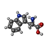 ChemComp-TRP: |
-Experimental details
-Structure determination
| Method | cryo EM |
|---|---|
 Processing Processing | single particle reconstruction |
| Aggregation state | particle |
-Sample preparation
| Buffer | pH: 7.5 |
|---|---|
| Vitrification | Cryogen name: NITROGEN |
- Electron microscopy
| Microscope | FEI POLARA 300 |
|---|---|
| Image recording | Film or detector model: DIRECT ELECTRON APOLLO (4k x 4k) / Average electron dose: 1.5625 e/Å2 |
| Electron beam | Acceleration voltage: 300 kV / Electron source:  FIELD EMISSION GUN FIELD EMISSION GUN |
| Electron optics | Illumination mode: FLOOD BEAM / Imaging mode: BRIGHT FIELD / Nominal defocus max: 1.8 µm / Nominal defocus min: 1.4000000000000001 µm |
| Experimental equipment | 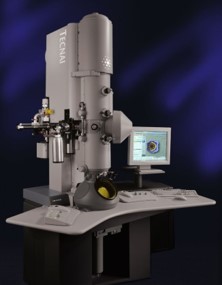 Model: Tecnai Polara / Image courtesy: FEI Company |
