[English] 日本語
Yorodumi
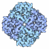
- EMDB-65615: Structure of adenosine A2AR with an agonist MRS3997 -

+
Open data


ID or keywords:

Loading...

-
Basic information

Entry
Database: EMDB / ID: EMD-65615
TitleStructure of adenosine A2AR with an agonist MRS3997
Map data
Sample
  • Complex: Structure of adenosine A2AR with an agonist MRS3997
    • Protein or peptide: Guanine nucleotide-binding protein G(s) subunit alpha isoforms short
    • Protein or peptide: Guanine nucleotide-binding protein G(I)/G(S)/G(O) subunit gamma-2
    • Protein or peptide: scFv16
    • Protein or peptide: Guanine nucleotide-binding protein G(I)/G(S)/G(T) subunit beta-1
    • Protein or peptide: Soluble cytochrome b562,Adenosine receptor A2a,Soluble cytochrome b562,Adenosine receptor A2a,LgBiT
  • Ligand: OLEIC ACID
  • Ligand: CHOLESTEROL
  • Ligand: (2R,3R,4S,5R)-2-[6-azanyl-2-[2-(6-bromanyl-1H-indol-3-yl)ethoxy]purin-9-yl]-5-(hydroxymethyl)oxolane-3,4-diol
  • Ligand: water
KeywordsGPCR / SIGNALING PROTEIN / MEMBRANE PROTEIN/IMMUNE SYSTEM / MEMBRANE PROTEIN-IMMUNE SYSTEM complex
Function / homology
Function and homology information


regulation of norepinephrine secretion / positive regulation of circadian sleep/wake cycle, sleep / Adenosine P1 receptors / positive regulation of acetylcholine secretion, neurotransmission / G protein-coupled adenosine receptor activity / response to purine-containing compound / G protein-coupled adenosine receptor signaling pathway / NGF-independant TRKA activation / Surfactant metabolism / type 5 metabotropic glutamate receptor binding ...regulation of norepinephrine secretion / positive regulation of circadian sleep/wake cycle, sleep / Adenosine P1 receptors / positive regulation of acetylcholine secretion, neurotransmission / G protein-coupled adenosine receptor activity / response to purine-containing compound / G protein-coupled adenosine receptor signaling pathway / NGF-independant TRKA activation / Surfactant metabolism / type 5 metabotropic glutamate receptor binding / negative regulation of vascular permeability / positive regulation of urine volume / intermediate filament / response to caffeine / blood circulation / presynaptic active zone / sensory perception / eating behavior / positive regulation of glutamate secretion / synaptic transmission, cholinergic / regulation of calcium ion transport / alpha-actinin binding / synaptic transmission, dopaminergic / membrane depolarization / asymmetric synapse / axolemma / prepulse inhibition / cellular defense response / phagocytosis / neuron projection morphogenesis / positive regulation of synaptic transmission, glutamatergic / presynaptic modulation of chemical synaptic transmission / astrocyte activation / regulation of mitochondrial membrane potential / excitatory postsynaptic potential / positive regulation of protein secretion / locomotory behavior / central nervous system development / positive regulation of long-term synaptic potentiation / electron transport chain / synaptic transmission, glutamatergic / vasodilation / blood coagulation / adenylate cyclase-modulating G protein-coupled receptor signaling pathway / phospholipase C-activating G protein-coupled receptor signaling pathway / Olfactory Signaling Pathway / Activation of the phototransduction cascade / G protein-coupled acetylcholine receptor signaling pathway / G beta:gamma signalling through PLC beta / Presynaptic function of Kainate receptors / Thromboxane signalling through TP receptor / Activation of G protein gated Potassium channels / Inhibition of voltage gated Ca2+ channels via Gbeta/gamma subunits / G-protein activation / Glucagon signaling in metabolic regulation / G beta:gamma signalling through CDC42 / Prostacyclin signalling through prostacyclin receptor / Synthesis, secretion, and inactivation of Glucagon-like Peptide-1 (GLP-1) / G beta:gamma signalling through BTK / photoreceptor disc membrane / ADP signalling through P2Y purinoceptor 12 / Glucagon-type ligand receptors / Sensory perception of sweet, bitter, and umami (glutamate) taste / Adrenaline,noradrenaline inhibits insulin secretion / cell-cell signaling / Vasopressin regulates renal water homeostasis via Aquaporins / Glucagon-like Peptide-1 (GLP1) regulates insulin secretion / G alpha (z) signalling events / cellular response to catecholamine stimulus / ADP signalling through P2Y purinoceptor 1 / G beta:gamma signalling through PI3Kgamma / ADORA2B mediated anti-inflammatory cytokines production / adenylate cyclase-activating dopamine receptor signaling pathway / cellular response to prostaglandin E stimulus / Cooperation of PDCL (PhLP1) and TRiC/CCT in G-protein beta folding / GPER1 signaling / heterotrimeric G-protein complex / Inactivation, recovery and regulation of the phototransduction cascade / G alpha (12/13) signalling events / G-protein beta-subunit binding / extracellular vesicle / Thrombin signalling through proteinase activated receptors (PARs) / signaling receptor complex adaptor activity / adenylate cyclase-activating G protein-coupled receptor signaling pathway / GTPase binding / presynaptic membrane / Ca2+ pathway / negative regulation of neuron apoptotic process / High laminar flow shear stress activates signaling by PIEZO1 and PECAM1:CDH5:KDR in endothelial cells / G alpha (i) signalling events / G alpha (s) signalling events / G alpha (q) signalling events / Ras protein signal transduction / positive regulation of ERK1 and ERK2 cascade / calmodulin binding / electron transfer activity / periplasmic space / postsynaptic membrane / Extra-nuclear estrogen signaling / response to xenobiotic stimulus
Similarity search - Function
Adenosine A2A receptor / Adenosine receptor / Cytochrome b562 / Cytochrome b562 / Cytochrome c/b562 / Serpentine type 7TM GPCR chemoreceptor Srsx / G-protein, gamma subunit / G-protein gamma subunit domain profile. / G-protein gamma-like domain / G-protein gamma-like domain superfamily ...Adenosine A2A receptor / Adenosine receptor / Cytochrome b562 / Cytochrome b562 / Cytochrome c/b562 / Serpentine type 7TM GPCR chemoreceptor Srsx / G-protein, gamma subunit / G-protein gamma subunit domain profile. / G-protein gamma-like domain / G-protein gamma-like domain superfamily / GGL domain / G protein gamma subunit-like motifs / GGL domain / G protein beta WD-40 repeat protein / Guanine nucleotide-binding protein, beta subunit / G-protein, beta subunit / G-protein coupled receptors family 1 signature. / 7 transmembrane receptor (rhodopsin family) / G protein-coupled receptor, rhodopsin-like / GPCR, rhodopsin-like, 7TM / G-protein coupled receptors family 1 profile. / G-protein beta WD-40 repeat / WD40 repeat, conserved site / Trp-Asp (WD) repeats signature. / Trp-Asp (WD) repeats profile. / Trp-Asp (WD) repeats circular profile. / WD40 repeats / WD40 repeat / WD40-repeat-containing domain superfamily / WD40/YVTN repeat-like-containing domain superfamily
Similarity search - Domain/homology
Soluble cytochrome b562 / Adenosine receptor A2a / Guanine nucleotide-binding protein G(I)/G(S)/G(O) subunit gamma-2 / Guanine nucleotide-binding protein G(I)/G(S)/G(T) subunit beta-1
Similarity search - Component
Biological speciesHomo sapiens (human) / Mus musculus (house mouse)
Methodsingle particle reconstruction / cryo EM / Resolution: 2.71 Å
AuthorsWeng Y / Chen Y / Pavan M / Gao ZG / Jacobson K / Song GJ
Funding support China, 1 items
OrganizationGrant numberCountry
National Natural Science Foundation of China (NSFC)32171215 China
CitationJournal: To Be Published
Title: Structure of adenosine A2AR with an agonist MRS3997
Authors: Song GJ / Weng Y / Chen Y
History
DepositionJul 30, 2025-
Header (metadata) releaseAug 5, 2026-
Map releaseAug 5, 2026-
UpdateAug 5, 2026-
Current statusAug 5, 2026Processing site: PDBc / Status: Released

-
Structure visualization

Supplemental images

Downloads & links

-
Map

FileDownload / File: emd_65615.map.gz / Format: CCP4 / Size: 178 MB / Type: IMAGE STORED AS FLOATING POINT NUMBER (4 BYTES)
Projections & slices

Image control

Size
Brightness
Contrast
Others
AxesZ (Sec.)Y (Row.)X (Col.)
0.66 Å/pix.
x 360 pix.
= 237.6 Å
0.66 Å/pix.
x 360 pix.
= 237.6 Å
0.66 Å/pix.
x 360 pix.
= 237.6 Å

Surface

Projections

Slices (1/3)

Slices (1/2)

Slices (2/3)

Images are generated by Spider.

Voxel sizeX=Y=Z: 0.66 Å
Density
Contour LevelBy AUTHOR: 0.134
Minimum - Maximum-0.8368168 - 1.297586
Average (Standard dev.)-0.00033422498 (±0.025015498)
SymmetrySpace group: 1
Details

EMDB XML:

Map geometry
Axis orderXYZ
Origin000
Dimensions360360360
Spacing360360360
CellA=B=C: 237.6 Å
α=β=γ: 90.0 °

-
Supplemental data

-
Half map: #2

Fileemd_65615_half_map_1.map
Projections & Slices
AxesZYX

Projections

Slices (1/2)
Density Histograms

-
Half map: #1

Fileemd_65615_half_map_2.map
Projections & Slices
AxesZYX

Projections

Slices (1/2)
Density Histograms

-
Sample components

+
Entire : Structure of adenosine A2AR with an agonist MRS3997

EntireName: Structure of adenosine A2AR with an agonist MRS3997
Components
  • Complex: Structure of adenosine A2AR with an agonist MRS3997
    • Protein or peptide: Guanine nucleotide-binding protein G(s) subunit alpha isoforms short
    • Protein or peptide: Guanine nucleotide-binding protein G(I)/G(S)/G(O) subunit gamma-2
    • Protein or peptide: scFv16
    • Protein or peptide: Guanine nucleotide-binding protein G(I)/G(S)/G(T) subunit beta-1
    • Protein or peptide: Soluble cytochrome b562,Adenosine receptor A2a,Soluble cytochrome b562,Adenosine receptor A2a,LgBiT
  • Ligand: OLEIC ACID
  • Ligand: CHOLESTEROL
  • Ligand: (2R,3R,4S,5R)-2-[6-azanyl-2-[2-(6-bromanyl-1H-indol-3-yl)ethoxy]purin-9-yl]-5-(hydroxymethyl)oxolane-3,4-diol
  • Ligand: water

+
Supramolecule #1: Structure of adenosine A2AR with an agonist MRS3997

SupramoleculeName: Structure of adenosine A2AR with an agonist MRS3997 / type: complex / ID: 1 / Parent: 0 / Macromolecule list: #1-#5
Source (natural)Organism: Homo sapiens (human)

+
Macromolecule #1: Guanine nucleotide-binding protein G(s) subunit alpha isoforms short

MacromoleculeName: Guanine nucleotide-binding protein G(s) subunit alpha isoforms short
type: protein_or_peptide / ID: 1 / Number of copies: 1 / Enantiomer: LEVO
Source (natural)Organism: Homo sapiens (human)
Molecular weightTheoretical: 28.146844 KDa
Recombinant expressionOrganism: Spodoptera frugiperda (fall armyworm)

SequenceString: TVSAEDKAAA ERSKMIEKQL QKDKQVYRAT HRLLLLGADN SGKSTIVKQM RIYHGGSGGS GGTSGIFETK FQVDKVNFHM FDVGGQRDE RRKWIQCFND VTAIIFVVDS SDYNRLQEAL NDFKSIWNNR WLRTISVILF LNKQDLLAEK VLAGKSKIED Y FPEFARYT ...String:
TVSAEDKAAA ERSKMIEKQL QKDKQVYRAT HRLLLLGADN SGKSTIVKQM RIYHGGSGGS GGTSGIFETK FQVDKVNFHM FDVGGQRDE RRKWIQCFND VTAIIFVVDS SDYNRLQEAL NDFKSIWNNR WLRTISVILF LNKQDLLAEK VLAGKSKIED Y FPEFARYT TPEDATPEPG EDPRVTRAKY FIRDEFLRIS TASGDGRHYC YPHFTCAVDT ENARRIFNDC RDIIQRMHLR QY ELL

+
Macromolecule #2: Guanine nucleotide-binding protein G(I)/G(S)/G(O) subunit gamma-2

MacromoleculeName: Guanine nucleotide-binding protein G(I)/G(S)/G(O) subunit gamma-2
type: protein_or_peptide / ID: 2 / Number of copies: 1 / Enantiomer: LEVO
Source (natural)Organism: Homo sapiens (human)
Molecular weightTheoretical: 7.891022 KDa
Recombinant expressionOrganism: Spodoptera frugiperda (fall armyworm)
SequenceString:
MASNNTASIA QARKLVEQLK MEANIDRIKV SKAAADLMAY CEAHAKEDPL LTPVPASENP FREKKFFGSA GSA

UniProtKB: Guanine nucleotide-binding protein G(I)/G(S)/G(O) subunit gamma-2

+
Macromolecule #3: scFv16

MacromoleculeName: scFv16 / type: protein_or_peptide / ID: 3 / Number of copies: 1 / Enantiomer: LEVO
Source (natural)Organism: Mus musculus (house mouse)
Molecular weightTheoretical: 30.765471 KDa
Recombinant expressionOrganism: Spodoptera frugiperda (fall armyworm)
SequenceString: MLLVNQSHQG FNKEHTSKMV SAIVLYVLLA AAAHSAFADV QLVESGGGLV QPGGSRKLSC SASGFAFSSF GMHWVRQAPE KGLEWVAYI SSGSGTIYYA DTVKGRFTIS RDDPKNTLFL QMTSLRSEDT AMYYCVRSIY YYGSSPFDFW GQGTTLTVSS G GGGSGGGG ...String:
MLLVNQSHQG FNKEHTSKMV SAIVLYVLLA AAAHSAFADV QLVESGGGLV QPGGSRKLSC SASGFAFSSF GMHWVRQAPE KGLEWVAYI SSGSGTIYYA DTVKGRFTIS RDDPKNTLFL QMTSLRSEDT AMYYCVRSIY YYGSSPFDFW GQGTTLTVSS G GGGSGGGG SGGGGSDIVM TQATSSVPVT PGESVSISCR SSKSLLHSNG NTYLYWFLQR PGQSPQLLIY RMSNLASGVP DR FSGSGSG TAFTLTISRL EAEDVGVYYC MQHLEYPLTF GAGTKLELKA AA

+
Macromolecule #4: Guanine nucleotide-binding protein G(I)/G(S)/G(T) subunit beta-1

MacromoleculeName: Guanine nucleotide-binding protein G(I)/G(S)/G(T) subunit beta-1
type: protein_or_peptide / ID: 4 / Number of copies: 1 / Enantiomer: LEVO
Source (natural)Organism: Homo sapiens (human)
Molecular weightTheoretical: 39.728426 KDa
Recombinant expressionOrganism: Spodoptera frugiperda (fall armyworm)
SequenceString: MSELDQLRQE AEQLKNQIRD ARKACADATL SQITNNIDPV GRIQMRTRRT LRGHLAKIYA MHWGTDSRLL VSASQDGKLI IWDSYTTNK VHAIPLRSSW VMTCAYAPSG NYVACGGLDN ICSIYNLKTR EGNVRVSREL AGHTGYLSCC RFLDDNQIVT S SGDTTCAL ...String:
MSELDQLRQE AEQLKNQIRD ARKACADATL SQITNNIDPV GRIQMRTRRT LRGHLAKIYA MHWGTDSRLL VSASQDGKLI IWDSYTTNK VHAIPLRSSW VMTCAYAPSG NYVACGGLDN ICSIYNLKTR EGNVRVSREL AGHTGYLSCC RFLDDNQIVT S SGDTTCAL WDIETGQQTT TFTGHTGDVM SLSLAPDTRL FVSGACDASA KLWDVREGMC RQTFTGHESD INAICFFPNG NA FATGSDD ATCRLFDLRA DQELMTYSHD NIICGITSVS FSKSGRLLLA GYDDFNCNVW DALKADRAGV LAGHDNRVSC LGV TDDGMA VATGSWDSFL KIWNGSSGGG GSGGGGSSGV SGWRLFKKIS

UniProtKB: Guanine nucleotide-binding protein G(I)/G(S)/G(T) subunit beta-1

+
Macromolecule #5: Soluble cytochrome b562,Adenosine receptor A2a,Soluble cytochrome...

MacromoleculeName: Soluble cytochrome b562,Adenosine receptor A2a,Soluble cytochrome b562,Adenosine receptor A2a,LgBiT
type: protein_or_peptide / ID: 5 / Number of copies: 1 / Enantiomer: LEVO
Source (natural)Organism: Homo sapiens (human)
Molecular weightTheoretical: 67.350023 KDa
Recombinant expressionOrganism: Spodoptera frugiperda (fall armyworm)
SequenceString: ADLEDNWETL NDNLKVIEKA DNAAQVKDAL TKMRAAALDA QKATPPKLED KSPDSPEMKD FRHGFDILVG QIDDALKLAN EGKVKEAQA AAEQLKTTRN AYIQKYLENL YFQSPIMGSS VYITVELAIA VLAILGNVLV CWAVWLNSNL QNVTNYFVVS L AAADIAVG ...String:
ADLEDNWETL NDNLKVIEKA DNAAQVKDAL TKMRAAALDA QKATPPKLED KSPDSPEMKD FRHGFDILVG QIDDALKLAN EGKVKEAQA AAEQLKTTRN AYIQKYLENL YFQSPIMGSS VYITVELAIA VLAILGNVLV CWAVWLNSNL QNVTNYFVVS L AAADIAVG VLAIPFAITI STGFCAACHG CLFIACFVLV LTQSSIFSLL AIAIDRYIAI RIPLRYNGLV TGTRAKGIIA IC WVLSFAI GLTPMLGWNN CGQPKEGKNH SQGCGEGQVA CLFEDVVPMN YMVYFNFFAC VLVPLLLMLG VYLRIFLAAR RQL KQMESQ PLPGERARST LQKEVHAAKS LAIIVGLFAL CWLPLHIINC FTFFCPDCSH APLWLMYLAI VLSHTNSVVN PFIY AYRIR EFRQTFRKII RSHVLRQQEP FKAAGTSARV LAAHGSDGEQ VSLRLVFTLE DFVGDWEQTA AYNLDQVLEQ GGVSS LLQN LAVSVTPIQR IVRSGENALK IDIHVIIPYE GLSADQMAQI EEVFKVVYPV DDHHFKVILP YGTLVIDGVT PNMLNY FGR PYEGIAVFDG KKITVTGTLW NGNKIIDERL ITPDGSMLFR VTIN

UniProtKB: Soluble cytochrome b562, Adenosine receptor A2a

+
Macromolecule #6: OLEIC ACID

MacromoleculeName: OLEIC ACID / type: ligand / ID: 6 / Number of copies: 2 / Formula: OLA
Molecular weightTheoretical: 282.461 Da
Chemical component information

ChemComp-OLA:
OLEIC ACID

+
Macromolecule #7: CHOLESTEROL

MacromoleculeName: CHOLESTEROL / type: ligand / ID: 7 / Number of copies: 3 / Formula: CLR
Molecular weightTheoretical: 386.654 Da
Chemical component information

ChemComp-CLR:
CHOLESTEROL

+
Macromolecule #8: (2R,3R,4S,5R)-2-[6-azanyl-2-[2-(6-bromanyl-1H-indol-3-yl)ethoxy]p...

MacromoleculeName: (2R,3R,4S,5R)-2-[6-azanyl-2-[2-(6-bromanyl-1H-indol-3-yl)ethoxy]purin-9-yl]-5-(hydroxymethyl)oxolane-3,4-diol
type: ligand / ID: 8 / Number of copies: 1 / Formula: A1EUU
Molecular weightTheoretical: 505.322 Da

+
Macromolecule #9: water

MacromoleculeName: water / type: ligand / ID: 9 / Number of copies: 4 / Formula: HOH
Molecular weightTheoretical: 18.015 Da
Chemical component information

ChemComp-HOH:
WATER

-
Experimental details

-
Structure determination

Methodcryo EM
Processingsingle particle reconstruction
Aggregation stateparticle

-
Sample preparation

BufferpH: 7.5
VitrificationCryogen name: ETHANE

-
Electron microscopy

MicroscopeTFS KRIOS
Image recordingFilm or detector model: GATAN K3 BIOQUANTUM (6k x 4k) / Average electron dose: 60.0 e/Å2
Electron beamAcceleration voltage: 300 kV / Electron source: FIELD EMISSION GUN
Electron opticsIllumination mode: FLOOD BEAM / Imaging mode: BRIGHT FIELD / Nominal defocus max: 2.0 µm / Nominal defocus min: 1.0 µm
Experimental equipment
Model: Titan Krios / Image courtesy: FEI Company

+
Image processing

CTF correctionType: PHASE FLIPPING AND AMPLITUDE CORRECTION
Startup modelType of model: INSILICO MODEL
Final reconstructionResolution.type: BY AUTHOR / Resolution: 2.71 Å / Resolution method: FSC 0.143 CUT-OFF / Software - Name: PHENIX / Number images used: 734128
Initial angle assignmentType: MAXIMUM LIKELIHOOD
Final angle assignmentType: MAXIMUM LIKELIHOOD

+
About Yorodumi

-
News

-
Feb 9, 2022. New format data for meta-information of EMDB entries

New format data for meta-information of EMDB entries

  • Version 3 of the EMDB header file is now the official format.
  • The previous official version 1.9 will be removed from the archive.

Related info.:EMDB header

External links:wwPDB to switch to version 3 of the EMDB data model

-
Aug 12, 2020. Covid-19 info

Covid-19 info

URL: https://pdbj.org/emnavi/covid19.php

New page: Covid-19 featured information page in EM Navigator.

Related info.:Covid-19 info / Mar 5, 2020. Novel coronavirus structure data

+
Mar 5, 2020. Novel coronavirus structure data

Novel coronavirus structure data

Related info.:Yorodumi Speices / Aug 12, 2020. Covid-19 info

External links:COVID-19 featured content - PDBj / Molecule of the Month (242):Coronavirus Proteases

+
Jan 31, 2019. EMDB accession codes are about to change! (news from PDBe EMDB page)

EMDB accession codes are about to change! (news from PDBe EMDB page)

  • The allocation of 4 digits for EMDB accession codes will soon come to an end. Whilst these codes will remain in use, new EMDB accession codes will include an additional digit and will expand incrementally as the available range of codes is exhausted. The current 4-digit format prefixed with “EMD-” (i.e. EMD-XXXX) will advance to a 5-digit format (i.e. EMD-XXXXX), and so on. It is currently estimated that the 4-digit codes will be depleted around Spring 2019, at which point the 5-digit format will come into force.
  • The EM Navigator/Yorodumi systems omit the EMD- prefix.

Related info.:Q: What is EMD? / ID/Accession-code notation in Yorodumi/EM Navigator

External links:EMDB Accession Codes are Changing Soon! / Contact to PDBj

+
Jul 12, 2017. Major update of PDB

Major update of PDB

  • wwPDB released updated PDB data conforming to the new PDBx/mmCIF dictionary.
  • This is a major update changing the version number from 4 to 5, and with Remediation, in which all the entries are updated.
  • In this update, many items about electron microscopy experimental information are reorganized (e.g. em_software).
  • Now, EM Navigator and Yorodumi are based on the updated data.

External links:wwPDB Remediation / Enriched Model Files Conforming to OneDep Data Standards Now Available in the PDB FTP Archive

-
Yorodumi

Thousand views of thousand structures

  • Yorodumi is a browser for structure data from EMDB, PDB, SASBDB, etc.
  • This page is also the successor to EM Navigator detail page, and also detail information page/front-end page for Omokage search.
  • The word "yorodu" (or yorozu) is an old Japanese word meaning "ten thousand". "mi" (miru) is to see.

Related info.:EMDB / PDB / SASBDB / Comparison of 3 databanks / Yorodumi Search / Aug 31, 2016. New EM Navigator & Yorodumi / Yorodumi Papers / Jmol/JSmol / Function and homology information / Changes in new EM Navigator and Yorodumi

Read more