 Movie
Movie Controller


+ Open data

- Basic information
| Entry |  | |||||||||
|---|---|---|---|---|---|---|---|---|---|---|
| Title | The structure of ITN bound OXGR1 | |||||||||
 Map data Map data | ||||||||||
 Sample Sample |
| |||||||||
 Keywords Keywords | GPCR / MEMBRANE PROTEIN | |||||||||
| Function / homology |  Function and homology information Function and homology informationClass A/1 (Rhodopsin-like receptors) / detection of maltose stimulus / carbohydrate transport / carbohydrate transmembrane transporter activity / maltose binding / maltose transport / maltodextrin transmembrane transport / ATP-binding cassette (ABC) transporter complex, substrate-binding subunit-containing / ATP-binding cassette (ABC) transporter complex / electron transport chain ...Class A/1 (Rhodopsin-like receptors) / detection of maltose stimulus / carbohydrate transport / carbohydrate transmembrane transporter activity / maltose binding / maltose transport / maltodextrin transmembrane transport / ATP-binding cassette (ABC) transporter complex, substrate-binding subunit-containing / ATP-binding cassette (ABC) transporter complex / electron transport chain / cell chemotaxis / G protein-coupled receptor activity / Olfactory Signaling Pathway / phospholipase C-activating G protein-coupled receptor signaling pathway / Activation of the phototransduction cascade / G protein-coupled acetylcholine receptor signaling pathway / G beta:gamma signalling through PLC beta / Presynaptic function of Kainate receptors / Thromboxane signalling through TP receptor / Activation of G protein gated Potassium channels / Inhibition of voltage gated Ca2+ channels via Gbeta/gamma subunits / G-protein activation / Glucagon signaling in metabolic regulation / G beta:gamma signalling through CDC42 / Prostacyclin signalling through prostacyclin receptor / Synthesis, secretion, and inactivation of Glucagon-like Peptide-1 (GLP-1) / photoreceptor disc membrane / G beta:gamma signalling through BTK / ADP signalling through P2Y purinoceptor 12 / Sensory perception of sweet, bitter, and umami (glutamate) taste / Glucagon-type ligand receptors / Adrenaline,noradrenaline inhibits insulin secretion / Vasopressin regulates renal water homeostasis via Aquaporins / G alpha (z) signalling events / Glucagon-like Peptide-1 (GLP1) regulates insulin secretion / ADP signalling through P2Y purinoceptor 1 / cellular response to catecholamine stimulus / G beta:gamma signalling through PI3Kgamma / ADORA2B mediated anti-inflammatory cytokines production / adenylate cyclase-activating dopamine receptor signaling pathway / cellular response to prostaglandin E stimulus / Cooperation of PDCL (PhLP1) and TRiC/CCT in G-protein beta folding / GPER1 signaling / outer membrane-bounded periplasmic space / heterotrimeric G-protein complex / Inactivation, recovery and regulation of the phototransduction cascade / G alpha (12/13) signalling events / G-protein beta-subunit binding / extracellular vesicle / Thrombin signalling through proteinase activated receptors (PARs) / signaling receptor complex adaptor activity / signaling receptor activity / GTPase binding / Ca2+ pathway / High laminar flow shear stress activates signaling by PIEZO1 and PECAM1:CDH5:KDR in endothelial cells / G alpha (i) signalling events / G alpha (s) signalling events / G alpha (q) signalling events / Ras protein signal transduction / electron transfer activity / periplasmic space / Extra-nuclear estrogen signaling / iron ion binding / G protein-coupled receptor signaling pathway / innate immune response / lysosomal membrane / heme binding / GTPase activity / DNA damage response / synapse / protein-containing complex binding / signal transduction / extracellular exosome / membrane / plasma membrane / cytosol / cytoplasm Similarity search - Function | |||||||||
| Biological species |  Homo sapiens (human) / Homo sapiens (human) /   Escherichia coli K-12 (bacteria) / Escherichia coli K-12 (bacteria) /   Mus musculus (house mouse) Mus musculus (house mouse) | |||||||||
| Method | single particle reconstruction / cryo EM / Resolution: 2.62 Å | |||||||||
 Authors Authors | Su Q / Liang EY / Ma TS / Chen ZY / Tang XF | |||||||||
| Funding support |  China, 1 items China, 1 items
| |||||||||
 Citation Citation | Journal: To Be Published Title: The structure of ITN bound OXGR1 Authors: Su Q / Liang EY / Ma TS / Chen ZY / Tang XF | |||||||||
| History |
|
- Structure visualization
| Supplemental images |
|---|

- Downloads & links
-EMDB archive
| Map data | emd_65376.map.gz | 114.4 MB | EMDB map data format | |
|---|---|---|---|---|
| Header (meta data) | emd-65376-v30.xmlemd-65376.xml | 22 KB 22 KB | Display Display | EMDB header |
| Images |  emd_65376.png emd_65376.png | 122.5 KB | ||
| Filedesc metadata | emd-65376.cif.gz | 7.5 KB | ||
| Others | emd_65376_half_map_1.map.gzemd_65376_half_map_2.map.gz | 120.6 MB 120.6 MB | ||
| Archive directory |  https://data.pdbj.org/pub/emdb/structures/EMD-65376ftp://data.pdbj.org/pub/emdb/structures/EMD-65376 https://data.pdbj.org/pub/emdb/structures/EMD-65376ftp://data.pdbj.org/pub/emdb/structures/EMD-65376 | HTTPS FTP |
-Related structure data
| Related structure data |  9vv6MC M: atomic model generated by this map C: citing same article ( |
|---|---|
| Similar structure data |
-Links
| EMDB pages | EMDB (EBI/PDBe) / EMDataResource |
|---|---|
| Related items in Molecule of the Month |

















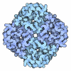





-Map
| File | Download / File: emd_65376.map.gz / Format: CCP4 / Size: 129.7 MB / Type: IMAGE STORED AS FLOATING POINT NUMBER (4 BYTES) | ||||||||||||||||||||||||||||||||||||
|---|---|---|---|---|---|---|---|---|---|---|---|---|---|---|---|---|---|---|---|---|---|---|---|---|---|---|---|---|---|---|---|---|---|---|---|---|---|
| Projections & slices | Image control
Images are generated by Spider. | ||||||||||||||||||||||||||||||||||||
| Voxel size | X=Y=Z: 0.936 Å | ||||||||||||||||||||||||||||||||||||
| Density |
| ||||||||||||||||||||||||||||||||||||
| Symmetry | Space group: 1 | ||||||||||||||||||||||||||||||||||||
| Details | EMDB XML:
|
 Z (Sec.)
 Y (Row.)
 X (Col.)


-Supplemental data
-Half map: #2
| File | emd_65376_half_map_1.map | ||||||||||||
|---|---|---|---|---|---|---|---|---|---|---|---|---|---|
| Projections & Slices |
| ||||||||||||
| Density Histograms |


-Half map: #1
| File | emd_65376_half_map_2.map | ||||||||||||
|---|---|---|---|---|---|---|---|---|---|---|---|---|---|
| Projections & Slices |
| ||||||||||||
| Density Histograms |


- Sample components
-Entire : ITN bound OXGR1
| Entire | Name: ITN bound OXGR1 |
|---|---|
| Components |
|
-Supramolecule #1: ITN bound OXGR1
| Supramolecule | Name: ITN bound OXGR1 / type: complex / ID: 1 / Parent: 0 / Macromolecule list: #1-#5 |
|---|---|
| Source (natural) | Organism: Homo sapiens (human) |
-Macromolecule #1: Soluble cytochrome b562,2-oxoglutarate receptor 1,Soluble cytochr...
| Macromolecule | Name: Soluble cytochrome b562,2-oxoglutarate receptor 1,Soluble cytochrome b562,2-oxoglutarate receptor 1,Soluble cytochrome b562,2-oxoglutarate receptor 1,Fusion protein,Maltose/maltodextrin-binding ...Name: Soluble cytochrome b562,2-oxoglutarate receptor 1,Soluble cytochrome b562,2-oxoglutarate receptor 1,Soluble cytochrome b562,2-oxoglutarate receptor 1,Fusion protein,Maltose/maltodextrin-binding periplasmic protein,Maltose/maltodextrin-binding periplasmic protein,Maltose/maltodextrin-binding periplasmic protein type: protein_or_peptide / ID: 1 / Number of copies: 1 / Enantiomer: LEVO |
|---|---|
| Source (natural) | Organism: Escherichia coli K-12 (bacteria) |
| Molecular weight | Theoretical: 154.29775 KDa |
| Recombinant expression | Organism: Homo sapiens (human) |
| Sequence | String: MDMRVPAQLL GLLLLWLSGA RCMDYKDDDD KGGSADLEDN WETLNDNLKV IEKADNAAQV KDALTKMRAA ALDAQKATPP KLEDKSPDS PEMKDFRHGF DILVGQIDDA LKLANEGKVK EAQAAAEQLK TTRNAYIQKY LGSMNEPLDY LANASDFPDY A AAFGNCTD ...String: MDMRVPAQLL GLLLLWLSGA RCMDYKDDDD KGGSADLEDN WETLNDNLKV IEKADNAAQV KDALTKMRAA ALDAQKATPP KLEDKSPDS PEMKDFRHGF DILVGQIDDA LKLANEGKVK EAQAAAEQLK TTRNAYIQKY LGSMNEPLDY LANASDFPDY A AAFGNCTD ENIPLKMHYL PVIYGIIFLV GFPGNAVVIS TYIFKMRPWK SSTIIMLNLA CTDLLYLTSL PFLIHYYASG EN WIFGDFM CKFIRFSFHF NLYSSILFLT CFSIFRYCVI IHPMSCFSIH KTRCAVVACA VVWIISLVAV IPMTFLITST NRT NRSACL DLTSSDELNT IKWYNLILTA TTFCLPLVIV TLCYTTIIHT LTHGLQTDSC LKQKARRLTI LLLLAFYVCF LPFH ILRVI RIESRLLSIS CSIENQIHEA YIVSRPLAAL NTFGNLLLYV VVSDNFQQAV CSTVRCKVSG NLEQAKKISY SNNPM VFTL EDFVGDWEQT AAYNLDQVLE QGGVSSLLQN LAVSVTPIQR IVRSGENALK IDIHVIIPYE GLSADQMAQI EEVFKV VYP VDDHHFKVIL PYGTLVIDGV TPNMLNYFGR PYEGIAVFDG KKITVTGTLW NGNKIIDERL ITPDGSMLFR VTINSGG GS GGDEVDAGGS MAKIEEGKLV IWINGDKGYN GLAEVGKKFE KDTGIKVTVE HPDKLEEKFP QVAATGDGPD IIFWAHDR F GGYAQSGLLA EITPDKAFQD KLYPFTWDAV RYNGKLIAYP IAVEALSLIY NKDLLPNPPK TWEEIPALDK ELKAKGKSA LMFNLQEPYF TWPLIAADGG YAFKYENGKY DIKDVGVDNA GAKAGLTFLV DLIKNKHMNA DTDYSIAEAA FNKGETAMTI NGPWAWSNI DTSKVNYGVT VLPTFKGQPS KPFVGVLSAG INAASPNKEL AKEFLENYLL TDEGLEAVNK DKPLGAVALK S YEEELAKD PRIAATMENA QKGEIMPNIP QMSAFWYAVR TAVINAASGR QTVDEALKDA QTGGGGSGGG GSMAKIEEGK LV IWINGDK GYNGLAEVGK KFEKDTGIKV TVEHPDKLEE KFPQVAATGD GPDIIFWAHD RFGGYAQSGL LAEITPDKAF QDK LYPFTW DAVRYNGKLI AYPIAVEALS LIYNKDLLPN PPKTWEEIPA LDKELKAKGK SALMFNLQEP YFTWPLIAAD GGYA FKYEN GKYDIKDVGV DNAGAKAGLT FLVDLIKNKH MNADTDYSIA EAAFNKGETA MTINGPWAWS NIDTSKVNYG VTVLP TFKG QPSKPFVGVL SAGINAASPN KELAKEFLEN YLLTDEGLEA VNKDKPLGAV ALKSYEEELA KDPRIAATME NAQKGE IMP NIPQMSAFWY AVRTAVINAA SGRQTVDEAL KDAQT UniProtKB: Soluble cytochrome b562, 2-oxoglutarate receptor 1, Maltose/maltodextrin-binding periplasmic protein |
-Macromolecule #2: Guanine nucleotide-binding protein G(I)/G(S)/G(O) subunit gamma-2
| Macromolecule | Name: Guanine nucleotide-binding protein G(I)/G(S)/G(O) subunit gamma-2 type: protein_or_peptide / ID: 2 / Number of copies: 1 / Enantiomer: LEVO |
|---|---|
| Source (natural) | Organism: Homo sapiens (human) |
| Molecular weight | Theoretical: 7.861143 KDa |
| Recombinant expression | Organism: Homo sapiens (human) |
| Sequence | String: MASNNTASIA QARKLVEQLK MEANIDRIKV SKAAADLMAY CEAHAKEDPL LTPVPASENP FREKKFFCAI L UniProtKB: Guanine nucleotide-binding protein G(I)/G(S)/G(O) subunit gamma-2 |
-Macromolecule #3: Fusion protein,Guanine nucleotide-binding protein G(I)/G(S)/G(T) ...
| Macromolecule | Name: Fusion protein,Guanine nucleotide-binding protein G(I)/G(S)/G(T) subunit beta-1,Guanine nucleotide-binding protein G(I)/G(S)/G(T) subunit beta-1,Guanine nucleotide-binding protein G(I)/G(S)/G(T) subunit beta-1 type: protein_or_peptide / ID: 3 / Number of copies: 1 / Enantiomer: LEVO |
|---|---|
| Source (natural) | Organism: Homo sapiens (human) |
| Molecular weight | Theoretical: 66.900766 KDa |
| Recombinant expression | Organism: Homo sapiens (human) |
| Sequence | String: MVSKGEEENM ASTPFKFQLK GTINGKSFTV EGEGEGNSHE GSHKGKYVCT SGKLPMSWAA LGTTFGYGMK YYTKYPSGLK NWFREVMPG GFTYDRHIQY KGDGSIHAKH QHFMKNGTYH NIVEFTGQDF KENSPVLTGD MNVSLPNEVP QIPRDDGVEC P VTLLYPLL ...String: MVSKGEEENM ASTPFKFQLK GTINGKSFTV EGEGEGNSHE GSHKGKYVCT SGKLPMSWAA LGTTFGYGMK YYTKYPSGLK NWFREVMPG GFTYDRHIQY KGDGSIHAKH QHFMKNGTYH NIVEFTGQDF KENSPVLTGD MNVSLPNEVP QIPRDDGVEC P VTLLYPLL SDKSKYVEAH QYTICKPLHN QPAPDVPYHW IRKQYTQSKD DAEERDHICQ SETLEAHLKG MDELYKGGGG SG GGSMSEL DQLRQEAEQL KNQIRDARKA CADATLSQIT NNIDPVGRIQ MRTRRTLRGH LAKIYAMHWG TDSRLLVSAS QDG KLIIWD SYTTNKVHAI PLRSSWVMTC AYAPSGNYVA CGGLDNICSI YNLKTREGNV RVSRELAGHT GYLSCCRFLD DNQI VTSSG DTTCALWDIE TGQQTTTFTG HTGDVMSLSL APDTRLFVSG ACDASAKLWD VREGMCRQTF TGHESDINAI CFFPN GNAF ATGSDDATCR LFDLRADQEL MTYSHDNIIC GITSVSFSKS GRLLLAGYDD FNCNVWDALK ADRAGVLAGH DNRVSC LGV TDDGMAVATG SWDSFLKIWN GSSGGGGSGG GGSSGVSGWR LFKKIS UniProtKB: Guanine nucleotide-binding protein G(I)/G(S)/G(T) subunit beta-1 |
-Macromolecule #4: Gq alpha
| Macromolecule | Name: Gq alpha / type: protein_or_peptide / ID: 4 / Number of copies: 1 / Enantiomer: LEVO |
|---|---|
| Source (natural) | Organism: Homo sapiens (human) |
| Molecular weight | Theoretical: 41.724383 KDa |
| Recombinant expression | Organism: Homo sapiens (human) |
| Sequence | String: MGCTLSAEDK AAVERSKMIE KQLQKDKQVY RRTLRLLLLG ADNSGKSTIV KQMRIYHVNG YSEEECKQYK AVVYSNTIQS IIAIIRAMG RLKIDFGDSA RADDARQLFV LAGAAEEGFM TAELAGVIKR LWKDSGVQAC FNRSREYQLN DSAAYYLNDL D RIAQPNYI ...String: MGCTLSAEDK AAVERSKMIE KQLQKDKQVY RRTLRLLLLG ADNSGKSTIV KQMRIYHVNG YSEEECKQYK AVVYSNTIQS IIAIIRAMG RLKIDFGDSA RADDARQLFV LAGAAEEGFM TAELAGVIKR LWKDSGVQAC FNRSREYQLN DSAAYYLNDL D RIAQPNYI PTQQDVLRTR VKTSGIFETK FQVDKVNFHM FDVGAQRDER RKWIQCFNDV TAIIFVVDSS DYNRLQEALN DF KSIWNNR WLRTISVILF LNKQDLLAEK VLAGKSKIED YFPEFARYTT PEDATPEPGE DPRVTRAKYF IRKEFVDIST ASG DGRHIC YPHFTCSVDT ENARRIFNDC KDIILQMNLR EYNLV |
-Macromolecule #5: scFv-16
| Macromolecule | Name: scFv-16 / type: protein_or_peptide / ID: 5 / Number of copies: 1 / Enantiomer: LEVO |
|---|---|
| Source (natural) | Organism: Mus musculus (house mouse) |
| Molecular weight | Theoretical: 59.466527 KDa |
| Recombinant expression | Organism: Homo sapiens (human) |
| Sequence | String: MDMRVPAQLL GLLLLWLSGA RCDVQLVESG GGLVQPGGSR KLSCSASGFA FSSFGMHWVR QAPEKGLEWV AYISSGSGTI YYADTVKGR FTISRDDPKN TLFLQMTSLR SEDTAMYYCV RSIYYYGSSP FDFWGQGTTL TVSAGGGGSG GGGSGGGGSA D IVMTQATS ...String: MDMRVPAQLL GLLLLWLSGA RCDVQLVESG GGLVQPGGSR KLSCSASGFA FSSFGMHWVR QAPEKGLEWV AYISSGSGTI YYADTVKGR FTISRDDPKN TLFLQMTSLR SEDTAMYYCV RSIYYYGSSP FDFWGQGTTL TVSAGGGGSG GGGSGGGGSA D IVMTQATS SVPVTPGESV SISCRSSKSL LHSNGNTYLY WFLQRPGQSP QLLIYRMSNL ASGVPDRFSG SGSGTAFTLT IS RLEAEDV GVYYCMQHLE YPLTFGAGTK LELGGGSMDS TEAVIKEFMR FKVHMEGSMN GHEFEIEGEG EGRPYEGTQT AKL KVTKGG PLPFSWDILS PQFMYGSRAF IKHPADIPDY WKQSFPEGFK WERVMIFEDG GTVSVTQDTS LEDGTLIYKV KLRG GNFPP DGPVMQKRTM GWEASTERLY PEDVVLKGDI KMALRLKDGG RYLADFKTTY KAKKPVQMPG AFNIDRKLDI TSHNE DYTV VEQYERSVAR HSTGGSGGSA AAWSHPQFEK GGGSGGGSGG SAWSHPQFEK GGGGSHHHHH HHH |
-Macromolecule #6: 2-methylidenebutanedioic acid
| Macromolecule | Name: 2-methylidenebutanedioic acid / type: ligand / ID: 6 / Number of copies: 1 / Formula: ITN |
|---|---|
| Molecular weight | Theoretical: 130.099 Da |
| Chemical component information | 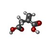 ChemComp-ITN: |
-Experimental details
-Structure determination
| Method | cryo EM |
|---|---|
 Processing Processing | single particle reconstruction |
| Aggregation state | particle |
-Sample preparation
| Buffer | pH: 7.4 |
|---|---|
| Vitrification | Cryogen name: NITROGEN |
- Electron microscopy
| Microscope | FEI POLARA 300 |
|---|---|
| Image recording | Film or detector model: FEI FALCON I (4k x 4k) / Average electron dose: 50.0 e/Å2 |
| Electron beam | Acceleration voltage: 300 kV / Electron source:  FIELD EMISSION GUN FIELD EMISSION GUN |
| Electron optics | Illumination mode: FLOOD BEAM / Imaging mode: BRIGHT FIELD / Nominal defocus max: 2.0 µm / Nominal defocus min: 1.0 µm |
| Experimental equipment | 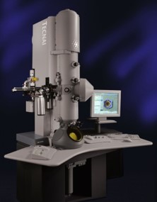 Model: Tecnai Polara / Image courtesy: FEI Company |
