[English] 日本語
Yorodumi
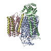
- EMDB-64127: Ubiquinol Binding Site of Cytochrome bo3 from Acinetobacter baumannii -

+
Open data


ID or keywords:

Loading...

-
Basic information

Entry
Database: EMDB / ID: EMD-64127
TitleUbiquinol Binding Site of Cytochrome bo3 from Acinetobacter baumannii
Map data
Sample
  • Complex: Cytochrome bo(3) ubiquinol oxidase
    • Protein or peptide: Cytochrome bo(3) ubiquinol oxidase subunit 1
    • Protein or peptide: Ubiquinol oxidase subunit 2
    • Protein or peptide: Cytochrome bo(3) ubiquinol oxidase subunit 3
    • Protein or peptide: Cytochrome bo(3) ubiquinol oxidase subunit 4
  • Ligand: PROTOPORPHYRIN IX CONTAINING FE
  • Ligand: HEME O
  • Ligand: COPPER (II) ION
  • Ligand: 1,2-Distearoyl-sn-glycerophosphoethanolamine
  • Ligand: 1,2-DISTEAROYL-MONOGALACTOSYL-DIGLYCERIDE
  • Ligand: Ubiquinone-8
Keywordsprotein pump / ubiquinol / oxidase / OXIDOREDUCTASE
Function / homology
Function and homology information


cytochrome o ubiquinol oxidase complex / ubiquinol oxidase (H+-transporting) / cytochrome bo3 ubiquinol oxidase activity / aerobic electron transport chain / oxidoreductase activity, acting on diphenols and related substances as donors, oxygen as acceptor / cytochrome-c oxidase activity / proton transmembrane transporter activity / electron transport coupled proton transport / ATP synthesis coupled electron transport / aerobic respiration ...cytochrome o ubiquinol oxidase complex / ubiquinol oxidase (H+-transporting) / cytochrome bo3 ubiquinol oxidase activity / aerobic electron transport chain / oxidoreductase activity, acting on diphenols and related substances as donors, oxygen as acceptor / cytochrome-c oxidase activity / proton transmembrane transporter activity / electron transport coupled proton transport / ATP synthesis coupled electron transport / aerobic respiration / respiratory electron transport chain / copper ion binding / heme binding / metal ion binding / plasma membrane
Similarity search - Function
Cytochrome o ubiquinol oxidase, subunit I / : / Cytochrome o ubiquinol oxidase, subunit III / Cytochrome o ubiquinol oxidase subunit IV / Ubiquinol oxidase subunit III domain / Cytochrome C oxidase subunit IV, prokaryotes / COX aromatic rich motif / Prokaryotic Cytochrome C oxidase subunit IV / COX Aromatic Rich Motif / Cytochrome o ubiquinol oxidase subunit II ...Cytochrome o ubiquinol oxidase, subunit I / : / Cytochrome o ubiquinol oxidase, subunit III / Cytochrome o ubiquinol oxidase subunit IV / Ubiquinol oxidase subunit III domain / Cytochrome C oxidase subunit IV, prokaryotes / COX aromatic rich motif / Prokaryotic Cytochrome C oxidase subunit IV / COX Aromatic Rich Motif / Cytochrome o ubiquinol oxidase subunit II / Ubiquinol oxidase subunit 2, cupredoxin domain / Cytochrome C oxidase subunit II, transmembrane domain / Cytochrome oxidase subunit II transmembrane region profile. / Cytochrome c oxidase subunit III / Cytochrome c oxidase subunit III-like / Cytochrome c oxidase, subunit III, 4-helical bundle / Cytochrome c oxidase subunit III / Heme-copper oxidase subunit III family profile. / Cytochrome c oxidase subunit III-like superfamily / Cytochrome c/quinol oxidase subunit II / Cytochrome C oxidase subunit II, transmembrane domain superfamily / Cytochrome c oxidase, subunit I, copper-binding site / Heme-copper oxidase catalytic subunit, copper B binding region signature. / Cytochrome c oxidase-like, subunit I domain / Cytochrome oxidase subunit I profile. / Cytochrome C oxidase subunit II, periplasmic domain / Cytochrome c oxidase subunit I / Cytochrome c oxidase-like, subunit I superfamily / Cytochrome C and Quinol oxidase polypeptide I / Cytochrome c oxidase subunit II-like C-terminal / Cytochrome oxidase subunit II copper A binding domain profile. / Cupredoxin / Prokaryotic membrane lipoprotein lipid attachment site profile.
Similarity search - Domain/homology
Ubiquinol oxidase subunit 2 / Cytochrome bo(3) ubiquinol oxidase subunit 1 / Cytochrome bo(3) ubiquinol oxidase subunit 4 / Cytochrome bo(3) ubiquinol oxidase subunit 3
Similarity search - Component
Biological speciesAcinetobacter baumannii (bacteria)
Methodsingle particle reconstruction / cryo EM / Resolution: 3.28 Å
AuthorsLi J / Zhu JP
Funding support China, 1 items
OrganizationGrant numberCountry
National Natural Science Foundation of China (NSFC) China
CitationJournal: J Biol Chem / Year: 2026
Title: Structure of Acinetobacter baumannii cytochrome bo ubiquinol oxidase.
Authors: Quan Li / Rui Hao / Jiapeng Zhu / Jiao Li /
Abstract: Heme-copper oxidases (heme-copper oxidoreductases) are terminal oxidases that couple oxygen reduction to proton pumping for ATP synthesis. Although our previous work has elucidated the structure and ...Heme-copper oxidases (heme-copper oxidoreductases) are terminal oxidases that couple oxygen reduction to proton pumping for ATP synthesis. Although our previous work has elucidated the structure and proton transfer mechanism of the Escherichia coli cytochrome bo ubiquinol oxidase, the quinone dynamics and structural diversity across heme-copper oxidoreductases remain unclear. Here, we report the high-resolution cryo-EM structures of cytochrome bo ubiquinol oxidase from the pathogen Acinetobacter baumannii. We captured four distinct conformational states of its native ubiquinone-8 substrate within the binding pocket. Comparative analysis revealed that conformational transitions of the substrate are directly coupled to movements of the transmembrane 0 helix. Notably, in the locked state, the substrate headgroup is stabilized by specific hydrogen bonds and adopts a distinct depth and orientation. In addition, a unique hairpin-like loop was identified in subunit II, a specific feature absent in the homologs. Our observations not only provide structural details of a pathogenic respiratory terminal oxidase but also reveal a dynamic substrate catalytic mechanism, highlighting potential avenues for targeting bacterial energy metabolism.
History
DepositionApr 10, 2025-
Header (metadata) releaseMay 13, 2026-
Map releaseMay 13, 2026-
UpdateMay 13, 2026-
Current statusMay 13, 2026Processing site: PDBc / Status: Released

-
Structure visualization

Supplemental images

Downloads & links

-
Map

FileDownload / File: emd_64127.map.gz / Format: CCP4 / Size: 103 MB / Type: IMAGE STORED AS FLOATING POINT NUMBER (4 BYTES)
Projections & slices

Image control

Size
Brightness
Contrast
Others
AxesZ (Sec.)Y (Row.)X (Col.)
0.82 Å/pix.
x 300 pix.
= 246. Å
0.82 Å/pix.
x 300 pix.
= 246. Å
0.82 Å/pix.
x 300 pix.
= 246. Å

Surface

Projections

Slices (1/3)

Slices (1/2)

Slices (2/3)

Images are generated by Spider.

Voxel sizeX=Y=Z: 0.82 Å
Density
Contour LevelBy AUTHOR: 0.15
Minimum - Maximum-0.790519 - 1.2048899
Average (Standard dev.)0.00035867622 (±0.027470773)
SymmetrySpace group: 1
Details

EMDB XML:

Map geometry
Axis orderXYZ
Origin000
Dimensions300300300
Spacing300300300
CellA=B=C: 246.0 Å
α=β=γ: 90.0 °

-
Supplemental data

-
Half map: #2

Fileemd_64127_half_map_1.map
Projections & Slices
AxesZYX

Projections

Slices (1/2)
Density Histograms

-
Half map: #1

Fileemd_64127_half_map_2.map
Projections & Slices
AxesZYX

Projections

Slices (1/2)
Density Histograms

-
Sample components

+
Entire : Cytochrome bo(3) ubiquinol oxidase

EntireName: Cytochrome bo(3) ubiquinol oxidase
Components
  • Complex: Cytochrome bo(3) ubiquinol oxidase
    • Protein or peptide: Cytochrome bo(3) ubiquinol oxidase subunit 1
    • Protein or peptide: Ubiquinol oxidase subunit 2
    • Protein or peptide: Cytochrome bo(3) ubiquinol oxidase subunit 3
    • Protein or peptide: Cytochrome bo(3) ubiquinol oxidase subunit 4
  • Ligand: PROTOPORPHYRIN IX CONTAINING FE
  • Ligand: HEME O
  • Ligand: COPPER (II) ION
  • Ligand: 1,2-Distearoyl-sn-glycerophosphoethanolamine
  • Ligand: 1,2-DISTEAROYL-MONOGALACTOSYL-DIGLYCERIDE
  • Ligand: Ubiquinone-8

+
Supramolecule #1: Cytochrome bo(3) ubiquinol oxidase

SupramoleculeName: Cytochrome bo(3) ubiquinol oxidase / type: complex / ID: 1 / Parent: 0 / Macromolecule list: #1-#4
Source (natural)Organism: Acinetobacter baumannii (bacteria)
Molecular weightTheoretical: 120 KDa

+
Macromolecule #1: Cytochrome bo(3) ubiquinol oxidase subunit 1

MacromoleculeName: Cytochrome bo(3) ubiquinol oxidase subunit 1 / type: protein_or_peptide / ID: 1 / Number of copies: 1 / Enantiomer: LEVO / EC number: ubiquinol oxidase (H+-transporting)
Source (natural)Organism: Acinetobacter baumannii (bacteria)
Molecular weightTheoretical: 74.200289 KDa
Recombinant expressionOrganism: Escherichia coli (E. coli)
SequenceString: IFGKLGWDSI PTEPIVLVTM VFMALGAIAV LGGITYFKKW GYLWKEWFTT VDHKKIGIMY IIVSVVMLLR GFADAIMMRL QLFLAKGGG EGYLHPDHYD QIFTAHGVIM IFFVAMGLVV GMMNISVPLQ IGARDVAFPL LNSLSFWLFA GAAGLMMLSL V LGEFAATG ...String:
IFGKLGWDSI PTEPIVLVTM VFMALGAIAV LGGITYFKKW GYLWKEWFTT VDHKKIGIMY IIVSVVMLLR GFADAIMMRL QLFLAKGGG EGYLHPDHYD QIFTAHGVIM IFFVAMGLVV GMMNISVPLQ IGARDVAFPL LNSLSFWLFA GAAGLMMLSL V LGEFAATG WMAYPPLSGI QYSPGVGVDY YIWALQVSGL GTLLSGVNFF VTIIKMRAPG MKLMDMPIFT WTSLCTAVLI IA SFPVLTG TLAMLTLDRY FGFHFFTNEL GGSPMLYVNL IWTWGHPEVY ILVLPAFGLY SEIVATFSRK ALFGYKSMVY ATI AITVLA FVVWLHHFFT MGAGANVNAF FGIMTMVIAI PTGVKIFSWL FTMYKGRITF TTPMLWTLGF LVTFGIGGLT GVLM AVPPA DFLVHNSLFL IAHFHNVIIG GVVFGMFAGI IYYWPKMFGW KLNEAWGKAA FWFWFFGFYF AFMPLYILGF LGMTR RLNT YDNPEWDPYL AIALFGAVLV AIGIACFLMQ IIVGFLQRHQ NMDYTGDPWD ARTLEWATSS PAPFYNFAHE PDASGI DRF WTDKENGVAY ARNTKYEDIH MPTDRAAGFV IAMFITLLGF ALIWHIWWLV VVSFVAAVVS LIVSSFTKNV DYYVPAA EV ERIENERYAL LEKHLKKD

UniProtKB: Cytochrome bo(3) ubiquinol oxidase subunit 1

+
Macromolecule #2: Ubiquinol oxidase subunit 2

MacromoleculeName: Ubiquinol oxidase subunit 2 / type: protein_or_peptide / ID: 2 / Number of copies: 1 / Enantiomer: LEVO
Source (natural)Organism: Acinetobacter baumannii (bacteria)
Molecular weightTheoretical: 31.121656 KDa
Recombinant expressionOrganism: Escherichia coli (E. coli)
SequenceString: GDMVLLNSKG PVGQGQSDLM MTAIYLMLLV VIPSIIMALW FGWKYRASNK DADYKPTWAH STAIEVVVWG IPVIIIGILA WLTWWGSHK YDPYRPLESD KAPLTIQVIA EQFKWIFIYP EQNIATVNEV RFPEKTPLSF KITSNFTMNS FFIPQLGGQI Y AMAGMQTH ...String:
GDMVLLNSKG PVGQGQSDLM MTAIYLMLLV VIPSIIMALW FGWKYRASNK DADYKPTWAH STAIEVVVWG IPVIIIGILA WLTWWGSHK YDPYRPLESD KAPLTIQVIA EQFKWIFIYP EQNIATVNEV RFPEKTPLSF KITSNFTMNS FFIPQLGGQI Y AMAGMQTH LHLLANETGV YRGFSSNYSG YGFSQMRFKA HSVTEQQFNE WVAAVKAGNG TTINPEAVQK TTLDQAELAT LR DGDRSKH QIEHLVNRAK AAGDQEALAK AEAMKPFPTK

UniProtKB: Ubiquinol oxidase subunit 2

+
Macromolecule #3: Cytochrome bo(3) ubiquinol oxidase subunit 3

MacromoleculeName: Cytochrome bo(3) ubiquinol oxidase subunit 3 / type: protein_or_peptide / ID: 3 / Number of copies: 1 / Enantiomer: LEVO
Source (natural)Organism: Acinetobacter baumannii (bacteria)
Molecular weightTheoretical: 21.569061 KDa
Recombinant expressionOrganism: Escherichia coli (E. coli)
SequenceString:
HEHDDTDLTV FGFWTYLMSD LILFGSLFIA FAVLSSHVPP GTPSAYDLFH ESLGYVLTET FALLISSVTF GFAVLASYKK NVNQVITWL FITFLFGATF IGMELYEFHH LVEEGHGPTH SAFLSSFFTL VGTHGIHVTS GLVWMLVLMY QIKTKGLTLP N TRRLACLS LFWHFLDIVW ICVFSVVYLL GVL

UniProtKB: Cytochrome bo(3) ubiquinol oxidase subunit 3

+
Macromolecule #4: Cytochrome bo(3) ubiquinol oxidase subunit 4

MacromoleculeName: Cytochrome bo(3) ubiquinol oxidase subunit 4 / type: protein_or_peptide / ID: 4 / Number of copies: 1 / Enantiomer: LEVO
Source (natural)Organism: Acinetobacter baumannii (bacteria)
Molecular weightTheoretical: 10.882164 KDa
Recombinant expressionOrganism: Escherichia coli (E. coli)
SequenceString:
GSVKTYMTGF ILSIILTVIP FGMVMAGGFS RGLLVTVIAI TAVAQVLVQL VYFLHMNTSS EQRWNMIAFI YTILCIAVLL IGSVWIMNY LHYNMMIH

UniProtKB: Cytochrome bo(3) ubiquinol oxidase subunit 4

+
Macromolecule #5: PROTOPORPHYRIN IX CONTAINING FE

MacromoleculeName: PROTOPORPHYRIN IX CONTAINING FE / type: ligand / ID: 5 / Number of copies: 1 / Formula: HEM
Molecular weightTheoretical: 616.487 Da
Chemical component information

ChemComp-HEM:
PROTOPORPHYRIN IX CONTAINING FE

+
Macromolecule #6: HEME O

MacromoleculeName: HEME O / type: ligand / ID: 6 / Number of copies: 1 / Formula: HEO
Molecular weightTheoretical: 838.854 Da
Chemical component information

ChemComp-HEO:
HEME O

+
Macromolecule #7: COPPER (II) ION

MacromoleculeName: COPPER (II) ION / type: ligand / ID: 7 / Number of copies: 1 / Formula: CU
Molecular weightTheoretical: 63.546 Da
Chemical component information

ChemComp-CU:
COPPER (II) ION

+
Macromolecule #8: 1,2-Distearoyl-sn-glycerophosphoethanolamine

MacromoleculeName: 1,2-Distearoyl-sn-glycerophosphoethanolamine / type: ligand / ID: 8 / Number of copies: 6 / Formula: 3PE
Molecular weightTheoretical: 748.065 Da
Chemical component information

ChemComp-3PE:
1,2-Distearoyl-sn-glycerophosphoethanolamine / phospholipid*YM

+
Macromolecule #9: 1,2-DISTEAROYL-MONOGALACTOSYL-DIGLYCERIDE

MacromoleculeName: 1,2-DISTEAROYL-MONOGALACTOSYL-DIGLYCERIDE / type: ligand / ID: 9 / Number of copies: 3 / Formula: LMG
Molecular weightTheoretical: 787.158 Da
Chemical component information

ChemComp-LMG:
1,2-DISTEAROYL-MONOGALACTOSYL-DIGLYCERIDE

+
Macromolecule #10: Ubiquinone-8

MacromoleculeName: Ubiquinone-8 / type: ligand / ID: 10 / Number of copies: 1 / Formula: UQ8
Molecular weightTheoretical: 727.109 Da
Chemical component information

ChemComp-UQ8:
Ubiquinone-8

-
Experimental details

-
Structure determination

Methodcryo EM
Processingsingle particle reconstruction
Aggregation stateparticle

-
Sample preparation

BufferpH: 8
VitrificationCryogen name: ETHANE

-
Electron microscopy

MicroscopeTFS KRIOS
Image recordingFilm or detector model: GATAN K3 (6k x 4k) / Average electron dose: 1.25 e/Å2
Electron beamAcceleration voltage: 300 kV / Electron source: FIELD EMISSION GUN
Electron opticsIllumination mode: FLOOD BEAM / Imaging mode: BRIGHT FIELD / Nominal defocus max: 2.2 µm / Nominal defocus min: 1.2 µm
Experimental equipment
Model: Titan Krios / Image courtesy: FEI Company

+
Image processing

CTF correctionType: PHASE FLIPPING AND AMPLITUDE CORRECTION
Startup modelType of model: PDB ENTRY
PDB model - PDB ID:
Final reconstructionResolution.type: BY AUTHOR / Resolution: 3.28 Å / Resolution method: FSC 0.143 CUT-OFF / Software - Name: cryoSPARC / Number images used: 48128
Initial angle assignmentType: MAXIMUM LIKELIHOOD
Final angle assignmentType: MAXIMUM LIKELIHOOD

+
About Yorodumi

-
News

-
Feb 9, 2022. New format data for meta-information of EMDB entries

New format data for meta-information of EMDB entries

  • Version 3 of the EMDB header file is now the official format.
  • The previous official version 1.9 will be removed from the archive.

Related info.:EMDB header

External links:wwPDB to switch to version 3 of the EMDB data model

-
Aug 12, 2020. Covid-19 info

Covid-19 info

URL: https://pdbj.org/emnavi/covid19.php

New page: Covid-19 featured information page in EM Navigator.

Related info.:Covid-19 info / Mar 5, 2020. Novel coronavirus structure data

+
Mar 5, 2020. Novel coronavirus structure data

Novel coronavirus structure data

Related info.:Yorodumi Speices / Aug 12, 2020. Covid-19 info

External links:COVID-19 featured content - PDBj / Molecule of the Month (242):Coronavirus Proteases

+
Jan 31, 2019. EMDB accession codes are about to change! (news from PDBe EMDB page)

EMDB accession codes are about to change! (news from PDBe EMDB page)

  • The allocation of 4 digits for EMDB accession codes will soon come to an end. Whilst these codes will remain in use, new EMDB accession codes will include an additional digit and will expand incrementally as the available range of codes is exhausted. The current 4-digit format prefixed with “EMD-” (i.e. EMD-XXXX) will advance to a 5-digit format (i.e. EMD-XXXXX), and so on. It is currently estimated that the 4-digit codes will be depleted around Spring 2019, at which point the 5-digit format will come into force.
  • The EM Navigator/Yorodumi systems omit the EMD- prefix.

Related info.:Q: What is EMD? / ID/Accession-code notation in Yorodumi/EM Navigator

External links:EMDB Accession Codes are Changing Soon! / Contact to PDBj

+
Jul 12, 2017. Major update of PDB

Major update of PDB

  • wwPDB released updated PDB data conforming to the new PDBx/mmCIF dictionary.
  • This is a major update changing the version number from 4 to 5, and with Remediation, in which all the entries are updated.
  • In this update, many items about electron microscopy experimental information are reorganized (e.g. em_software).
  • Now, EM Navigator and Yorodumi are based on the updated data.

External links:wwPDB Remediation / Enriched Model Files Conforming to OneDep Data Standards Now Available in the PDB FTP Archive

-
Yorodumi

Thousand views of thousand structures

  • Yorodumi is a browser for structure data from EMDB, PDB, SASBDB, etc.
  • This page is also the successor to EM Navigator detail page, and also detail information page/front-end page for Omokage search.
  • The word "yorodu" (or yorozu) is an old Japanese word meaning "ten thousand". "mi" (miru) is to see.

Related info.:EMDB / PDB / SASBDB / Comparison of 3 databanks / Yorodumi Search / Aug 31, 2016. New EM Navigator & Yorodumi / Yorodumi Papers / Jmol/JSmol / Function and homology information / Changes in new EM Navigator and Yorodumi

Read more