 Movie
Movie Controller


+ Open data

- Basic information
| Entry |  | |||||||||
|---|---|---|---|---|---|---|---|---|---|---|
| Title | structure of the FliD cap | |||||||||
 Map data Map data | ||||||||||
 Sample Sample |
| |||||||||
 Keywords Keywords | Complex / STRUCTURAL PROTEIN | |||||||||
| Function / homology |  Function and homology information Function and homology informationbacterial-type flagellum filament cap / bacterial-type flagellum hook / bacterial-type flagellum-dependent cell motility / cell adhesion / extracellular region Similarity search - Function | |||||||||
| Biological species |  Salmonella enterica subsp. enterica serovar Typhimurium (bacteria) Salmonella enterica subsp. enterica serovar Typhimurium (bacteria) | |||||||||
| Method | single particle reconstruction / cryo EM / Resolution: 3.06 Å | |||||||||
 Authors Authors | Xing Q / Cheng XQ / Jiang WX | |||||||||
| Funding support |  China, 1 items China, 1 items
| |||||||||
 Citation Citation | Journal: Nat Commun / Year: 2025 Title: Ultraweak interactions drive cap-mediated positioning and elongation of the bacterial flagellar filament. Authors: Lixia Chen / Xiaoqi Cheng / Fangfang Zhang / Xu Dong / Xin Wang / Wenxue Jiang / Lixin Ma / Qiong Xing / Abstract: The bacterial flagellum, essential for motility and pathogenesis, requires the filament cap (FliD) to polymerize flagellin (FliC). However, the mechanisms governing the transition from the hook ...The bacterial flagellum, essential for motility and pathogenesis, requires the filament cap (FliD) to polymerize flagellin (FliC). However, the mechanisms governing the transition from the hook junction to the filament elongation remain elusive, obscured by stoichiometric mismatches and barely detectable interactions. To resolve this, we deploy solution NMR to characterize ultra-weak interactions, quantify affinities (K ≈ 0.1 mM for junction protein FlgL; 1.65 mM for FliC). These data enable rational complex stabilization for cryo-EM structure determination of Salmonella FliD pentamers complexed with FlgL or FliC, revealing that both substrates engage an identical conserved surface in a 5:5 stoichiometry. Integrating these structures into native flagellar tip densities reveal a 5:11 FliD:FlgL/FliC architecture, where six additional subunits barely detected by NMR dock at secondary sites. Mutations that disrupt or enhance these interfaces impair motility and filament integrity, while disulfide-locked FliD pentamers confirm that cap rigidity is crucial for elongation. These findings support a rotary cap mechanism where ultra-weak binding and structural fidelity of the cap ensure efficient flagellin polymerization. Our study resolves the long-standing paradox of stoichiometric mismatch in flagellar filament biogenesis, providing a blueprint for the assembly of dynamic macromolecular machines. | |||||||||
| History |
|
- Structure visualization
| Supplemental images |
|---|

- Downloads & links
-EMDB archive
| Map data | emd_63663.map.gz | 397.9 MB | EMDB map data format | |
|---|---|---|---|---|
| Header (meta data) | emd-63663-v30.xmlemd-63663.xml | 18.4 KB 18.4 KB | Display Display | EMDB header |
| FSC (resolution estimation) | emd_63663_fsc.xml | 15.9 KB | Display | FSC data file |
| Images |  emd_63663.png emd_63663.png | 71.3 KB | ||
| Filedesc metadata | emd-63663.cif.gz | 5.9 KB | ||
| Others | emd_63663_half_map_1.map.gzemd_63663_half_map_2.map.gz | 391.8 MB 391.9 MB | ||
| Archive directory |  http://ftp.pdbj.org/pub/emdb/structures/EMD-63663ftp://ftp.pdbj.org/pub/emdb/structures/EMD-63663 http://ftp.pdbj.org/pub/emdb/structures/EMD-63663ftp://ftp.pdbj.org/pub/emdb/structures/EMD-63663 | HTTPS FTP |
-Related structure data
| Related structure data |  9m6iMC  9m67C  9m68C  9m6hC M: atomic model generated by this map C: citing same article ( |
|---|---|
| Similar structure data |


-Links
| EMDB pages | EMDB (EBI/PDBe) / EMDataResource |
|---|
-Map
| File | Download / File: emd_63663.map.gz / Format: CCP4 / Size: 421.9 MB / Type: IMAGE STORED AS FLOATING POINT NUMBER (4 BYTES) | ||||||||||||||||||||||||||||||||||||
|---|---|---|---|---|---|---|---|---|---|---|---|---|---|---|---|---|---|---|---|---|---|---|---|---|---|---|---|---|---|---|---|---|---|---|---|---|---|
| Projections & slices | Image control
Images are generated by Spider. | ||||||||||||||||||||||||||||||||||||
| Voxel size | X=Y=Z: 0.567 Å | ||||||||||||||||||||||||||||||||||||
| Density |
| ||||||||||||||||||||||||||||||||||||
| Symmetry | Space group: 1 | ||||||||||||||||||||||||||||||||||||
| Details | EMDB XML:
|
 Z (Sec.)
 Y (Row.)
 X (Col.)


-Supplemental data
-Half map: #2
| File | emd_63663_half_map_1.map | ||||||||||||
|---|---|---|---|---|---|---|---|---|---|---|---|---|---|
| Projections & Slices |
| ||||||||||||
| Density Histograms |


-Half map: #1
| File | emd_63663_half_map_2.map | ||||||||||||
|---|---|---|---|---|---|---|---|---|---|---|---|---|---|
| Projections & Slices |
| ||||||||||||
| Density Histograms |


- Sample components
-Entire : the FliD cap
| Entire | Name: the FliD cap |
|---|---|
| Components |
|
-Supramolecule #1: the FliD cap
| Supramolecule | Name: the FliD cap / type: complex / ID: 1 / Parent: 0 / Macromolecule list: all |
|---|---|
| Source (natural) | Organism: Salmonella enterica subsp. enterica serovar Typhimurium (bacteria) |
-Macromolecule #1: Flagellar hook-associated protein 2
| Macromolecule | Name: Flagellar hook-associated protein 2 / type: protein_or_peptide / ID: 1 Details: Sequence reference for strain 'Salmonella enterica subsp. enterica serovar Typhimurium' is not available in UniProt at the time of biocuration. Current sequence reference is from UniProt id P16328. Number of copies: 5 / Enantiomer: LEVO |
|---|---|
| Source (natural) | Organism: Salmonella enterica subsp. enterica serovar Typhimurium (bacteria) |
| Molecular weight | Theoretical: 49.951723 KDa |
| Recombinant expression | Organism:  Escherichia coli (E. coli) Escherichia coli (E. coli) |
| Sequence | String: MASISSLGVG SNLPLDQLLT DLTKNEKGRL TPITKQQSAN SAKLTAYGTL KSALEKFQTA NTALNKADLF KSTVASSTTE DLKVSTTAG ACAGTYKINV TQLAAAQSLA TKTTFATTKE QLGDTSVTSR TIKIEQPGRK EPLEIKLDKG DTSMEAIRDA I NDADSGIC ...String: MASISSLGVG SNLPLDQLLT DLTKNEKGRL TPITKQQSAN SAKLTAYGTL KSALEKFQTA NTALNKADLF KSTVASSTTE DLKVSTTAG ACAGTYKINV TQLAAAQSLA TKTTFATTKE QLGDTSVTSR TIKIEQPGRK EPLEIKLDKG DTSMEAIRDA I NDADSGIC ASIVKVKENE FQLVLTANSG TDNTMKITVE GDTKLNDLLA YDSTTNTGNM QELVKAENAK LNVNGIDIER QS NTVTDAP QGITLTLTKK VTDATVTVTK DDTKAKEAIK SWVDAYNSLV DTFSSLTKYT AVEPGEEASD KNGALLGDSV VRT IQTGIR AQFANSGSNS AFKTMAEIGI TQDGTSGKLK IDDDKLTKVL KDNTAAAREL LVGDGKETGI TTKIATEVKS YLAD DGIID NAQDNVNATL KSLTKQYLSV SNSIDETVAR YKAQFTQLDT MMSKLNNTSS YLTQQFTAMN KS UniProtKB: Flagellar hook-associated protein 2 |
-Experimental details
-Structure determination
| Method | cryo EM |
|---|---|
 Processing Processing | single particle reconstruction |
| Aggregation state | particle |
-Sample preparation
| Buffer | pH: 8 |
|---|---|
| Vitrification | Cryogen name: ETHANE |
- Electron microscopy
| Microscope | FEI TALOS ARCTICA |
|---|---|
| Image recording | Film or detector model: GATAN K3 (6k x 4k) / Average electron dose: 30.0 e/Å2 |
| Electron beam | Acceleration voltage: 300 kV / Electron source:  FIELD EMISSION GUN FIELD EMISSION GUN |
| Electron optics | Illumination mode: OTHER / Imaging mode: BRIGHT FIELD / Nominal defocus max: 2.0 µm / Nominal defocus min: 1.0 µm |
| Experimental equipment | 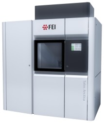 Model: Talos Arctica / Image courtesy: FEI Company |

