 Movie
Movie Controller


+ Open data

- Basic information
| Entry | 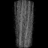 | |||||||||
|---|---|---|---|---|---|---|---|---|---|---|
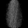
| Title | BTA-2-bound E46K alpha-synuclein fibrils | |||||||||
 Map data Map data | ||||||||||
 Sample Sample |
| |||||||||
 Keywords Keywords | amyloid fibril / complex / PROTEIN FIBRIL | |||||||||
| Function / homology |  Function and homology information Function and homology informationnegative regulation of mitochondrial electron transport, NADH to ubiquinone / neutral lipid metabolic process / regulation of acyl-CoA biosynthetic process / negative regulation of dopamine uptake involved in synaptic transmission / negative regulation of norepinephrine uptake / response to desipramine / positive regulation of SNARE complex assembly / positive regulation of hydrogen peroxide catabolic process / supramolecular fiber / regulation of synaptic vesicle recycling ...negative regulation of mitochondrial electron transport, NADH to ubiquinone / neutral lipid metabolic process / regulation of acyl-CoA biosynthetic process / negative regulation of dopamine uptake involved in synaptic transmission / negative regulation of norepinephrine uptake / response to desipramine / positive regulation of SNARE complex assembly / positive regulation of hydrogen peroxide catabolic process / supramolecular fiber / regulation of synaptic vesicle recycling / negative regulation of chaperone-mediated autophagy / regulation of reactive oxygen species biosynthetic process / positive regulation of protein localization to cell periphery / mitochondrial membrane organization / negative regulation of exocytosis / regulation of glutamate secretion / dopamine biosynthetic process / positive regulation of neurotransmitter secretion / response to iron(II) ion / regulation of macrophage activation / negative regulation of dopamine metabolic process / negative regulation of platelet-derived growth factor receptor signaling pathway / SNARE complex assembly / negative regulation of thrombin-activated receptor signaling pathway / Lewy body / regulation of locomotion / negative regulation of microtubule polymerization / synaptic vesicle priming / regulation of norepinephrine uptake / transporter regulator activity / protein kinase inhibitor activity / positive regulation of inositol phosphate biosynthetic process / synaptic vesicle transport / dopamine uptake involved in synaptic transmission / regulation of dopamine secretion / positive regulation of receptor recycling / cuprous ion binding / positive regulation of exocytosis / nuclear outer membrane / mitochondrial ATP synthesis coupled electron transport / dynein complex binding / synaptic vesicle exocytosis / response to magnesium ion / positive regulation of endocytosis / negative regulation of serotonin uptake / response to type II interferon / cysteine-type endopeptidase inhibitor activity / kinesin binding / regulation of presynapse assembly / synaptic vesicle endocytosis / alpha-tubulin binding / beta-tubulin binding / phospholipase binding / phospholipid metabolic process / supramolecular fiber organization / behavioral response to cocaine / cellular response to fibroblast growth factor stimulus / cellular response to epinephrine stimulus / inclusion body / Hsp70 protein binding / enzyme inhibitor activity / response to interleukin-1 / axon terminus / cellular response to copper ion / regulation of microtubule cytoskeleton organization / positive regulation of release of sequestered calcium ion into cytosol / SNARE binding / adult locomotory behavior / glutathione metabolic process / protein tetramerization / protein sequestering activity / tubulin binding / phosphoprotein binding / excitatory postsynaptic potential / microglial cell activation / ferrous iron binding / fatty acid metabolic process / phospholipid binding / PKR-mediated signaling / synapse organization / receptor internalization / regulation of long-term neuronal synaptic plasticity / protein destabilization / tau protein binding / enzyme activator activity / positive regulation of inflammatory response / terminal bouton / long-term synaptic potentiation / synaptic vesicle membrane / actin cytoskeleton / growth cone / actin binding / cellular response to oxidative stress / neuron apoptotic process / cell cortex / histone binding / response to lipopolysaccharide / microtubule binding / amyloid fibril formation / chemical synaptic transmission Similarity search - Function | |||||||||
| Biological species |  Homo sapiens (human) Homo sapiens (human) | |||||||||
| Method | helical reconstruction / cryo EM / Resolution: 3.4 Å | |||||||||
 Authors Authors | Liu KE / Tao YQ / Li D / Liu C | |||||||||
| Funding support | 1 items
| |||||||||
 Citation Citation | Journal: Proc Natl Acad Sci U S A / Year: 2024 Title: Binding adaptability of chemical ligands to polymorphic α-synuclein amyloid fibrils. Authors: Kaien Liu / Youqi Tao / Qinyue Zhao / Wencheng Xia / Xiang Li / Shenqing Zhang / Yuxuan Yao / Huaijiang Xiang / Chao Han / Li Tan / Bo Sun / Dan Li / Ang Li / Cong Liu /  Abstract: α-synuclein (α-syn) assembles into structurally distinct fibril polymorphs seen in different synucleinopathies, such as Parkinson's disease and multiple system atrophy. Targeting these unique ...α-synuclein (α-syn) assembles into structurally distinct fibril polymorphs seen in different synucleinopathies, such as Parkinson's disease and multiple system atrophy. Targeting these unique fibril structures using chemical ligands holds diagnostic significance for different disease subtypes. However, the molecular mechanisms governing small molecules interacting with different fibril polymorphs remain unclear. Here, we investigated the interactions of small molecules belonging to four distinct scaffolds, with different α-syn fibril polymorphs. Using cryo-electron microscopy, we determined the structures of these molecules when bound to the fibrils formed by E46K mutant α-syn and compared them to those bound with wild-type α-syn fibrils. Notably, we observed that these ligands exhibit remarkable binding adaptability, as they engage distinct binding sites across different fibril polymorphs. While the molecular scaffold primarily steered the binding locations and geometries on specific sites, the conjugated functional groups further refined this adaptable binding by fine-tuning the geometries and binding sites. Overall, our finding elucidates the adaptability of small molecules binding to different fibril structures, which sheds light on the diagnostic tracer and drug developments tailored to specific pathological fibril polymorphs. | |||||||||
| History |
|
- Structure visualization
| Supplemental images |
|---|

- Downloads & links
-EMDB archive
| Map data | emd_60226.map.gz | 22.8 MB | EMDB map data format | |
|---|---|---|---|---|
| Header (meta data) | emd-60226-v30.xmlemd-60226.xml | 14 KB 14 KB | Display Display | EMDB header |
| FSC (resolution estimation) | emd_60226_fsc.xml | 9.2 KB | Display | FSC data file |
| Images |  emd_60226.png emd_60226.png | 59.6 KB | ||
| Filedesc metadata | emd-60226.cif.gz | 5 KB | ||
| Others | emd_60226_half_map_1.map.gzemd_60226_half_map_2.map.gz | 49.6 MB 49.6 MB | ||
| Archive directory |  http://ftp.pdbj.org/pub/emdb/structures/EMD-60226ftp://ftp.pdbj.org/pub/emdb/structures/EMD-60226 http://ftp.pdbj.org/pub/emdb/structures/EMD-60226ftp://ftp.pdbj.org/pub/emdb/structures/EMD-60226 | HTTPS FTP |
-Related structure data
| Related structure data |  8zliMC  8x7bC  8x7lC  8x7mC  8x7oC  8x7pC  8x7qC  8x7rC  8zloC 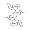 8zlpC  8zmyC M: atomic model generated by this map C: citing same article ( |
|---|---|
| Similar structure data |

-Links
| EMDB pages | EMDB (EBI/PDBe) / EMDataResource |
|---|
-Map
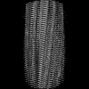
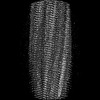
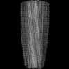
| File | Download / File: emd_60226.map.gz / Format: CCP4 / Size: 64 MB / Type: IMAGE STORED AS FLOATING POINT NUMBER (4 BYTES) | ||||||||||||||||||||||||||||||||||||
|---|---|---|---|---|---|---|---|---|---|---|---|---|---|---|---|---|---|---|---|---|---|---|---|---|---|---|---|---|---|---|---|---|---|---|---|---|---|
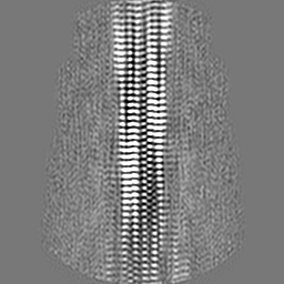
| Projections & slices | Image control
Images are generated by Spider. | ||||||||||||||||||||||||||||||||||||
| Voxel size | X=Y=Z: 0.83 Å | ||||||||||||||||||||||||||||||||||||
| Density |
| ||||||||||||||||||||||||||||||||||||
| Symmetry | Space group: 1 | ||||||||||||||||||||||||||||||||||||
| Details | EMDB XML:
|
 Z (Sec.)
 Y (Row.)
 X (Col.)


-Supplemental data
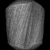
-Half map: #1
| File | emd_60226_half_map_1.map | ||||||||||||
|---|---|---|---|---|---|---|---|---|---|---|---|---|---|
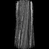
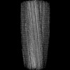
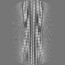
| Projections & Slices |
| ||||||||||||
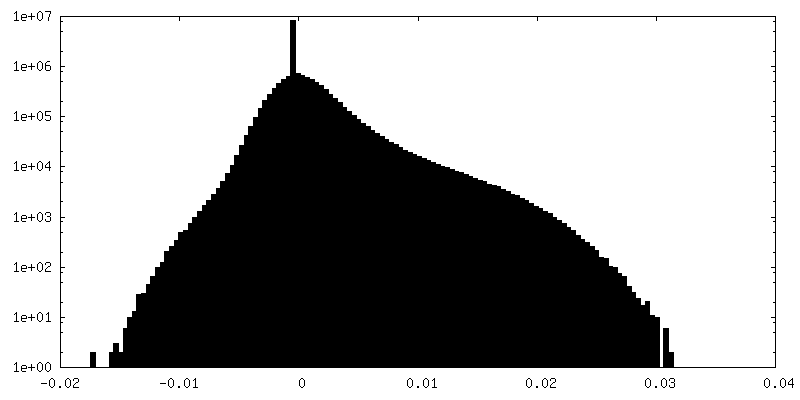
| Density Histograms |


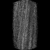
-Half map: #2
| File | emd_60226_half_map_2.map | ||||||||||||
|---|---|---|---|---|---|---|---|---|---|---|---|---|---|
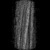
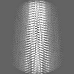
| Projections & Slices |
| ||||||||||||
| Density Histograms |


- Sample components
-Entire : BTA-2-bound E46K alpha-synuclein fibril
| Entire | Name: BTA-2-bound E46K alpha-synuclein fibril |
|---|---|
| Components |
|
-Supramolecule #1: BTA-2-bound E46K alpha-synuclein fibril
| Supramolecule | Name: BTA-2-bound E46K alpha-synuclein fibril / type: complex / ID: 1 / Parent: 0 / Macromolecule list: #1 |
|---|---|
| Source (natural) | Organism: Homo sapiens (human) |
-Macromolecule #1: Alpha-synuclein
| Macromolecule | Name: Alpha-synuclein / type: protein_or_peptide / ID: 1 / Number of copies: 10 / Enantiomer: LEVO |
|---|---|
| Source (natural) | Organism: Homo sapiens (human) |
| Molecular weight | Theoretical: 5.402157 KDa |
| Recombinant expression | Organism:  |
| Sequence | String: KKGVVHGVAT VAEKTKEQVT NVGGAVVTGV TAVAQKTVEG AGSIAAATGF VKKDQ UniProtKB: Alpha-synuclein |
-Macromolecule #2: ~{N},~{N}-dimethyl-4-(6-methyl-1,3-benzothiazol-2-yl)aniline
| Macromolecule | Name: ~{N},~{N}-dimethyl-4-(6-methyl-1,3-benzothiazol-2-yl)aniline type: ligand / ID: 2 / Number of copies: 10 / Formula: A1L13 |
|---|---|
| Molecular weight | Theoretical: 268.377 Da |
-Experimental details
-Structure determination
| Method | cryo EM |
|---|---|
 Processing Processing | helical reconstruction |
| Aggregation state | helical array |
-Sample preparation
| Buffer | pH: 7.5 |
|---|---|
| Vitrification | Cryogen name: ETHANE |
- Electron microscopy
| Microscope | FEI TITAN KRIOS |
|---|---|
| Image recording | Film or detector model: GATAN K3 BIOQUANTUM (6k x 4k) / Average electron dose: 55.0 e/Å2 |
| Electron beam | Acceleration voltage: 300 kV / Electron source:  FIELD EMISSION GUN FIELD EMISSION GUN |
| Electron optics | Illumination mode: FLOOD BEAM / Imaging mode: BRIGHT FIELD / Nominal defocus max: 2.0 µm / Nominal defocus min: 1.0 µm |
| Experimental equipment |  Model: Titan Krios / Image courtesy: FEI Company |
-Image processing
| Final reconstruction | Applied symmetry - Helical parameters - Δz: 2.40514 Å Applied symmetry - Helical parameters - Δ&Phi: -179.403 ° Applied symmetry - Helical parameters - Axial symmetry: C1 (asymmetric) Resolution.type: BY AUTHOR / Resolution: 3.4 Å / Resolution method: FSC 0.143 CUT-OFF / Number images used: 16191 |
|---|---|
| Startup model | Type of model: PDB ENTRY PDB model - PDB ID: |
| Final angle assignment | Type: NOT APPLICABLE |
| FSC plot (resolution estimation) |  |

