 Movie
Movie Controller


+ Open data

- Basic information
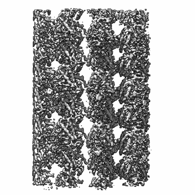
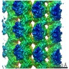
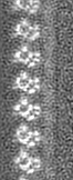
| Entry | Database: EMDB / ID: EMD-5895 | |||||||||
|---|---|---|---|---|---|---|---|---|---|---|
| Title | Cryo EM density of microtubules stabilized by GMPCPP | |||||||||
 Map data Map data | cryo-EM reconstruction of microtubule stabilized by GMPCPP | |||||||||
 Sample Sample |
| |||||||||
 Keywords Keywords | microtubule / gmpcpp | |||||||||
| Function / homology |  Function and homology information Function and homology informationstructural constituent of cytoskeleton / microtubule cytoskeleton organization / neuron migration / mitotic cell cycle / microtubule / Hydrolases; Acting on acid anhydrides; Acting on GTP to facilitate cellular and subcellular movement / hydrolase activity / GTPase activity / GTP binding / metal ion binding / cytoplasm Similarity search - Function | |||||||||
| Biological species |   Homo sapiens (human) Homo sapiens (human) | |||||||||
| Method | single particle reconstruction / cryo EM / Resolution: 4.7 Å | |||||||||
 Authors Authors | Alushin GM / Lander GC / Kellogg EH / Zhang R / Baker D / Nogales E | |||||||||
 Citation Citation | Journal: Cell / Year: 2014 Title: High-resolution microtubule structures reveal the structural transitions in αβ-tubulin upon GTP hydrolysis. Authors: Gregory M Alushin / Gabriel C Lander / Elizabeth H Kellogg / Rui Zhang / David Baker / Eva Nogales /  Abstract: Dynamic instability, the stochastic switching between growth and shrinkage, is essential for microtubule function. This behavior is driven by GTP hydrolysis in the microtubule lattice and is ...Dynamic instability, the stochastic switching between growth and shrinkage, is essential for microtubule function. This behavior is driven by GTP hydrolysis in the microtubule lattice and is inhibited by anticancer agents like Taxol. We provide insight into the mechanism of dynamic instability, based on high-resolution cryo-EM structures (4.7-5.6 Å) of dynamic microtubules and microtubules stabilized by GMPCPP or Taxol. We infer that hydrolysis leads to a compaction around the E-site nucleotide at longitudinal interfaces, as well as movement of the α-tubulin intermediate domain and H7 helix. Displacement of the C-terminal helices in both α- and β-tubulin subunits suggests an effect on interactions with binding partners that contact this region. Taxol inhibits most of these conformational changes, allosterically inducing a GMPCPP-like state. Lateral interactions are similar in all conditions we examined, suggesting that microtubule lattice stability is primarily modulated at longitudinal interfaces. | |||||||||
| History |
|
- Structure visualization
| Movie |
Movie viewer |
|---|---|
| Structure viewer | EM map: SurfViewMolmilJmol/JSmol |
| Supplemental images |
- Downloads & links
-EMDB archive
| Map data | emd_5895.map.gz | 4.1 MB | EMDB map data format | |
|---|---|---|---|---|
| Header (meta data) | emd-5895-v30.xmlemd-5895.xml | 13.5 KB 13.5 KB | Display Display | EMDB header |
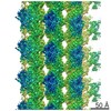
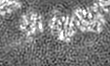
| Images |  400_5895.gif 400_5895.gif 80_5895.gif 80_5895.gif | 58.4 KB 4.2 KB | ||
| Archive directory |  http://ftp.pdbj.org/pub/emdb/structures/EMD-5895ftp://ftp.pdbj.org/pub/emdb/structures/EMD-5895 http://ftp.pdbj.org/pub/emdb/structures/EMD-5895ftp://ftp.pdbj.org/pub/emdb/structures/EMD-5895 | HTTPS FTP |
-Related structure data
| Related structure data |  3j6eMC  5896C  5897C  5898C 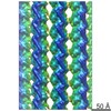 5899C  3j6fC  3j6gC M: atomic model generated by this map C: citing same article ( |
|---|---|
| Similar structure data |

-Links
| EMDB pages | EMDB (EBI/PDBe) / EMDataResource |
|---|---|
| Related items in Molecule of the Month |


-Map
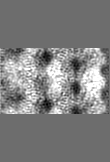
| File | Download / File: emd_5895.map.gz / Format: CCP4 / Size: 4.4 MB / Type: IMAGE STORED AS FLOATING POINT NUMBER (4 BYTES) | ||||||||||||||||||||||||||||||||||||||||||||||||||||||||||||||||||||
|---|---|---|---|---|---|---|---|---|---|---|---|---|---|---|---|---|---|---|---|---|---|---|---|---|---|---|---|---|---|---|---|---|---|---|---|---|---|---|---|---|---|---|---|---|---|---|---|---|---|---|---|---|---|---|---|---|---|---|---|---|---|---|---|---|---|---|---|---|---|
| Annotation | cryo-EM reconstruction of microtubule stabilized by GMPCPP | ||||||||||||||||||||||||||||||||||||||||||||||||||||||||||||||||||||
| Projections & slices | Image control
Images are generated by Spider. generated in cubic-lattice coordinate | ||||||||||||||||||||||||||||||||||||||||||||||||||||||||||||||||||||
| Voxel size | X=Y=Z: 1.74 Å | ||||||||||||||||||||||||||||||||||||||||||||||||||||||||||||||||||||
| Density |
| ||||||||||||||||||||||||||||||||||||||||||||||||||||||||||||||||||||
| Symmetry | Space group: 1 | ||||||||||||||||||||||||||||||||||||||||||||||||||||||||||||||||||||
| Details | EMDB XML:
CCP4 map header:
| ||||||||||||||||||||||||||||||||||||||||||||||||||||||||||||||||||||
 Z (Sec.)
 Y (Row.)
 X (Col.)


-Supplemental data
- Sample components
-Entire : microtubule stabilized by GMPCPP
| Entire | Name: microtubule stabilized by GMPCPP |
|---|---|
| Components |
|
-Supramolecule #1000: microtubule stabilized by GMPCPP
| Supramolecule | Name: microtubule stabilized by GMPCPP / type: sample / ID: 1000 / Number unique components: 3 |
|---|
-Macromolecule #1: Alpha tubulin
| Macromolecule | Name: Alpha tubulin / type: protein_or_peptide / ID: 1 / Oligomeric state: heterodimer / Recombinant expression: No / Database: NCBI |
|---|---|
| Source (natural) | Organism: |
| Molecular weight | Theoretical: 55 KDa |
-Macromolecule #2: Beta tubulin
| Macromolecule | Name: Beta tubulin / type: protein_or_peptide / ID: 2 / Details: Beta subunit is bound to GMPCPP / Oligomeric state: heterodimer / Recombinant expression: No / Database: NCBI |
|---|---|
| Source (natural) | Organism: |
| Molecular weight | Theoretical: 55 KDa |
-Macromolecule #3: kinesin
| Macromolecule | Name: kinesin / type: protein_or_peptide / ID: 3 Details: Human monomeric kinesin K349 cys-lite described in: Rice, S., Lin, A.W., Safer, D., Hart, C.L., Naber, N., Carragher, B.O., Cain, S.M., Pechatnikova, E., Wilson-Kubalek, E.M., Whittaker, M., ...Details: Human monomeric kinesin K349 cys-lite described in: Rice, S., Lin, A.W., Safer, D., Hart, C.L., Naber, N., Carragher, B.O., Cain, S.M., Pechatnikova, E., Wilson-Kubalek, E.M., Whittaker, M., et al. (1999). A structural change in the kinesin motor protein that drives motility. Nature 402, 778-784. Oligomeric state: monomer / Recombinant expression: Yes |
|---|---|
| Source (natural) | Organism: Homo sapiens (human) / synonym: human / Organelle: cytoplasm / Location in cell: cytoskeleton |
| Molecular weight | Theoretical: 36 KDa |
| Recombinant expression | Organism:  |
-Experimental details
-Structure determination
| Method | cryo EM |
|---|---|
 Processing Processing | single particle reconstruction |
| Aggregation state | helical array |
-Sample preparation
| Concentration | 0.25 mg/mL |
|---|---|
| Buffer | pH: 6.8 Details: 80mM PIPES, 1mM EGTA, 1mM MgCl2, 1mM DTT, 0.05% Nonidet P-40 |
| Grid | Details: 400 mesh C-flat 1.2/1.3, glow discharged in Edwards carbon evaporator |
| Vitrification | Cryogen name: ETHANE / Chamber humidity: 90 % / Chamber temperature: 90.4 K / Instrument: FEI VITROBOT MARK II Method: 4 uL microtubules were applied to grid for 30-60 seconds, 4 uL kinesin was added and incubated for 30 seconds, 4 uL buffer was removed, then another 4 uL kinesin was added and incubated for ...Method: 4 uL microtubules were applied to grid for 30-60 seconds, 4 uL kinesin was added and incubated for 30 seconds, 4 uL buffer was removed, then another 4 uL kinesin was added and incubated for 30 seconds. 4 uL buffer was removed, then the grid was blotted for 2 seconds before plunging. |
- Electron microscopy
| Microscope | FEI TITAN |
|---|---|
| Alignment procedure | Legacy - Astigmatism: Objective lens astigmatism was corrected at 100,000 times magnifacation. |
| Date | Apr 26, 2012 |
| Image recording | Category: FILM / Film or detector model: KODAK SO-163 FILM / Digitization - Scanner: OTHER / Digitization - Sampling interval: 6.35 µm / Number real images: 252 / Average electron dose: 25.0 e/Å2 / Bits/pixel: 16 |
| Tilt angle min | 0 |
| Tilt angle max | 0 |
| Electron beam | Acceleration voltage: 300 kV / Electron source:  FIELD EMISSION GUN FIELD EMISSION GUN |
| Electron optics | Calibrated magnification: 72000 / Illumination mode: FLOOD BEAM / Imaging mode: BRIGHT FIELD / Cs: 2.7 mm / Nominal defocus max: 3.5 µm / Nominal defocus min: 1.4 µm / Nominal magnification: 72000 |
| Sample stage | Specimen holder: Gatan 626 holder / Specimen holder model: GATAN LIQUID NITROGEN |
-Image processing
| Details | Initial alignments performed with EMAN2/SPARX, including refinement of helical parameters with the IHRSR programs of Egelman. Final alignment and reconstruction performed with FREALIGN. |
|---|---|
| CTF correction | Details: ctftilt |
| Final reconstruction | Applied symmetry - Helical parameters - Δz: 8.9 Å Applied symmetry - Helical parameters - Δ&Phi: 25.75 ° Applied symmetry - Helical parameters - Axial symmetry: C1 (asymmetric) Algorithm: OTHER / Resolution.type: BY AUTHOR / Resolution: 4.7 Å / Resolution method: OTHER / Software - Name: FREALIGN / Number images used: 57451 |
-Atomic model buiding 1
| Initial model | PDB ID: |
|---|---|
| Software | Name: ROSETTA |
| Details | Structure represents the minimized average structure of the 1% lowest energy structures from the refinement run. |
| Refinement | Space: REAL / Protocol: FLEXIBLE FIT |
| Output model | PDB-3j6e: |

