 Movie
Movie Controller


+ Open data

- Basic information
| Entry |  | |||||||||
|---|---|---|---|---|---|---|---|---|---|---|
| Title | UapA (Q408E) in MSP1E3D1 nanodiscs, Xanthine-bound state | |||||||||
 Map data Map data | Unsharpened map | |||||||||
 Sample Sample |
| |||||||||
 Keywords Keywords | UapA / DDM / Transporter / Inward-facing / Uric Acid / Xanthine / MEMBRANE PROTEIN | |||||||||
| Function / homology |  Function and homology information Function and homology informationxanthine transport / xanthine transmembrane transporter activity / autophagosome lumen / purine nucleobase binding / urate transport / urate transmembrane transporter activity / fungal-type vacuole / endosome membrane / plasma membrane Similarity search - Function | |||||||||
| Biological species |   Aspergillus nidulans FGSC A4 (mold) Aspergillus nidulans FGSC A4 (mold) | |||||||||
| Method | single particle reconstruction / cryo EM / Resolution: 3.48 Å | |||||||||
 Authors Authors | Broutzakis G / Gatsogiannis C | |||||||||
| Funding support |  Germany, 2 items Germany, 2 items
| |||||||||
 Citation Citation | Journal: Proc Natl Acad Sci U S A / Year: 2026 Title: Cryo-EM of the eukaryotic purine transporter UapA demonstrates intramolecular and lipid regulation of transport. Authors: George Broutzakis / Yiannis Pyrris / Ifigeneia Akrani / Alexander Neuhaus / Emmanuel Mikros / George Diallinas / Christos Gatsogiannis /  Abstract: Members of the nucleobase ascorbate transporter (NAT) family (SLC23) are elevator-type transporters that are responsible for the uptake of nucleobases and ascorbate. In fungi, NAT members are also ...Members of the nucleobase ascorbate transporter (NAT) family (SLC23) are elevator-type transporters that are responsible for the uptake of nucleobases and ascorbate. In fungi, NAT members are also responsible for the specific uptake of antifungal nucleobase analogues, such as oxypurinol, allopurinol, or 8-azaguanine. Here, we report nearly full-length cryo-EM structures of UapA, a high-affinity purine transporter from the model fungus , in inward-facing apo- and substrate-loaded conformations at 2.06 to 3.5 Å in detergent and lipid nanodiscs. The high-resolution structures reveal the role of water molecules and lipids in substrate binding, specificity, transporter dimerization, and activity. Notably, the N-tail of UapA is found to be structured, interacting with both the core and scaffold domains, which in combination with functional data suggests a dual role in trafficking and transport dynamics. Overall, our study provides unprecedented structural and functional insights into an elevator-type fungal transporter, which may well contribute to the exploitation of NAT transporters as specific gateways for targeted pharmacological antifungal approaches. | |||||||||
| History |
|
- Structure visualization
| Supplemental images |
|---|

- Downloads & links
-EMDB archive
| Map data | emd_58354.map.gz | 11.3 MB | EMDB map data format | |
|---|---|---|---|---|
| Header (meta data) | emd-58354-v30.xmlemd-58354.xml | 24 KB 24 KB | Display Display | EMDB header |
| FSC (resolution estimation) | emd_58354_fsc.xml | 5.9 KB | Display | FSC data file |
| Images |  emd_58354.png emd_58354.png | 116.7 KB | ||
| Masks | emd_58354_msk_1.map | 22.2 MB | Mask map | |
| Filedesc metadata | emd-58354.cif.gz | 6.8 KB | ||
| Others | emd_58354_additional_1.map.gzemd_58354_half_map_1.map.gzemd_58354_half_map_2.map.gz | 21 MB 20.7 MB 20.7 MB | ||
| Archive directory |  https://data.pdbj.org/pub/emdb/structures/EMD-58354ftp://data.pdbj.org/pub/emdb/structures/EMD-58354 https://data.pdbj.org/pub/emdb/structures/EMD-58354ftp://data.pdbj.org/pub/emdb/structures/EMD-58354 | HTTPS FTP |
-Related structure data
| Related structure data |  31ezMC  31ewC  31eyC M: atomic model generated by this map C: citing same article ( |
|---|---|
| Similar structure data |


-Links
| EMDB pages | EMDB (EBI/PDBe) / EMDataResource |
|---|
-Map
| File | Download / File: emd_58354.map.gz / Format: CCP4 / Size: 22.2 MB / Type: IMAGE STORED AS FLOATING POINT NUMBER (4 BYTES) | ||||||||||||||||||||||||||||||||||||
|---|---|---|---|---|---|---|---|---|---|---|---|---|---|---|---|---|---|---|---|---|---|---|---|---|---|---|---|---|---|---|---|---|---|---|---|---|---|
| Annotation | Unsharpened map | ||||||||||||||||||||||||||||||||||||
| Projections & slices | Image control
Images are generated by Spider. | ||||||||||||||||||||||||||||||||||||
| Voxel size | X=Y=Z: 1.38 Å | ||||||||||||||||||||||||||||||||||||
| Density |
| ||||||||||||||||||||||||||||||||||||
| Symmetry | Space group: 1 | ||||||||||||||||||||||||||||||||||||
| Details | EMDB XML:
|
 Z (Sec.)
 Y (Row.)
 X (Col.)


-Supplemental data
-Mask #1
| File | emd_58354_msk_1.map | ||||||||||||
|---|---|---|---|---|---|---|---|---|---|---|---|---|---|
| Projections & Slices |
| ||||||||||||
| Density Histograms |


-Additional map: Sharpened map
| File | emd_58354_additional_1.map | ||||||||||||
|---|---|---|---|---|---|---|---|---|---|---|---|---|---|
| Annotation | Sharpened map | ||||||||||||
| Projections & Slices |
| ||||||||||||
| Density Histograms |


-Half map: Half map B
| File | emd_58354_half_map_1.map | ||||||||||||
|---|---|---|---|---|---|---|---|---|---|---|---|---|---|
| Annotation | Half map B | ||||||||||||
| Projections & Slices |
| ||||||||||||
| Density Histograms |


-Half map: Half map A
| File | emd_58354_half_map_2.map | ||||||||||||
|---|---|---|---|---|---|---|---|---|---|---|---|---|---|
| Annotation | Half map A | ||||||||||||
| Projections & Slices |
| ||||||||||||
| Density Histograms |


- Sample components
-Entire : UapA (Q408E) in MSP1E3D1 nanodiscs, the in xanthine-bound state.
| Entire | Name: UapA (Q408E) in MSP1E3D1 nanodiscs, the in xanthine-bound state. |
|---|---|
| Components |
|
-Supramolecule #1: UapA (Q408E) in MSP1E3D1 nanodiscs, the in xanthine-bound state.
| Supramolecule | Name: UapA (Q408E) in MSP1E3D1 nanodiscs, the in xanthine-bound state. type: complex / ID: 1 / Parent: 0 / Macromolecule list: #1 |
|---|---|
| Source (natural) | Organism: Aspergillus nidulans FGSC A4 (mold) / Strain: A4 |
| Molecular weight | Theoretical: 120 KDa |
-Macromolecule #1: Uric acid-xanthine permease
| Macromolecule | Name: Uric acid-xanthine permease / type: protein_or_peptide / ID: 1 / Number of copies: 2 / Enantiomer: LEVO |
|---|---|
| Source (natural) | Organism: Aspergillus nidulans FGSC A4 (mold) |
| Molecular weight | Theoretical: 55.353102 KDa |
| Recombinant expression | Organism:   Saccharomyces cerevisiae (brewer's yeast) Saccharomyces cerevisiae (brewer's yeast) |
| Sequence | String: QRVRLLARHL TTREGLIGDY DYGFLFRPEL PFMKKDPRAP PFFGLNEKIP VLLAFILGLQ HALAMLAGVV TPPLIISSSL SLPSDLQQY LVSTSLIVCG LLSMVQITRF HIYKTPYYIG SGVLSVMGVS FSIISVASGA FNQMYSNGFC QLDEAGNRLP C PEAYGALI ...String: QRVRLLARHL TTREGLIGDY DYGFLFRPEL PFMKKDPRAP PFFGLNEKIP VLLAFILGLQ HALAMLAGVV TPPLIISSSL SLPSDLQQY LVSTSLIVCG LLSMVQITRF HIYKTPYYIG SGVLSVMGVS FSIISVASGA FNQMYSNGFC QLDEAGNRLP C PEAYGALI GTSACCALVE ILLAFVPPKV IQKIFPPIVT GPTVMLIGIS LIGTGFKDWA GGSACMDDGM LCPSATAPRP LP WGSPEFI GLGFLVFVSI ILCERFGAPI MKSCSVVIGL LVGCIVAAAC GYFSHADIDA APAASFIWVK TFPLSVYGPM VLP IIAVFI ICACECIGDV TATCDVSRLE VRGGTFESRI QGAVLADGIN SVVAALATMT PMTTFAENNG VIALTRCANR WAGY CCCLI LIVAGIFAKF AAAIVAIPNS VMGGMKTFLF ASVVISGQAI VAKAPFTRRN RFILTASMAL GYGATLVPTW FGNVF PQTE NRDLEGFENA IELVLETGFA VTAFVAMLLN AIMPAE UniProtKB: Uric acid-xanthine permease |
-Macromolecule #2: MYRISTIC ACID
| Macromolecule | Name: MYRISTIC ACID / type: ligand / ID: 2 / Number of copies: 36 / Formula: MYR |
|---|---|
| Molecular weight | Theoretical: 228.371 Da |
| Chemical component information | 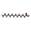 ChemComp-MYR: |
-Macromolecule #3: ERGOSTEROL
| Macromolecule | Name: ERGOSTEROL / type: ligand / ID: 3 / Number of copies: 10 / Formula: ERG |
|---|---|
| Molecular weight | Theoretical: 396.648 Da |
| Chemical component information |  ChemComp-ERG: |
-Macromolecule #4: XANTHINE
| Macromolecule | Name: XANTHINE / type: ligand / ID: 4 / Number of copies: 2 / Formula: XAN |
|---|---|
| Molecular weight | Theoretical: 152.111 Da |
| Chemical component information | 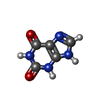 ChemComp-XAN: |
-Experimental details
-Structure determination
| Method | cryo EM |
|---|---|
 Processing Processing | single particle reconstruction |
| Aggregation state | particle |
-Sample preparation
| Concentration | 5 mg/mL | ||||||||||||
|---|---|---|---|---|---|---|---|---|---|---|---|---|---|
| Buffer | pH: 7.5 Component:
| ||||||||||||
| Vitrification | Cryogen name: ETHANE / Chamber humidity: 100 % / Chamber temperature: 286.15 K / Instrument: FEI VITROBOT MARK IV Details: Vitrification chamber was allowed to equilibrate for 15 min at 100% humidity.. |
- Electron microscopy
| Microscope | TFS KRIOS |
|---|---|
| Image recording | Film or detector model: FEI FALCON IV (4k x 4k) / Average electron dose: 50.0 e/Å2 |
| Electron beam | Acceleration voltage: 300 kV / Electron source:  FIELD EMISSION GUN FIELD EMISSION GUN |
| Electron optics | Illumination mode: FLOOD BEAM / Imaging mode: BRIGHT FIELD / Nominal defocus max: 2.2 µm / Nominal defocus min: 0.8 µm |
| Experimental equipment |  Model: Titan Krios / Image courtesy: FEI Company |

