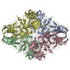
Movie
Movie Controller


+ Open data

- Basic information
| Entry |  | |||||||||
|---|---|---|---|---|---|---|---|---|---|---|
| Title | E. coli beta-galactosidase with galacto-cyclophellitol | |||||||||
 Map data Map data | ||||||||||
 Sample Sample |
| |||||||||
 Keywords Keywords | BETA-GALACTOSIDASE / COMPLEX / ACTIVITY-BASED PROBE / HYDROLASE | |||||||||
| Function / homology |  Function and homology information Function and homology informationalkali metal ion binding / lactose catabolic process / beta-galactosidase complex / beta-galactosidase / beta-galactosidase activity / carbohydrate binding / magnesium ion binding / identical protein binding Similarity search - Function | |||||||||
| Biological species |   Escherichia coli (E. coli) Escherichia coli (E. coli) | |||||||||
| Method | single particle reconstruction / cryo EM / Resolution: 2.7 Å | |||||||||
 Authors Authors | Pham PT / Alzahrani MM / Agirre J | |||||||||
| Funding support |  Saudi Arabia, Saudi Arabia,  United Kingdom, 2 items United Kingdom, 2 items
| |||||||||
 Citation Citation | Journal: To Be Published Title: E. coli beta-galactosidase with galacto-cyclophellitol Authors: Alzahrani MM / Pham PT / Agirre J | |||||||||
| History |
|
- Structure visualization
| Supplemental images |
|---|

- Downloads & links
-EMDB archive
| Header (meta data) | emd-57206-v30.xmlemd-57206.xml | 20.2 KB 20.2 KB | Display Display | EMDB header |
|---|---|---|---|---|
| FSC (resolution estimation) | emd_57206_fsc.xml | 15.8 KB | Display | FSC data file |
| Images |  emd_57206.png emd_57206.png | 84.9 KB | ||
| Map data | emd_57206.map.gz | 276.2 MB | EMDB map data format | |
| Masks | emd_57206_msk_1.map | 347.6 MB | Mask map | |
| Filedesc metadata | emd-57206.cif.gz | 7.1 KB | ||
| Others | emd_57206_half_map_1.map.gzemd_57206_half_map_2.map.gz | 276.1 MB 275.6 MB | ||
| Archive directory |  http://ftp.pdbj.org/pub/emdb/structures/EMD-57206ftp://ftp.pdbj.org/pub/emdb/structures/EMD-57206 http://ftp.pdbj.org/pub/emdb/structures/EMD-57206ftp://ftp.pdbj.org/pub/emdb/structures/EMD-57206 | HTTPS FTP |
-Related structure data
| Related structure data |  29jfMC M: atomic model generated by this map C: citing same article ( |
|---|---|
| Similar structure data |
-Links
| EMDB pages | EMDB (EBI/PDBe) / EMDataResource |
|---|---|
| Related items in Molecule of the Month |
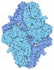

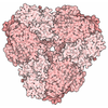
-Map
| File | Released | ||||||||||||||||||||||||||||||||||||
|---|---|---|---|---|---|---|---|---|---|---|---|---|---|---|---|---|---|---|---|---|---|---|---|---|---|---|---|---|---|---|---|---|---|---|---|---|---|
| Projections & slices | Image control
Images are generated by Spider. | ||||||||||||||||||||||||||||||||||||
| Voxel size | X=Y=Z: 0.88978 Å | ||||||||||||||||||||||||||||||||||||
| Density |
| ||||||||||||||||||||||||||||||||||||
| Symmetry | Space group: 1 | ||||||||||||||||||||||||||||||||||||
| Details | EMDB XML:
|
 Z (Sec.)
 Y (Row.)
 X (Col.)


-Supplemental data
-Mask #1
| File | emd_57206_msk_1.map | ||||||||||||
|---|---|---|---|---|---|---|---|---|---|---|---|---|---|
| Projections & Slices |
| ||||||||||||
| Density Histograms |


-Half map: #2
| File | emd_57206_half_map_1.map | ||||||||||||
|---|---|---|---|---|---|---|---|---|---|---|---|---|---|
| Projections & Slices |
| ||||||||||||
| Density Histograms |


-Half map: #1
| File | emd_57206_half_map_2.map | ||||||||||||
|---|---|---|---|---|---|---|---|---|---|---|---|---|---|
| Projections & Slices |
| ||||||||||||
| Density Histograms |


- Sample components
-Entire : E. coli beta-Galactosidase in a covalent complex with galacto-cyc...
| Entire | Name: E. coli beta-Galactosidase in a covalent complex with galacto-cyclophellitol |
|---|---|
| Components |
|
-Supramolecule #1: E. coli beta-Galactosidase in a covalent complex with galacto-cyc...
| Supramolecule | Name: E. coli beta-Galactosidase in a covalent complex with galacto-cyclophellitol type: complex / ID: 1 / Parent: 0 / Macromolecule list: #1 |
|---|---|
| Source (natural) | Organism: Escherichia coli (E. coli) |
| Molecular weight | Theoretical: 116 KDa |
-Macromolecule #1: Beta-galactosidase
| Macromolecule | Name: Beta-galactosidase / type: protein_or_peptide / ID: 1 Details: adapted from the sequence of PDB accession code 4V40 Number of copies: 4 / Enantiomer: LEVO / EC number: beta-galactosidase |
|---|---|
| Source (natural) | Organism: Escherichia coli (E. coli) |
| Molecular weight | Theoretical: 116.471297 KDa |
| Recombinant expression | Organism: Escherichia coli (E. coli) |
| Sequence | String: TMITDSLAVV LQRRDWENPG VTQLNRLAAH PPFASWRNSE EARTDRPSQQ LRSLNGEWRF AWFPAPEAVP ESWLECDLPE ADTVVVPSN WQMHGYDAPI YTNVTYPITV NPPFVPTENP TGCYSLTFNV DESWLQEGQT RIIFDGVNSA FHLWCNGRWV G YGQDSRLP ...String: TMITDSLAVV LQRRDWENPG VTQLNRLAAH PPFASWRNSE EARTDRPSQQ LRSLNGEWRF AWFPAPEAVP ESWLECDLPE ADTVVVPSN WQMHGYDAPI YTNVTYPITV NPPFVPTENP TGCYSLTFNV DESWLQEGQT RIIFDGVNSA FHLWCNGRWV G YGQDSRLP SEFDLSAFLR AGENRLAVMV LRWSDGSYLE DQDMWRMSGI FRDVSLLHKP TTQISDFHVA TRFNDDFSRA VL EAEVQMC GELRDYLRVT VSLWQGETQV ASGTAPFGGE IIDERGGYAD RVTLRLNVEN PKLWSAEIPN LYRAVVELHT ADG TLIEAE ACDVGFREVR IENGLLLLNG KPLLIRGVNR HEHHPLHGQV MDEQTMVQDI LLMKQNNFNA VRCSHYPNHP LWYT LCDRY GLYVVDEANI ETHGMVPMNR LTDDPRWLPA MSERVTRMVQ RDRNHPSVII WSLGNESGHG ANHDALYRWI KSVDP SRPV QYEGGGADTT ATDIICPMYA RVDEDQPFPA VPKWSIKKWL SLPGETRPLI LCEYAHAMGN SLGGFAKYWQ AFRQYP RLQ GGFVWDWVDQ SLIKYDENGN PWSAYGGDFG DTPNDRQFCM NGLVFADRTP HPALTEAKHQ QQFFQFRLSG QTIEVTS EY LFRHSDNELL HWMVALDGKP LASGEVPLDV APQGKQLIEL PELPQPESAG QLWLTVRVVQ PNATAWSEAG HISAWQQW R LAENLSVTLP AASHAIPHLT TSEMDFCIEL GNKRWQFNRQ SGFLSQMWIG DKKQLLTPLR DQFTRAPLDN DIGVSEATR IDPNAWVERW KAAGHYQAEA ALLQCTADTL ADAVLITTAH AWQHQGKTLF ISRKTYRIDG SGQMAITVDV EVASDTPHPA RIGLNCQLA QVAERVNWLG LGPQENYPDR LTAACFDRWD LPLSDMYTPY VFPSENGLRC GTRELNYGPH QWRGDFQFNI S RYSQQQLM ETSHRHLLHA EEGTWLNIDG FHMGIGGDDS WSPSVSAEFQ LSAGRYHYQL VWCQK UniProtKB: Beta-galactosidase |
-Macromolecule #2: MAGNESIUM ION
| Macromolecule | Name: MAGNESIUM ION / type: ligand / ID: 2 / Number of copies: 4 / Formula: MG |
|---|---|
| Molecular weight | Theoretical: 24.305 Da |
-Macromolecule #3: (1S,2S,4S,5R)-6-(hydroxymethyl)cyclohexane-1,2,3,4,5-pentol
| Macromolecule | Name: (1S,2S,4S,5R)-6-(hydroxymethyl)cyclohexane-1,2,3,4,5-pentol type: ligand / ID: 3 / Number of copies: 4 / Formula: YGX |
|---|---|
| Molecular weight | Theoretical: 194.182 Da |
-Macromolecule #4: water
| Macromolecule | Name: water / type: ligand / ID: 4 / Number of copies: 170 / Formula: HOH |
|---|---|
| Molecular weight | Theoretical: 18.015 Da |
| Chemical component information |  ChemComp-HOH: |
-Experimental details
-Structure determination
| Method | cryo EM |
|---|---|
 Processing Processing | single particle reconstruction |
| Aggregation state | particle |
-Sample preparation
| Concentration | 2 mg/mL | |||||||||
|---|---|---|---|---|---|---|---|---|---|---|
| Buffer | pH: 7.5 Component:
| |||||||||
| Grid | Model: Quantifoil R2/2 / Material: COPPER / Mesh: 300 / Pretreatment - Type: GLOW DISCHARGE | |||||||||
| Vitrification | Cryogen name: ETHANE / Chamber humidity: 100 % / Chamber temperature: 295 K / Instrument: FEI VITROBOT MARK IV |
- Electron microscopy
| Microscope | TFS GLACIOS |
|---|---|
| Specialist optics | Phase plate: VOLTA PHASE PLATE |
| Image recording | Film or detector model: FEI FALCON IV (4k x 4k) / Number real images: 5931 / Average exposure time: 5.12 sec. / Average electron dose: 50.0 e/Å2 |
| Electron beam | Acceleration voltage: 200 kV / Electron source:  FIELD EMISSION GUN FIELD EMISSION GUN |
| Electron optics | Illumination mode: FLOOD BEAM / Imaging mode: BRIGHT FIELD / Nominal defocus max: 2.0 µm / Nominal defocus min: 1.0 µm / Nominal magnification: 240000 |
| Sample stage | Cooling holder cryogen: NITROGEN |