Movie
Movie Controller

[English] 日本語
 Yorodumi
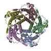
- EMDB-56772: split variant of Aquifex aeolicus lumazine synthase-derived nucle... -
+ Open data

- Basic information
| Entry |  | |||||||||
|---|---|---|---|---|---|---|---|---|---|---|
| Title | split variant of Aquifex aeolicus lumazine synthase-derived nucleocapsid variant NC-4 | |||||||||
 Map data Map data | ||||||||||
 Sample Sample |
| |||||||||
 Keywords Keywords | capsid / design / virus mimic / VIRUS LIKE PARTICLE | |||||||||
| Function / homology |  Function and homology information Function and homology information6,7-dimethyl-8-ribityllumazine synthase / 6,7-dimethyl-8-ribityllumazine synthase activity / riboflavin synthase complex / riboflavin biosynthetic process / cytosol / cytoplasm Similarity search - Function | |||||||||
| Biological species |  Escherichia virus lambda / Escherichia virus lambda /   Aquifex aeolicus (bacteria) Aquifex aeolicus (bacteria) | |||||||||
| Method | single particle reconstruction / cryo EM / Resolution: 3.1 Å | |||||||||
 Authors Authors | Tetter S / Hilvert D | |||||||||
| Funding support | European Union, 1 items
| |||||||||
 Citation Citation | Journal: Proc Natl Acad Sci U S A / Year: 2026 Title: An engineered closed-shell, two-component, 480-subunit nucleocapsid. Authors: Mikail D Levasseur / Naohiro Terasaka / Angela Steinauer / Stephan Tetter / Sara Pfister / Beat H Meier / Donald Hilvert /  Abstract: Self-assembling protein cages are valuable nanoscale containers for biotechnology and medical applications. Two-component systems are especially attractive due to their potential for functional ...Self-assembling protein cages are valuable nanoscale containers for biotechnology and medical applications. Two-component systems are especially attractive due to their potential for functional complexity. In this study, we demonstrate that the subunits of the 240-subunit nucleocapsid NC-4, which was previously evolved to package and protect its encoding mRNA, can be split into two fragments without disrupting cage assembly or structure, generating a two-component, 480-subunit capsid. This modification introduces additional termini on the cage's exterior surface, creating opportunities for functionalization. We exploited these new sites by genetically appending peptide and protein tags to the exterior surface of split NC-4 (spNC-4), enabling site-specific glycosylation via posttranslational modification and cell-specific delivery by targeted antibody recruitment. Our findings broaden the utility of the NC-4 nucleocapsid. By extension, splitting related protein compartments that bind diverse cargoes could offer a robust platform for biotechnological applications requiring simultaneous encapsulation and customizable surface modification. #1: Journal: Biorxiv / Year: 2025Title: An engineered closed-shell, two-component, 480-subunit nucleocapsid Authors: Levasseur MD / Terasaka N / Steinauer A / Tetter S / Pfister S / Meier BH / Hilvert D | |||||||||
| History |
|
- Structure visualization
| Supplemental images |
|---|

- Downloads & links
-EMDB archive
| Map data | emd_56772.map.gz | 59.1 MB | EMDB map data format | |
|---|---|---|---|---|
| Header (meta data) | emd-56772-v30.xmlemd-56772.xml | 22.1 KB 22.1 KB | Display Display | EMDB header |
| Images |  emd_56772.png emd_56772.png | 92.5 KB | ||
| Masks | emd_56772_msk_1.map | 476.8 MB | Mask map | |
| Filedesc metadata | emd-56772.cif.gz | 6.4 KB | ||
| Others | emd_56772_half_map_1.map.gzemd_56772_half_map_2.map.gz | 380.7 MB 380.7 MB | ||
| Archive directory |  https://data.pdbj.org/pub/emdb/structures/EMD-56772ftp://data.pdbj.org/pub/emdb/structures/EMD-56772 https://data.pdbj.org/pub/emdb/structures/EMD-56772ftp://data.pdbj.org/pub/emdb/structures/EMD-56772 | HTTPS FTP |
-Related structure data
| Related structure data |  28roMC M: atomic model generated by this map C: citing same article ( |
|---|---|
| Similar structure data |
-Links
| EMDB pages | EMDB (EBI/PDBe) / EMDataResource |
|---|
-Map
| File | Download / File: emd_56772.map.gz / Format: CCP4 / Size: 476.8 MB / Type: IMAGE STORED AS FLOATING POINT NUMBER (4 BYTES) | ||||||||||||||||||||||||||||||||||||
|---|---|---|---|---|---|---|---|---|---|---|---|---|---|---|---|---|---|---|---|---|---|---|---|---|---|---|---|---|---|---|---|---|---|---|---|---|---|
| Projections & slices | Image control
Images are generated by Spider. | ||||||||||||||||||||||||||||||||||||
| Voxel size | X=Y=Z: 1.1 Å | ||||||||||||||||||||||||||||||||||||
| Density |
| ||||||||||||||||||||||||||||||||||||
| Symmetry | Space group: 1 | ||||||||||||||||||||||||||||||||||||
| Details | EMDB XML:
|
 Z (Sec.)
 Y (Row.)
 X (Col.)


-Supplemental data
-Mask #1
| File | emd_56772_msk_1.map | ||||||||||||
|---|---|---|---|---|---|---|---|---|---|---|---|---|---|
| Projections & Slices |
| ||||||||||||
| Density Histograms |


-Half map: #2
| File | emd_56772_half_map_1.map | ||||||||||||
|---|---|---|---|---|---|---|---|---|---|---|---|---|---|
| Projections & Slices |
| ||||||||||||
| Density Histograms |


-Half map: #1
| File | emd_56772_half_map_2.map | ||||||||||||
|---|---|---|---|---|---|---|---|---|---|---|---|---|---|
| Projections & Slices |
| ||||||||||||
| Density Histograms |


- Sample components
-Entire : Nucleocapsid NC-4
| Entire | Name: Nucleocapsid NC-4 |
|---|---|
| Components |
|
-Supramolecule #1: Nucleocapsid NC-4
| Supramolecule | Name: Nucleocapsid NC-4 / type: complex / ID: 1 / Parent: 0 / Macromolecule list: all Details: Evolved and split variant of a virus-inspired nucleocapsid design based on the Aquifex aeolicus riboflavin synthase |
|---|
-Supramolecule #2: Nucleocapsid NC-4, N-terminal segments
| Supramolecule | Name: Nucleocapsid NC-4, N-terminal segments / type: complex / ID: 2 / Parent: 1 |
|---|---|
| Source (natural) | Organism: Escherichia virus lambda |
-Macromolecule #1: 6,7-dimethyl-8-ribityllumazine synthase
| Macromolecule | Name: 6,7-dimethyl-8-ribityllumazine synthase / type: protein_or_peptide / ID: 1 / Number of copies: 240 / Enantiomer: LEVO / EC number: 6,7-dimethyl-8-ribityllumazine synthase |
|---|---|
| Source (natural) | Organism: Aquifex aeolicus (bacteria) |
| Molecular weight | Theoretical: 12.324837 KDa |
| Recombinant expression | Organism:  Escherichia coli BL21(DE3) (bacteria) Escherichia coli BL21(DE3) (bacteria) |
| Sequence | String: MGNARTRRRE RRAEKQAQWK AANAGAGAGA MATPHFDYNA SVVSKGLANL SLELRKPVSF DIITADTLEQ AIERAGTKHG NKGWEAALS AIEMANLYKS LRGTEHHHHL HGSS UniProtKB: 6,7-dimethyl-8-ribityllumazine synthase |
-Macromolecule #2: 6,7-dimethyl-8-ribityllumazine synthase
| Macromolecule | Name: 6,7-dimethyl-8-ribityllumazine synthase / type: protein_or_peptide / ID: 2 / Number of copies: 240 / Enantiomer: LEVO / EC number: 6,7-dimethyl-8-ribityllumazine synthase |
|---|---|
| Source (natural) | Organism: Aquifex aeolicus (bacteria) |
| Molecular weight | Theoretical: 9.356705 KDa |
| Recombinant expression | Organism: Escherichia coli BL21(DE3) (bacteria) |
| Sequence | String: MGIEIYEGKL TAEGLRFGIV ASRFNHTLVD RLVEGAIDCI VRHGGRGEDI TLVRVPGAWE IPVAADELAR KEDIDAVIAF GDLIRG UniProtKB: 6,7-dimethyl-8-ribityllumazine synthase |
-Experimental details
-Structure determination
| Method | cryo EM |
|---|---|
 Processing Processing | single particle reconstruction |
| Aggregation state | particle |
-Sample preparation
| Concentration | 2 mg/mL |
|---|---|
| Buffer | pH: 7.4 |
| Grid | Model: Quantifoil R2/2 / Material: COPPER / Mesh: 400 / Pretreatment - Type: GLOW DISCHARGE |
| Vitrification | Cryogen name: ETHANE / Chamber humidity: 100 % / Chamber temperature: 22 K / Instrument: FEI VITROBOT MARK IV |
- Electron microscopy
| Microscope | TFS KRIOS |
|---|---|
| Image recording | Film or detector model: FEI FALCON III (4k x 4k) / Detector mode: COUNTING / Average electron dose: 60.0 e/Å2 |
| Electron beam | Acceleration voltage: 300 kV / Electron source:  FIELD EMISSION GUN FIELD EMISSION GUN |
| Electron optics | Illumination mode: FLOOD BEAM / Imaging mode: BRIGHT FIELD / Cs: 2.7 mm / Nominal defocus max: 2.7 µm / Nominal defocus min: 0.8 µm / Nominal magnification: 130000 |
| Sample stage | Cooling holder cryogen: NITROGEN |
| Experimental equipment |  Model: Titan Krios / Image courtesy: FEI Company |