 Movie
Movie Controller

[English] 日本語
 Yorodumi
- EMDB-56604: CryoEM structure of carbon monoxide dehydrogenase from Ruminococc... -
+ Open data

- Basic information
| Entry |  | ||||||||||||
|---|---|---|---|---|---|---|---|---|---|---|---|---|---|
| Title | CryoEM structure of carbon monoxide dehydrogenase from Ruminococcus flavefaciens | ||||||||||||
 Map data Map data | CryoSPARC non-uniform refinement map of RfCODH | ||||||||||||
 Sample Sample |
| ||||||||||||
 Keywords Keywords | carbon monoxide dehydrogenase / CODH / metalloenzyme / iron sulfur protein / anaerobic enzyme / OXIDOREDUCTASE | ||||||||||||
| Biological species |  Ruminococcus flavefaciens ATCC 19208 (bacteria) Ruminococcus flavefaciens ATCC 19208 (bacteria) | ||||||||||||
| Method | single particle reconstruction / cryo EM / Resolution: 2.55 Å | ||||||||||||
 Authors Authors | Srinivas V / Hogbom M | ||||||||||||
| Funding support |  Sweden, Sweden,  Denmark, 3 items Denmark, 3 items
| ||||||||||||
 Citation Citation | Journal: Angew Chem Int Ed Engl / Year: 2026 Title: Beyond Canonical CO Oxidation: Structural and Evolutionary Insights Into a Non-Canonical Carbon Monoxide Dehydrogenase. Authors: Maximilian Böhm / Vivek Srinivas / Benjamin Wiseman / Ping Huang / Moritz Senger / Martin Högbom / Henrik Land / Abstract: Carbon monoxide dehydrogenases (CODHs) catalyse the reversible oxidation of CO to CO and play central roles in microbial carbon metabolism. While well-characterised CODHs from different phylogenetic ...Carbon monoxide dehydrogenases (CODHs) catalyse the reversible oxidation of CO to CO and play central roles in microbial carbon metabolism. While well-characterised CODHs from different phylogenetic backgrounds exhibit high bidirectional activity, the enigmatic clade B remains functionally uncharacterised. Here, we present the first structural and biochemical characterisation of a clade B CODH from Ruminococcus flavefaciens (RfCODH). It reveals striking divergence from canonical enzymes. A new anaerobic cryo-EM workflow was developed, carried out entirely under anoxic conditions by manual blotting and plunge freezing. It resulted in a 2.53 Å RfCODH structure. The structure adopts the typical CODH fold, but exhibits blocked gas channels, a compromised proton transfer pathway and disrupted cofactor coordination. This provides a structural rationale for RfCODH's severely attenuated CO oxidation activity (13 mU/mg vs. 900 U/mg for the well-studied ChCODH-II). EPR spectroscopy reveals unique oxidised C-cluster states not previously characterised in CODHs. Mirror tree analysis hints to co-evolution between clade B CODHs and associated ABC transporter substrate-binding proteins, suggesting these enzymes function in metabolism of substrates imported via the ABC transporter module. All findings indicate evolutionary repurposing of the CODH scaffold for alternative physiological functions. | ||||||||||||
| History |
|
- Structure visualization
| Supplemental images |
|---|

- Downloads & links
-EMDB archive
| Map data | emd_56604.map.gz | 62.2 MB |  EMDB map data format EMDB map data format | |
|---|---|---|---|---|
| Header (meta data) | emd-56604-v30.xmlemd-56604.xml | 19.9 KB 19.9 KB | Display Display | EMDB header |
| FSC (resolution estimation) | emd_56604_fsc.xml | 10.6 KB | Display | FSC data file |
| Images |  emd_56604.png emd_56604.png | 113.1 KB | ||
| Masks | emd_56604_msk_1.map | 125 MB | Mask map | |
| Filedesc metadata | emd-56604.cif.gz | 6.7 KB | ||
| Others | emd_56604_half_map_1.map.gzemd_56604_half_map_2.map.gz | 116.2 MB 116.2 MB | ||
| Archive directory |  https://data.pdbj.org/pub/emdb/structures/EMD-56604ftp://data.pdbj.org/pub/emdb/structures/EMD-56604 https://data.pdbj.org/pub/emdb/structures/EMD-56604ftp://data.pdbj.org/pub/emdb/structures/EMD-56604 | HTTPS FTP |
-Related structure data
-Links
| EMDB pages | EMDB (EBI/PDBe) / EMDataResource |
|---|
-Map
| File | Download / File: emd_56604.map.gz / Format: CCP4 / Size: 125 MB / Type: IMAGE STORED AS FLOATING POINT NUMBER (4 BYTES) | ||||||||||||||||||||||||||||||||||||
|---|---|---|---|---|---|---|---|---|---|---|---|---|---|---|---|---|---|---|---|---|---|---|---|---|---|---|---|---|---|---|---|---|---|---|---|---|---|
| Annotation | CryoSPARC non-uniform refinement map of RfCODH | ||||||||||||||||||||||||||||||||||||
| Projections & slices | Image control
Images are generated by Spider. | ||||||||||||||||||||||||||||||||||||
| Voxel size | X=Y=Z: 0.65 Å | ||||||||||||||||||||||||||||||||||||
| Density |
| ||||||||||||||||||||||||||||||||||||
| Symmetry | Space group: 1 | ||||||||||||||||||||||||||||||||||||
| Details | EMDB XML:
|
 Z (Sec.)
 Y (Row.)
 X (Col.)


-Supplemental data
-Mask #1
| File | emd_56604_msk_1.map | ||||||||||||
|---|---|---|---|---|---|---|---|---|---|---|---|---|---|
| Projections & Slices |
| ||||||||||||
| Density Histograms |


-Half map: CryoSPARC non-uniform refinement half-map B of RfCODH
| File | emd_56604_half_map_1.map | ||||||||||||
|---|---|---|---|---|---|---|---|---|---|---|---|---|---|
| Annotation | CryoSPARC non-uniform refinement half-map B of RfCODH | ||||||||||||
| Projections & Slices |
| ||||||||||||
| Density Histograms |


-Half map: CryoSPARC non-uniform refinement half-map A of RfCODH
| File | emd_56604_half_map_2.map | ||||||||||||
|---|---|---|---|---|---|---|---|---|---|---|---|---|---|
| Annotation | CryoSPARC non-uniform refinement half-map A of RfCODH | ||||||||||||
| Projections & Slices |
| ||||||||||||
| Density Histograms |


- Sample components
-Entire : Homodimer of carbon-monoxide dehydrogenase from Ruminococcus flav...
| Entire | Name: Homodimer of carbon-monoxide dehydrogenase from Ruminococcus flavefaciens |
|---|---|
| Components |
|
-Supramolecule #1: Homodimer of carbon-monoxide dehydrogenase from Ruminococcus flav...
| Supramolecule | Name: Homodimer of carbon-monoxide dehydrogenase from Ruminococcus flavefaciens type: complex / ID: 1 / Parent: 0 / Macromolecule list: #1 |
|---|---|
| Source (natural) | Organism: Ruminococcus flavefaciens ATCC 19208 (bacteria) |
| Molecular weight | Theoretical: 158 KDa |
-Macromolecule #1: anaerobic carbon-monoxide dehydrogenase
| Macromolecule | Name: anaerobic carbon-monoxide dehydrogenase / type: protein_or_peptide / ID: 1 / Number of copies: 2 / Enantiomer: LEVO / EC number: anaerobic carbon monoxide dehydrogenase |
|---|---|
| Source (natural) | Organism: Ruminococcus flavefaciens ATCC 19208 (bacteria) |
| Molecular weight | Theoretical: 78.97725 KDa |
| Recombinant expression | Organism:  Escherichia coli (E. coli) Escherichia coli (E. coli) |
| Sequence | String: MSKKDHDHCH DHEHKHDHDH AHHEHSPAHI HSHRNIIGFD EHGIPTVILK ASHGEGEQDD PQAFIKDYMN AVSEYRKTFP TKQDVIEQT PDPAVREMLL RMEQLGIDTA FDRFDQQKPQ CNFGLAGVCC KICNMGPCRV TAKSPKGVCG ADADLIVARN L LRSAAAGA ...String: MSKKDHDHCH DHEHKHDHDH AHHEHSPAHI HSHRNIIGFD EHGIPTVILK ASHGEGEQDD PQAFIKDYMN AVSEYRKTFP TKQDVIEQT PDPAVREMLL RMEQLGIDTA FDRFDQQKPQ CNFGLAGVCC KICNMGPCRV TAKSPKGVCG ADADLIVARN L LRSAAAGA AQHGMHAREV MLALKWAAEG KLDVPILGEQ KIRATAEAFG IKQKNRQLKN VAKDLADALL EDLSRTVPDE YK TISACAV PERQQVWKDL DILPISAYHE VFEAYHKSGC ATDGDWKSVM QQFLRCGLAF TFSGVVGASI ATDSLFGVGD RVT SKVNIG ALEKGYVNIA VHGHLPLLVS QIVEAGKRED LIELAKSKGA KGIRFYGICC SGLSAMYRYA GVIPLSNAVS AELI LGTGA LDLWVADVQD VFPSIMEVAH CFKTTVVTTS ESARLPGAER YEYDHHHSNI DETKALAEKI VRRAIESFEA RKGIP VYIP PYEVEAEVGF SVEYIHKRFG SMKPLAEAIK DGRILGIVNM VGCNNPKVLY EKCIVDVADV LLKNNVLIIS NGCASF PLM KLGYCAVSGK EKAGDSLREF LEPDLPPVWH VGECIDNTRS SGIFAGVAGA LGHKMYEMPF AFSSPEWGNE KGIDAAL GF RLNGISSYHC VEAQIYGSKN VIEFLKYGTL ETLGSSMNVD TDPVKLGEKI VADMKAKRKA LGWDKSSGWS HPQFEK |
-Macromolecule #2: IRON/SULFUR CLUSTER
| Macromolecule | Name: IRON/SULFUR CLUSTER / type: ligand / ID: 2 / Number of copies: 3 / Formula: SF4 |
|---|---|
| Molecular weight | Theoretical: 351.64 Da |
| Chemical component information | 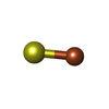 ChemComp-FS1: |
-Experimental details
-Structure determination
| Method | cryo EM |
|---|---|
 Processing Processing | single particle reconstruction |
| Aggregation state | particle |
-Sample preparation
| Concentration | 1 mg/mL |
|---|---|
| Buffer | pH: 8 / Details: 50 mM Tris-HCl pH 8, 20 mM NaCl, 5 % w/v glycerol, |
| Grid | Model: Quantifoil R1.2/1.3 / Material: COPPER / Mesh: 300 / Support film - Material: CARBON / Support film - topology: HOLEY / Pretreatment - Type: GLOW DISCHARGE |
| Vitrification | Cryogen name: ETHANE / Chamber humidity: 0 % Details: Vetrification carried out by manually plunge-freezing in a nitrogen glove box. |
- Electron microscopy
| Microscope | TFS KRIOS |
|---|---|
| Image recording | Film or detector model: GATAN K3 BIOQUANTUM (6k x 4k) / Average electron dose: 41.65 e/Å2 |
| Electron beam | Acceleration voltage: 300 kV / Electron source:  FIELD EMISSION GUN FIELD EMISSION GUN |
| Electron optics | Illumination mode: FLOOD BEAM / Imaging mode: BRIGHT FIELD / Cs: 2.7 mm / Nominal defocus max: 2.0 µm / Nominal defocus min: 0.6 µm / Nominal magnification: 130000 |
| Experimental equipment |  Model: Titan Krios / Image courtesy: FEI Company |
+Image processing

-Atomic model buiding 1
| Initial model | Chain - Source name: AlphaFold / Chain - Initial model type: in silico model |
|---|---|
| Refinement | Protocol: RIGID BODY FIT |
| Output model |  PDB-28lw: |
