 Movie
Movie Controller

[English] 日本語
 Yorodumi
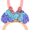
- EMDB-56388: Mature MoMLV capsid hexamer structure from capsid-like particles -
+ Open data

- Basic information
| Entry |  | ||||||||||||
|---|---|---|---|---|---|---|---|---|---|---|---|---|---|
| Title | Mature MoMLV capsid hexamer structure from capsid-like particles | ||||||||||||
 Map data Map data | |||||||||||||
 Sample Sample |
| ||||||||||||
 Keywords Keywords | MLV / capsid / mature / CA / IP6 / retrovirus / hexamer / capsomer / VIRAL PROTEIN | ||||||||||||
| Function / homology |  Function and homology information Function and homology informationretroviral 3' processing activity / host cell late endosome membrane / DNA catabolic process / ribonuclease H / Hydrolases; Acting on peptide bonds (peptidases); Aspartic endopeptidases / virion assembly / viral genome integration into host DNA / protein-DNA complex / establishment of integrated proviral latency / host multivesicular body ...retroviral 3' processing activity / host cell late endosome membrane / DNA catabolic process / ribonuclease H / Hydrolases; Acting on peptide bonds (peptidases); Aspartic endopeptidases / virion assembly / viral genome integration into host DNA / protein-DNA complex / establishment of integrated proviral latency / host multivesicular body / RNA-directed DNA polymerase / RNA-directed DNA polymerase activity / RNA-DNA hybrid ribonuclease activity / Transferases; Transferring phosphorus-containing groups; Nucleotidyltransferases / viral nucleocapsid / DNA recombination / DNA-directed DNA polymerase / aspartic-type endopeptidase activity / structural constituent of virion / Hydrolases; Acting on ester bonds / DNA-directed DNA polymerase activity / symbiont-mediated suppression of host gene expression / symbiont entry into host cell / host cell cytoplasm / host cell plasma membrane / proteolysis / DNA binding / RNA binding / zinc ion binding Similarity search - Function | ||||||||||||
| Biological species |   Escherichia coli (E. coli) / Escherichia coli (E. coli) /  Moloney murine leukemia virus Moloney murine leukemia virus | ||||||||||||
| Method | single particle reconstruction / cryo EM / Resolution: 3.5 Å | ||||||||||||
 Authors Authors | Klarhof JO / Stacey JCV / Briggs JAG / James LC | ||||||||||||
| Funding support |  United Kingdom, 3 items United Kingdom, 3 items
| ||||||||||||
 Citation Citation | Journal: Nat Commun / Year: 2026 Title: Retroviruses use different IP binding mechanisms to alter the properties of their capsids. Authors: J Ole Klarhof / Donna L Mallery / James C V Stacey / Davide Torre / Michaela Rumlova / Tomas Ruml / John A G Briggs / Leo C James /   Abstract: HIV-1 uses the metabolite inositol hexakisphosphate (IP) as a host factor to assemble its capsid, but whether this strategy is unique to lentiviruses or represents a common feature of retroviral ...HIV-1 uses the metabolite inositol hexakisphosphate (IP) as a host factor to assemble its capsid, but whether this strategy is unique to lentiviruses or represents a common feature of retroviral capsids remains unclear. Here we show that IP binding is conserved across diverse retroviruses but occurs through distinct capsid sites and mechanisms, and influences viral behaviour. In contrast to HIV-1, the beta-retrovirus Mason-Pfizer Monkey Virus (MPMV) and the gamma-retrovirus Murine Leukaemia Virus (MLV) bind IP at the threefold lattice interface between capsomers rather than within capsomer pores. Cryo-EM structures of core-like particles reveal that two lysine residues from each capsomer coordinate IP between either two discrete three-lysine rings (MPMV) or a single heterogeneous six-lysine ring (MLV). MPMV and MLV are largely insensitive to IP availability in producer cells, but this binding mode renders them highly dependent on IP6 in target cells - the opposite of the dependency pattern of HIV-1. The way in which retroviruses use IP to build their capsids alters their dependence on the metabolite at different stages of the replicative cycle and in key capsid behaviours, such as assembly and stability. | ||||||||||||
| History |
|
- Structure visualization
| Supplemental images |
|---|

- Downloads & links
-EMDB archive
| Map data | emd_56388.map.gz | 203.3 MB | EMDB map data format | |
|---|---|---|---|---|
| Header (meta data) | emd-56388-v30.xmlemd-56388.xml | 19 KB 19 KB | Display Display | EMDB header |
| FSC (resolution estimation) | emd_56388_fsc.xml | 14.3 KB | Display | FSC data file |
| Images |  emd_56388.png emd_56388.png | 176.5 KB | ||
| Masks | emd_56388_msk_1.map | 216 MB | Mask map | |
| Filedesc metadata | emd-56388.cif.gz | 6.3 KB | ||
| Others | emd_56388_half_map_1.map.gzemd_56388_half_map_2.map.gz | 200 MB 200 MB | ||
| Archive directory |  https://data.pdbj.org/pub/emdb/structures/EMD-56388ftp://data.pdbj.org/pub/emdb/structures/EMD-56388 https://data.pdbj.org/pub/emdb/structures/EMD-56388ftp://data.pdbj.org/pub/emdb/structures/EMD-56388 | HTTPS FTP |
-Related structure data
| Related structure data |  9tx5MC  9tx4C  9tx6C  9tx7C  9tx8C  9tx9C  9txaC  9txbC M: atomic model generated by this map C: citing same article ( |
|---|---|
| Similar structure data |


-Links
| EMDB pages | EMDB (EBI/PDBe) / EMDataResource |
|---|---|
| Related items in Molecule of the Month |
-Map
| File | Download / File: emd_56388.map.gz / Format: CCP4 / Size: 216 MB / Type: IMAGE STORED AS FLOATING POINT NUMBER (4 BYTES) | ||||||||||||||||||||||||||||||||||||
|---|---|---|---|---|---|---|---|---|---|---|---|---|---|---|---|---|---|---|---|---|---|---|---|---|---|---|---|---|---|---|---|---|---|---|---|---|---|
| Projections & slices | Image control
Images are generated by Spider. | ||||||||||||||||||||||||||||||||||||
| Voxel size | X=Y=Z: 0.84 Å | ||||||||||||||||||||||||||||||||||||
| Density |
| ||||||||||||||||||||||||||||||||||||
| Symmetry | Space group: 1 | ||||||||||||||||||||||||||||||||||||
| Details | EMDB XML:
|
 Z (Sec.)
 Y (Row.)
 X (Col.)


-Supplemental data
-Mask #1
| File | emd_56388_msk_1.map | ||||||||||||
|---|---|---|---|---|---|---|---|---|---|---|---|---|---|
| Projections & Slices |
| ||||||||||||
| Density Histograms |


-Half map: #2
| File | emd_56388_half_map_1.map | ||||||||||||
|---|---|---|---|---|---|---|---|---|---|---|---|---|---|
| Projections & Slices |
| ||||||||||||
| Density Histograms |


-Half map: #1
| File | emd_56388_half_map_2.map | ||||||||||||
|---|---|---|---|---|---|---|---|---|---|---|---|---|---|
| Projections & Slices |
| ||||||||||||
| Density Histograms |


- Sample components
-Entire : MPMV Mature Capsid E26A (5mM IP6)
| Entire | Name: MPMV Mature Capsid E26A (5mM IP6) |
|---|---|
| Components |
|
-Supramolecule #1: MPMV Mature Capsid E26A (5mM IP6)
| Supramolecule | Name: MPMV Mature Capsid E26A (5mM IP6) / type: complex / ID: 1 / Parent: 0 / Macromolecule list: all / Details: Hexamer |
|---|---|
| Source (natural) | Organism: Escherichia coli (E. coli) / Strain: C41 (DE3) |
| Molecular weight | Theoretical: 30 KDa |
-Macromolecule #1: Capsid protein p30
| Macromolecule | Name: Capsid protein p30 / type: protein_or_peptide / ID: 1 / Number of copies: 6 / Enantiomer: LEVO |
|---|---|
| Source (natural) | Organism: Moloney murine leukemia virus |
| Molecular weight | Theoretical: 30.657123 KDa |
| Recombinant expression | Organism: Escherichia coli BL21(DE3) (bacteria) |
| Sequence | String: PLRAGGNGQL QYWPFSSSDL YNWKNNNPSF SEDPGKLTAL IESVLITHQP TWDDCQQLLG TLLTGEEKQR VLLEARKAVR GDDGRPTQL PNEVDAAFPL ERPDWDYTTQ AGRNHLVHYR QLLLAGLQNA GRSPTNLAKV KGITQGPNES PSAFLERLKE A YRRYTPYD ...String: PLRAGGNGQL QYWPFSSSDL YNWKNNNPSF SEDPGKLTAL IESVLITHQP TWDDCQQLLG TLLTGEEKQR VLLEARKAVR GDDGRPTQL PNEVDAAFPL ERPDWDYTTQ AGRNHLVHYR QLLLAGLQNA GRSPTNLAKV KGITQGPNES PSAFLERLKE A YRRYTPYD PEDPGQETNV SMSFIWQSAP DIGRKLERLE DLKNKTLGDL VREAEKIFNK RETPEEREER IRRETEEKEE RR RTEDEQK EKERDRRRHR EMSKLL UniProtKB: Gag-Pol polyprotein |
-Experimental details
-Structure determination
| Method | cryo EM |
|---|---|
 Processing Processing | single particle reconstruction |
| Aggregation state | particle |
-Sample preparation
| Concentration | 15 mg/mL | ||||||||||||
|---|---|---|---|---|---|---|---|---|---|---|---|---|---|
| Buffer | pH: 8 Component:
| ||||||||||||
| Grid | Model: C-flat-2/2 / Material: COPPER / Mesh: 300 / Support film - Material: CARBON / Support film - topology: HOLEY / Support film - Film thickness: 20 | ||||||||||||
| Vitrification | Cryogen name: ETHANE / Chamber humidity: 100 % / Chamber temperature: 277 K / Instrument: FEI VITROBOT MARK IV | ||||||||||||
| Details | MPMV capsid-like particles assembled from recombinant capsid in 2.5mM IP6 |
- Electron microscopy
| Microscope | TFS KRIOS |
|---|---|
| Image recording | Film or detector model: FEI FALCON IV (4k x 4k) / Average electron dose: 40.0 e/Å2 |
| Electron beam | Acceleration voltage: 300 kV / Electron source:  FIELD EMISSION GUN FIELD EMISSION GUN |
| Electron optics | C2 aperture diameter: 50.0 µm / Illumination mode: FLOOD BEAM / Imaging mode: BRIGHT FIELD / Cs: 2.7 mm / Nominal defocus max: 3.2 µm / Nominal defocus min: 1.2 µm / Nominal magnification: 96000 |
| Sample stage | Specimen holder model: FEI TITAN KRIOS AUTOGRID HOLDER / Cooling holder cryogen: NITROGEN |
| Experimental equipment |  Model: Titan Krios / Image courtesy: FEI Company |
+Image processing

-Atomic model buiding 1
| Refinement | Space: REAL / Protocol: OTHER / Target criteria: Cross-correlation |
|---|---|
| Output model | PDB-9tx5: |
