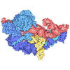
- EMDB-55100: Naked mole-rat 80S ribosome in post-translocation non-rotated state -
+
Open data
ID or keywords:
Loading...
-
Basic information
Entry
Database: EMDB / ID: EMD-55100
Title
Naked mole-rat 80S ribosome in post-translocation non-rotated state
Map data
Sample
Complex: 80S ribosome
Keywords
naked mole-rat / ribosome / 80S
Function / homology
Function and homology information
oxidized pyrimidine DNA binding / response to TNF agonist / positive regulation of base-excision repair / protein-DNA complex disassembly / protein-synthesizing GTPase / positive regulation of intrinsic apoptotic signaling pathway in response to DNA damage / positive regulation of DNA-templated transcription initiation / negative regulation of DNA repair / supercoiled DNA binding / oxidized purine DNA binding ...oxidized pyrimidine DNA binding / response to TNF agonist / positive regulation of base-excision repair / protein-DNA complex disassembly / protein-synthesizing GTPase / positive regulation of intrinsic apoptotic signaling pathway in response to DNA damage / positive regulation of DNA-templated transcription initiation / negative regulation of DNA repair / supercoiled DNA binding / oxidized purine DNA binding / NF-kappaB complex / ubiquitin-like protein conjugating enzyme binding / laminin receptor activity / protein kinase A binding / TOR signaling / gastrulation / protein localization to nucleus / protein targeting / positive regulation of intrinsic apoptotic signaling pathway by p53 class mediator / negative regulation of ubiquitin-dependent protein catabolic process / ubiquitin ligase inhibitor activity / 90S preribosome / positive regulation of signal transduction by p53 class mediator / positive regulation of microtubule polymerization / spindle assembly / protein-RNA complex assembly / translation regulator activity / rough endoplasmic reticulum / ribosomal small subunit export from nucleus / laminin binding / negative regulation of protein ubiquitination / positive regulation of cell cycle / negative regulation of proteasomal ubiquitin-dependent protein catabolic process / MDM2/MDM4 family protein binding / Hsp70 protein binding / DNA-(apurinic or apyrimidinic site) lyase / class I DNA-(apurinic or apyrimidinic site) endonuclease activity / maturation of SSU-rRNA from tricistronic rRNA transcript (SSU-rRNA, 5.8S rRNA, LSU-rRNA) / maturation of LSU-rRNA from tricistronic rRNA transcript (SSU-rRNA, 5.8S rRNA, LSU-rRNA) / maturation of SSU-rRNA / mRNA 3'-UTR binding / small-subunit processome / bone development / Hsp90 protein binding / positive regulation of non-canonical NF-kappaB signal transduction / base-excision repair / response to virus / mitotic spindle / cellular response to hydrogen peroxide / mRNA 5'-UTR binding / transcription coactivator binding / ruffle membrane / cytoplasmic ribonucleoprotein granule / innate immune response in mucosa / kinase activity / rRNA processing / cytosolic ribosome / large ribosomal subunit / ribosomal small subunit assembly / ribosome binding / ribosomal small subunit biogenesis / ribosome biogenesis / 5S rRNA binding / cell body / ribosomal large subunit assembly / small ribosomal subunit / cytosolic small ribosomal subunit / small ribosomal subunit rRNA binding / antimicrobial humoral immune response mediated by antimicrobial peptide / large ribosomal subunit rRNA binding / DNA-binding transcription activator activity, RNA polymerase II-specific / antibacterial humoral response / DNA-binding transcription factor binding / microtubule binding / cytosolic large ribosomal subunit / cell differentiation / cytoplasmic translation / protein stabilization / defense response to Gram-positive bacterium / postsynaptic density / negative regulation of translation / mitochondrial inner membrane / rRNA binding / positive regulation of apoptotic process / ribosome / translation / structural constituent of ribosome / mitochondrial matrix / ribonucleoprotein complex / ubiquitin protein ligase binding / apoptotic process / mRNA binding / centrosome / positive regulation of gene expression / positive regulation of cell population proliferation / negative regulation of apoptotic process / nucleolus / dendrite / synapse / protein kinase binding Similarity search - Function
40S ribosomal protein SA / 40S ribosomal protein SA, C-terminal domain / 40S ribosomal protein SA C-terminus / Ubiquitin-like protein FUBI / Ribosomal protein L30e / Ribosomal protein L28e / Ribosomal L15/L27a, N-terminal / : / Ribosomal L28e/Mak16 / Ribosomal L28e protein family ...40S ribosomal protein SA / 40S ribosomal protein SA, C-terminal domain / 40S ribosomal protein SA C-terminus / Ubiquitin-like protein FUBI / Ribosomal protein L30e / Ribosomal protein L28e / Ribosomal L15/L27a, N-terminal / : / Ribosomal L28e/Mak16 / Ribosomal L28e protein family / Ribosomal protein L23 / Ribosomal protein L2, archaeal-type / metallochaperone-like domain / TRASH domain / : / Ribosomal protein S26e signature. / Ribosomal protein L41 / Ribosomal protein L41 / Ribosomal protein L13e, conserved site / Ribosomal protein L13e signature. / Ribosomal protein S21e, conserved site / Ribosomal protein S21e signature. / Ribosomal protein S26e / Ribosomal protein S26e superfamily / Ribosomal protein S26e / Small (40S) ribosomal subunit Asc1/RACK1 / Ribosomal protein L27e, conserved site / Ribosomal protein L27e signature. / Ribosomal protein L22e / Ribosomal protein L22e superfamily / Ribosomal L22e protein family / Ribosomal protein L13e / Ribosomal protein L13e / Ribosomal protein L38e / Ribosomal protein L38e superfamily / Ribosomal L38e protein family / Ribosomal protein S5, eukaryotic/archaeal / Ribosomal protein S21e / Ribosomal protein S21e superfamily / Ribosomal protein S21e / Ribosomal protein L19, eukaryotic / Ribosomal protein S19e, conserved site / Ribosomal protein S19e signature. / Ribosomal protein S2, eukaryotic / 60S ribosomal protein L18a/ L20, eukaryotes / Ribosomal protein L10e, conserved site / 40S Ribosomal protein S10 / Ribosomal protein L10e signature. / Ribosomal protein L18/L18-A/B/e, conserved site / Ribosomal protein L18e signature. / Ribosomal protein L44e signature. / Plectin/S10, N-terminal / Ribosomal protein L24e, conserved site / Ribosomal protein L19/L19e conserved site / Plectin/S10 domain / Ribosomal protein L19e signature. / Ribosomal protein L24e signature. / Ribosomal protein L5 eukaryotic, C-terminal / Ribosomal L18 C-terminal region / Ribosomal protein L23/L25, N-terminal / Ribosomal protein L23, N-terminal domain / Ribosomal protein L10e / Ribosomal protein L34e, conserved site / Ribosomal protein L34e signature. / Ribosomal protein S30 / Ribosomal protein S30 / Ribosomal L40e family / Ribosomal protein L36e signature. / Ribosomal protein S10, eukaryotic/archaeal / Ribosomal protein L30e signature 1. / Eukaryotic Ribosomal Protein L27, KOW domain / Ribosomal protein S25 / S25 ribosomal protein / Ribosomal protein L27e / Ribosomal protein L27e superfamily / Ribosomal L27e protein family / 50S ribosomal protein L18Ae/60S ribosomal protein L20 and L18a / : / Ribosomal protein L35Ae, conserved site / Ribosomal protein S8e subdomain, eukaryotes / Ribosomal protein L35Ae signature. / Ribosomal protein 50S-L18Ae/60S-L20/60S-L18A / Ribosomal proteins 50S-L18Ae/60S-L20/60S-L18A / Ribosomal_L40e / Ribosomal protein L40e / Ribosomal protein L40e superfamily / : / Ribosomal protein L44e / : / Ribosomal protein L44 / Ribosomal protein S7e signature. / Ribosomal protein 60S L18 and 50S L18e / 60S ribosomal protein L35 / Ribosomal protein S17e, conserved site / Ribosomal protein S17e signature. / Ribosomal protein L7A/L8 / Ribosomal protein S2, eukaryotic/archaeal / Ribosomal protein L30e signature 2. / Ribosomal protein L30e, conserved site / : Similarity search - Domain/homology
Large ribosomal subunit protein eL31 / Small ribosomal subunit protein uS11 / Large ribosomal subunit protein eL22 / Large ribosomal subunit protein uL5 / Large ribosomal subunit protein uL3 / Large ribosomal subunit protein eL38 / Small ribosomal subunit protein eS10 / Small ribosomal subunit protein uS17 / 40S ribosomal protein S6 / Small ribosomal subunit protein RACK1 ...Large ribosomal subunit protein eL31 / Small ribosomal subunit protein uS11 / Large ribosomal subunit protein eL22 / Large ribosomal subunit protein uL5 / Large ribosomal subunit protein uL3 / Large ribosomal subunit protein eL38 / Small ribosomal subunit protein eS10 / Small ribosomal subunit protein uS17 / 40S ribosomal protein S6 / Small ribosomal subunit protein RACK1 / Large ribosomal subunit protein uL2 / Large ribosomal subunit protein eL33 / 60S ribosomal protein L41 / Large ribosomal subunit protein eL20 / 40S ribosomal protein S4 / Large ribosomal subunit protein uL22 / Ribosomal protein L37 / Small ribosomal subunit protein uS2 / 60S ribosomal protein L7a / 60S ribosomal protein L37a / 40S ribosomal protein S26 / 40S ribosomal protein S27 / 40S ribosomal protein S7 / 40S ribosomal protein S29 / Large ribosomal subunit protein eL39 / Large ribosomal subunit protein uL18 / 40S ribosomal protein S23 / 60S ribosomal protein L13 / 40S ribosomal protein S24 / 40S ribosomal protein S18 / Large ribosomal subunit protein uL29 / 60S ribosomal protein L27a / 40S ribosomal protein S13 / Ubiquitin-like protein fubi and ribosomal protein S30 / 40S ribosomal protein S17 / 40S ribosomal protein S15a isoform X1 / 40S ribosomal protein S25 / 60S ribosomal protein L15 / Large ribosomal subunit protein uL23 / 60S ribosomal protein L26 / 60S ribosomal protein L24 / 40S ribosomal protein S16 / 60S ribosomal protein L23 / 60S ribosomal protein L36 / 60S ribosomal protein L34 / 60S ribosomal protein L13a isoform X1 / 40S ribosomal protein S9 / 40S ribosomal protein S15 / 60S ribosomal protein L32 / 40S ribosomal protein S20 / 40S ribosomal protein S19 / Small ribosomal subunit protein uS5 / 60S ribosomal protein L10 / 40S ribosomal protein S8 / Small ribosomal subunit protein uS3 / Large ribosomal subunit protein eL21 / Small ribosomal subunit protein eS28 / Small ribosomal subunit protein uS7 / 60S ribosomal protein L18 / Large ribosomal subunit protein eL30 / 60S ribosomal protein L27 / Ribosomal protein L19 / Large ribosomal subunit protein eL28 / 60S ribosomal protein L36a / Small ribosomal subunit protein eS1 / Large ribosomal subunit protein uL6 / Ubiquitin-ribosomal protein eL40 fusion protein / 40S ribosomal protein S21 Similarity search - Component
Biological species
Heterocephalus glaber (naked mole-rat)
Method
single particle reconstruction / cryo EM / Resolution: 2.89 Å
Journal: Nat Commun / Year: 2026 Title: Cryo-EM structure of the naked mole-rat ribosome reveals a stabilized split 28S rRNA. Authors: Mehmet Gül / Alice Rossi / Christian M T Spahn / Gary R Lewin / Mikhail Kudryashev / Abstract: The naked mole-rat (Heterocephalus glaber) is a long-lived mammal with resistance to cancer and hypoxia, suggesting the evolution of robust proteostasis networks. The ribosome, central for protein ...The naked mole-rat (Heterocephalus glaber) is a long-lived mammal with resistance to cancer and hypoxia, suggesting the evolution of robust proteostasis networks. The ribosome, central for protein synthesis, is key to cellular stress responses and has an unusual feature: the 28S rRNA split; however, the details of its organization remain unknown. Here, we present high-resolution cryo-EM structures of the naked mole-rat 80S ribosome in four states of the elongation cycle. The structures reveal a conserved overall architecture and rRNA modification landscape compared to other mammals, and provide an atomic-level view of the distinct break in the 28S rRNA. This cleavage event, located in the D6 expansion segment, is structurally stabilized by a network of interactions with surrounding ribosomal proteins, maintaining the integrity of the large subunit. Our comparative analysis revealed that this compensatory network preserves a canonical architecture that is nearly indistinguishable from intact mouse and human ribosomes. These findings resolve the structural basis of this distinct cleavage, showing that it is a stable, integrated feature whose function is likely linked to more subtle regulatory mechanisms, rather than inducing major structural rearrangements.
In the structure databanks used in Yorodumi, some data are registered as the other names, "COVID-19 virus" and "2019-nCoV". Here are the details of the virus and the list of structure data.
Jan 31, 2019. EMDB accession codes are about to change! (news from PDBe EMDB page)
EMDB accession codes are about to change! (news from PDBe EMDB page)
The allocation of 4 digits for EMDB accession codes will soon come to an end. Whilst these codes will remain in use, new EMDB accession codes will include an additional digit and will expand incrementally as the available range of codes is exhausted. The current 4-digit format prefixed with “EMD-” (i.e. EMD-XXXX) will advance to a 5-digit format (i.e. EMD-XXXXX), and so on. It is currently estimated that the 4-digit codes will be depleted around Spring 2019, at which point the 5-digit format will come into force.
The EM Navigator/Yorodumi systems omit the EMD- prefix.
Related info.:Q: What is EMD? / ID/Accession-code notation in Yorodumi/EM Navigator
Yorodumi is a browser for structure data from EMDB, PDB, SASBDB, etc.
This page is also the successor to EM Navigator detail page, and also detail information page/front-end page for Omokage search.
The word "yorodu" (or yorozu) is an old Japanese word meaning "ten thousand". "mi" (miru) is to see.
Related info.:EMDB / PDB / SASBDB / Comparison of 3 databanks / Yorodumi Search / Aug 31, 2016. New EM Navigator & Yorodumi / Yorodumi Papers / Jmol/JSmol / Function and homology information / Changes in new EM Navigator and Yorodumi
 Movie
Movie Controller

 Yorodumi
 Open data
 Basic information
 Map data
 Sample
 Keywords
 Function and homology information
 Heterocephalus glaber (naked mole-rat)
 Authors
 Germany, 1 items
  Citation
 Structure visualization
 Downloads & links
 emd_55100.png
 http://ftp.pdbj.org/pub/emdb/structures/EMD-55100


 Z (Sec.)
 Y (Row.)
 X (Col.)


 Sample components
 Processing
 Electron microscopy
 FIELD EMISSION GUN

