 Movie
Movie Controller

[English] 日本語
 Yorodumi
- EMDB-54818: middle core protein of bacteriophage LUZ19 computed in C8 symmetry -
+ Open data

- Basic information
| Entry |  | |||||||||
|---|---|---|---|---|---|---|---|---|---|---|
| Title | middle core protein of bacteriophage LUZ19 computed in C8 symmetry | |||||||||
 Map data Map data | map middle core in C8 symmetry | |||||||||
 Sample Sample |
| |||||||||
 Keywords Keywords | core protein / octamer / ejectosome / STRUCTURAL PROTEIN | |||||||||
| Function / homology | Lysozyme domain superfamily / lysozyme activity / Lysozyme-like domain superfamily / killing of cells of another organism / defense response to bacterium / Internal virion protein Function and homology information Function and homology information | |||||||||
| Biological species |  Pseudomonas phage LUZ19 (virus) Pseudomonas phage LUZ19 (virus) | |||||||||
| Method | single particle reconstruction / cryo EM / Resolution: 3.49 Å | |||||||||
 Authors Authors | Stefanik Sobotkova A / Cienikova Z / Mironova Y / Fuzik T / Plevka P / Novacek J / Hlavenkova Z | |||||||||
| Funding support |  Czech Republic, European Union, 2 items Czech Republic, European Union, 2 items
| |||||||||
 Citation Citation | Journal: To Be Published Title: Structure of bacteriophage LUZ19 native particle Authors: Stefanik Sobotkova A / Cienikova Z / Mironova Y / Fuzik T / Plevka P / Novacek J / Hlavenkova Z | |||||||||
| History |
|
- Structure visualization
| Supplemental images |
|---|

- Downloads & links
-EMDB archive
| Map data | emd_54818.map.gz | 23.5 MB | EMDB map data format | |
|---|---|---|---|---|
| Header (meta data) | emd-54818-v30.xmlemd-54818.xml | 16.4 KB 16.4 KB | Display Display | EMDB header |
| FSC (resolution estimation) | emd_54818_fsc.xml | 17 KB | Display | FSC data file |
| Images |  emd_54818.png emd_54818.png | 68.4 KB | ||
| Masks | emd_54818_msk_1.map | 421.9 MB | Mask map | |
| Filedesc metadata | emd-54818.cif.gz | 5.9 KB | ||
| Others | emd_54818_half_map_1.map.gzemd_54818_half_map_2.map.gz | 327.7 MB 326.9 MB | ||
| Archive directory |  https://data.pdbj.org/pub/emdb/structures/EMD-54818ftp://data.pdbj.org/pub/emdb/structures/EMD-54818 https://data.pdbj.org/pub/emdb/structures/EMD-54818ftp://data.pdbj.org/pub/emdb/structures/EMD-54818 | HTTPS FTP |
-Related structure data
| Related structure data |  9sfdMC  28sdC  9s0yC  9s5jC  9s9bC  9sfnC  9sysC M: atomic model generated by this map C: citing same article ( |
|---|---|
| Similar structure data |


-Links
| EMDB pages | EMDB (EBI/PDBe) / EMDataResource |
|---|---|
| Related items in Molecule of the Month |

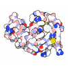



-Map
| File | Download / File: emd_54818.map.gz / Format: CCP4 / Size: 421.9 MB / Type: IMAGE STORED AS FLOATING POINT NUMBER (4 BYTES) | ||||||||||||||||||||||||||||||||||||
|---|---|---|---|---|---|---|---|---|---|---|---|---|---|---|---|---|---|---|---|---|---|---|---|---|---|---|---|---|---|---|---|---|---|---|---|---|---|
| Annotation | map middle core in C8 symmetry | ||||||||||||||||||||||||||||||||||||
| Projections & slices | Image control
Images are generated by Spider. | ||||||||||||||||||||||||||||||||||||
| Voxel size | X=Y=Z: 1.07 Å | ||||||||||||||||||||||||||||||||||||
| Density |
| ||||||||||||||||||||||||||||||||||||
| Symmetry | Space group: 1 | ||||||||||||||||||||||||||||||||||||
| Details | EMDB XML:
|
 Z (Sec.)
 Y (Row.)
 X (Col.)


-Supplemental data
-Mask #1
| File | emd_54818_msk_1.map | ||||||||||||
|---|---|---|---|---|---|---|---|---|---|---|---|---|---|
| Projections & Slices |
| ||||||||||||
| Density Histograms |


-Half map: half1 map middle core in C8 symmetry
| File | emd_54818_half_map_1.map | ||||||||||||
|---|---|---|---|---|---|---|---|---|---|---|---|---|---|
| Annotation | half1 map middle core in C8 symmetry | ||||||||||||
| Projections & Slices |
| ||||||||||||
| Density Histograms |


-Half map: half2 map middle core in C8 symmetry
| File | emd_54818_half_map_2.map | ||||||||||||
|---|---|---|---|---|---|---|---|---|---|---|---|---|---|
| Annotation | half2 map middle core in C8 symmetry | ||||||||||||
| Projections & Slices |
| ||||||||||||
| Density Histograms |


- Sample components
-Entire : Pseudomonas phage LUZ19
| Entire | Name: Pseudomonas phage LUZ19 (virus) |
|---|---|
| Components |
|
-Supramolecule #1: Pseudomonas phage LUZ19
| Supramolecule | Name: Pseudomonas phage LUZ19 / type: virus / ID: 1 / Parent: 0 / Macromolecule list: all / NCBI-ID: 484896 / Sci species name: Pseudomonas phage LUZ19 / Virus type: VIRION / Virus isolate: STRAIN / Virus enveloped: No / Virus empty: No |
|---|---|
| Molecular weight | Theoretical: 924 KDa |
-Macromolecule #1: Internal virion protein
| Macromolecule | Name: Internal virion protein / type: protein_or_peptide / ID: 1 / Number of copies: 1 / Enantiomer: LEVO |
|---|---|
| Source (natural) | Organism: Pseudomonas phage LUZ19 (virus) |
| Molecular weight | Theoretical: 98.368633 KDa |
| Sequence | String: MAESQRASQE LGINVGQTQL QPGQSARRGV RDSEINYSGP SVGSQILDGI LGAGQQIAGK WFEHNVQQEV LRGERARMAG EAEEAVDSN VLAKPFVKGG WRKQDYRIAQ ADFSLKMQRF IANKGREMTP EEFRKYLSQE ATHVLDSTEG MNPNDALQAL A QQQKAEEQ ...String: MAESQRASQE LGINVGQTQL QPGQSARRGV RDSEINYSGP SVGSQILDGI LGAGQQIAGK WFEHNVQQEV LRGERARMAG EAEEAVDSN VLAKPFVKGG WRKQDYRIAQ ADFSLKMQRF IANKGREMTP EEFRKYLSQE ATHVLDSTEG MNPNDALQAL A QQQKAEEQ LFGMQAKAYM DWSIDQAARG FRTQGNSILA KAVQAQATGD ELSRQLSLEE AGLFYTNIMT SEDIPLEVRD KV GMQFLAA SLDMNQRGIY EGLRDAGFLD SMSFDDRRAL NGLYEKSKAQ TRAKESMATL RADADFQQRV ANGAITDLAE VEA YSRGMV EEGRWSDAQA ISFMTKAMTG LGNAQRMQGI MAALEAGDIN ALHTLGTNVT EALEQWDKMQ AANGSSLTDR LVQG TQLGL RLGTFPKTYG ESVGSAVRMI QAAKEGEANP ELVNTLNSIF EQVASAQEIN PSAGNVMLSG IPEAEQGAVA WALKQ MKMG IAPAQALREF SANAEVVKQM DEFEKGQNTK AFKDNLGKQV NDKFVNNIFG RAWNMLTGES DLSNNEAVLS MYRRAT IDE ANWLASDRKH AGLLTSDTGR EALLEIAAAN VRNRTIQVGE GRNLKEGDLF SRRDSAPLIL PRGTTAEQLF GTNDTET IG TVLAEQHKPH VEGLLGYKSV VAFEYDRTSG SLLAVEYDEN GVALDRTRVD PQAVGNEVLK RNADKLNAMR GAEYGANV K ISGTDIRMNG GNSAGMLKQD VFNWRKELAQ FEAYRGEAYK DADGYSVGLG HYLGSGNAGA GTTVTPEQAA QWFAEDTDR ALDQGVRLAD ELGVTNNASI LGLAGMAFQM GEGRARQFRN TFQAIKDRNK EAFEAGVRNS KWYTQTPNRA EAFIKRMAPH FDTPSQIGV DWYSAATAE UniProtKB: Internal virion protein |
-Experimental details
-Structure determination
| Method | cryo EM |
|---|---|
 Processing Processing | single particle reconstruction |
| Aggregation state | particle |
-Sample preparation
| Buffer | pH: 8 |
|---|---|
| Vitrification | Cryogen name: ETHANE |
- Electron microscopy
| Microscope | TFS KRIOS |
|---|---|
| Image recording | Film or detector model: FEI FALCON III (4k x 4k) / Average electron dose: 94.0 e/Å2 |
| Electron beam | Acceleration voltage: 300 kV / Electron source:  FIELD EMISSION GUN FIELD EMISSION GUN |
| Electron optics | Illumination mode: FLOOD BEAM / Imaging mode: BRIGHT FIELD / Nominal defocus max: 2.0 µm / Nominal defocus min: 0.6 µm |
| Experimental equipment |  Model: Titan Krios / Image courtesy: FEI Company |

