Movie
Movie Controller

[English] 日本語
 Yorodumi
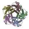
- EMDB-54389: Aquifex aeolicus lumazine synthase R29A mutant 11-pentamer cage -
+ Open data

- Basic information
| Entry |  | |||||||||||||||
|---|---|---|---|---|---|---|---|---|---|---|---|---|---|---|---|---|
| Title | Aquifex aeolicus lumazine synthase R29A mutant 11-pentamer cage | |||||||||||||||
 Map data Map data | Map combined from 2 independent halves and not post-processed | |||||||||||||||
 Sample Sample |
| |||||||||||||||
 Keywords Keywords | protein cage / protein engineering / self-assembly / geometry / pentamer / encapsulation / BIOSYNTHETIC PROTEIN | |||||||||||||||
| Function / homology |  Function and homology information Function and homology information6,7-dimethyl-8-ribityllumazine synthase / 6,7-dimethyl-8-ribityllumazine synthase activity / riboflavin synthase complex / riboflavin biosynthetic process / cytosol / cytoplasm Similarity search - Function | |||||||||||||||
| Biological species |   Aquifex aeolicus VF5 (bacteria) Aquifex aeolicus VF5 (bacteria) | |||||||||||||||
| Method | single particle reconstruction / cryo EM / Resolution: 2.08 Å | |||||||||||||||
 Authors Authors | Koziej L / Azuma Y | |||||||||||||||
| Funding support |  Poland, European Union, 4 items Poland, European Union, 4 items
| |||||||||||||||
 Citation Citation | Journal: Nat Commun / Year: 2026 Title: A molecular basis for stoichiometric enzyme encapsulation in the vitamin B2 biosynthesis compartment. Authors: Lukasz Koziej / Jedrzej Pankowski / Monika Stefanska / Daniel Jankowski / Agnieszka Gawin / V Vishal Malolan / Juha T Huiskonen / Takahiro Kosugi / Yusuke Azuma /   Abstract: Encapsulating metabolic enzymes within protein cages enhances catalytic efficiency through substrate channeling. The vitamin B2 biosynthesis system, in which a dodecahedral lumazine synthase (LS) ...Encapsulating metabolic enzymes within protein cages enhances catalytic efficiency through substrate channeling. The vitamin B2 biosynthesis system, in which a dodecahedral lumazine synthase (LS) cage encapsulates a homotrimeric riboflavin synthase (RS), exemplifies this strategy, yet the molecular basis for this stoichiometric enzyme encapsulation has remained elusive. Here, cryogenic electron microscopy structures reveal a hierarchical assembly mechanism that ensures the defined host-guest ratio. RS C-terminal cage-localization signal peptides anchor at LS pentamer-pentamer interfaces early during assembly, stabilizing open intermediates that, together with delayed later-stage cage closure, extend the loading window until guest incorporation is complete. RS spatial occupancy avoids overloading, while a molecular lock upon final closure prevents disassembly. The elucidated anchoring mechanism enabled structure-based phylogenetic analysis across diverse organisms, suggesting multiple independent evolutionary origins of this modular encapsulation strategy. This naturally occurring architecture provides design principles for engineering synthetic catalytic compartments with programmable stoichiometric control. | |||||||||||||||
| History |
|
- Structure visualization
| Supplemental images |
|---|

- Downloads & links
-EMDB archive
| Map data | emd_54389.map.gz | 99 MB | EMDB map data format | |
|---|---|---|---|---|
| Header (meta data) | emd-54389-v30.xmlemd-54389.xml | 26 KB 26 KB | Display Display | EMDB header |
| FSC (resolution estimation) | emd_54389_fsc.xml | 12.2 KB | Display | FSC data file |
| Images |  emd_54389.png emd_54389.png | 163.3 KB | ||
| Masks | emd_54389_msk_1.map | 196.4 MB | Mask map | |
| Filedesc metadata | emd-54389.cif.gz | 7 KB | ||
| Others | emd_54389_additional_1.map.gzemd_54389_half_map_1.map.gzemd_54389_half_map_2.map.gz | 185.7 MB 182.5 MB 182.5 MB | ||
| Archive directory |  http://ftp.pdbj.org/pub/emdb/structures/EMD-54389ftp://ftp.pdbj.org/pub/emdb/structures/EMD-54389 http://ftp.pdbj.org/pub/emdb/structures/EMD-54389ftp://ftp.pdbj.org/pub/emdb/structures/EMD-54389 | HTTPS FTP |
-Related structure data
| Related structure data |  9ryqMC  9ryiC  9ryjC  9rykC  9rymC  9rynC  9ryoC  9rypC  9ryuC  9ryvC  9rywC  9ryxC M: atomic model generated by this map C: citing same article ( |
|---|---|
| Similar structure data |


-Links
| EMDB pages | EMDB (EBI/PDBe) / EMDataResource |
|---|
-Map
| File | Download / File: emd_54389.map.gz / Format: CCP4 / Size: 196.4 MB / Type: IMAGE STORED AS FLOATING POINT NUMBER (4 BYTES) | ||||||||||||||||||||||||||||||||||||
|---|---|---|---|---|---|---|---|---|---|---|---|---|---|---|---|---|---|---|---|---|---|---|---|---|---|---|---|---|---|---|---|---|---|---|---|---|---|
| Annotation | Map combined from 2 independent halves and not post-processed | ||||||||||||||||||||||||||||||||||||
| Projections & slices | Image control
Images are generated by Spider. | ||||||||||||||||||||||||||||||||||||
| Voxel size | X=Y=Z: 0.8456 Å | ||||||||||||||||||||||||||||||||||||
| Density |
| ||||||||||||||||||||||||||||||||||||
| Symmetry | Space group: 1 | ||||||||||||||||||||||||||||||||||||
| Details | EMDB XML:
|
 Z (Sec.)
 Y (Row.)
 X (Col.)


-Supplemental data
-Mask #1
| File | emd_54389_msk_1.map | ||||||||||||
|---|---|---|---|---|---|---|---|---|---|---|---|---|---|
| Projections & Slices |
| ||||||||||||
| Density Histograms |


-Additional map: Combined half maps symmetrized and post-processed using sharpening...
| File | emd_54389_additional_1.map | ||||||||||||
|---|---|---|---|---|---|---|---|---|---|---|---|---|---|
| Annotation | Combined half maps symmetrized and post-processed using sharpening B factor 43.9 | ||||||||||||
| Projections & Slices |
| ||||||||||||
| Density Histograms |


-Half map: #1
| File | emd_54389_half_map_1.map | ||||||||||||
|---|---|---|---|---|---|---|---|---|---|---|---|---|---|
| Projections & Slices |
| ||||||||||||
| Density Histograms |


-Half map: #2
| File | emd_54389_half_map_2.map | ||||||||||||
|---|---|---|---|---|---|---|---|---|---|---|---|---|---|
| Projections & Slices |
| ||||||||||||
| Density Histograms |


- Sample components
-Entire : Aquifex aeolicus lumazine synthase R29A mutant 11-pentamer cage
| Entire | Name: Aquifex aeolicus lumazine synthase R29A mutant 11-pentamer cage |
|---|---|
| Components |
|
-Supramolecule #1: Aquifex aeolicus lumazine synthase R29A mutant 11-pentamer cage
| Supramolecule | Name: Aquifex aeolicus lumazine synthase R29A mutant 11-pentamer cage type: complex / ID: 1 / Parent: 0 / Macromolecule list: all |
|---|---|
| Source (natural) | Organism: Aquifex aeolicus VF5 (bacteria) |
| Molecular weight | Theoretical: 1 MDa |
-Macromolecule #1: 6,7-dimethyl-8-ribityllumazine synthase
| Macromolecule | Name: 6,7-dimethyl-8-ribityllumazine synthase / type: protein_or_peptide / ID: 1 / Details: AaLS R29A mutant / Number of copies: 55 / Enantiomer: LEVO / EC number: 6,7-dimethyl-8-ribityllumazine synthase |
|---|---|
| Source (natural) | Organism: Aquifex aeolicus VF5 (bacteria) |
| Molecular weight | Theoretical: 18.040594 KDa |
| Recombinant expression | Organism:  Escherichia coli (E. coli) Escherichia coli (E. coli) |
| Sequence | String: MEIYEGKLTA EGLRFGIVAS RFNHALVDAL VEGAIDCIVR HGGREEDITL VRVPGSWEIP VAAGELARKE DIDAVIAIGV LIRGATPHF DYIASEVSKG LANLSLELRK PITFGVITAD TLEQAIERAG TKHGNKGWEA ALSAIEMANL FKSLRLEGGW S HPQFEK UniProtKB: 6,7-dimethyl-8-ribityllumazine synthase |
-Experimental details
-Structure determination
| Method | cryo EM |
|---|---|
 Processing Processing | single particle reconstruction |
| Aggregation state | particle |
-Sample preparation
| Concentration | 1 mg/mL | ||||||||||||
|---|---|---|---|---|---|---|---|---|---|---|---|---|---|
| Buffer | pH: 8 Component:
| ||||||||||||
| Grid | Model: Quantifoil R2/1 / Material: COPPER / Mesh: 200 / Support film - Material: CARBON / Support film - topology: HOLEY / Pretreatment - Type: GLOW DISCHARGE / Pretreatment - Time: 5 sec. / Pretreatment - Atmosphere: AIR | ||||||||||||
| Vitrification | Cryogen name: ETHANE / Chamber humidity: 100 % / Chamber temperature: 277 K / Instrument: FEI VITROBOT MARK IV |
- Electron microscopy
| Microscope | TFS KRIOS |
|---|---|
| Specialist optics | Energy filter - Name: GIF Bioquantum / Energy filter - Slit width: 20 eV |
| Image recording | Film or detector model: GATAN K3 BIOCONTINUUM (6k x 4k) / Digitization - Dimensions - Width: 5760 pixel / Digitization - Dimensions - Height: 4092 pixel / Number grids imaged: 1 / Number real images: 5306 / Average electron dose: 40.0 e/Å2 |
| Electron beam | Acceleration voltage: 300 kV / Electron source:  FIELD EMISSION GUN FIELD EMISSION GUN |
| Electron optics | C2 aperture diameter: 50.0 µm / Illumination mode: FLOOD BEAM / Imaging mode: BRIGHT FIELD / Cs: 2.7 mm / Nominal defocus max: 2.1 µm / Nominal defocus min: 0.6 µm |
| Sample stage | Specimen holder model: FEI TITAN KRIOS AUTOGRID HOLDER / Cooling holder cryogen: NITROGEN |
| Experimental equipment |  Model: Titan Krios / Image courtesy: FEI Company |
+Image processing

-Atomic model buiding 1
| Initial model | PDB ID: Chain - Source name: PDB / Chain - Initial model type: experimental model / Details: lumazine synthase |
|---|---|
| Details | Initial fitting was done using ChimeraX. Flexible fitting was done using Isolde. |
| Refinement | Space: REAL / Protocol: RIGID BODY FIT |
| Output model | PDB-9ryq: |