 Movie
Movie Controller


+ Open data

- Basic information
| Entry |  | |||||||||
|---|---|---|---|---|---|---|---|---|---|---|
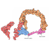
| Title | Structure of the ZAK-bound human disome | |||||||||
 Map data Map data | Composite map of the ZAK-bound disome complex | |||||||||
 Sample Sample |
| |||||||||
 Keywords Keywords | ZAK / collision / RSR / quality control / RIBOSOME | |||||||||
| Function / homology |  Function and homology information Function and homology informationpositive regulation of mitotic DNA damage checkpoint / negative regulation of stress-activated protein kinase signaling cascade / negative regulation of translation in response to endoplasmic reticulum stress / stalled ribosome sensor activity / GCN2-mediated signaling / cell death / mitogen-activated protein kinase kinase kinase / endothelial cell differentiation / JUN kinase kinase kinase activity / embryonic brain development ...positive regulation of mitotic DNA damage checkpoint / negative regulation of stress-activated protein kinase signaling cascade / negative regulation of translation in response to endoplasmic reticulum stress / stalled ribosome sensor activity / GCN2-mediated signaling / cell death / mitogen-activated protein kinase kinase kinase / endothelial cell differentiation / JUN kinase kinase kinase activity / embryonic brain development / male meiosis I / translation at presynapse / embryonic digit morphogenesis / response to insecticide / negative regulation of endoplasmic reticulum unfolded protein response / eukaryotic 80S initiation complex / ribosomal protein import into nucleus / regulation of G1 to G0 transition / stress-activated protein kinase signaling cascade / positive regulation of DNA binding / oxidized pyrimidine DNA binding / response to TNF agonist / positive regulation of base-excision repair / positive regulation of respiratory burst involved in inflammatory response / regulation of adenylate cyclase-activating G protein-coupled receptor signaling pathway / positive regulation of intrinsic apoptotic signaling pathway in response to DNA damage / positive regulation of gastrulation / protein tyrosine kinase inhibitor activity / positive regulation of programmed cell death / cellular response to UV-B / IRE1-RACK1-PP2A complex / TNFR1-mediated ceramide production / positive regulation of Golgi to plasma membrane protein transport / G1 to G0 transition / regulation of mitotic metaphase/anaphase transition / negative regulation of formation of translation preinitiation complex / nucleolus organization / positive regulation of ubiquitin-protein transferase activity / negative regulation of RNA splicing / GAIT complex / positive regulation of DNA-templated transcription initiation / negative regulation of DNA repair / TORC2 complex binding / erythrocyte homeostasis / supercoiled DNA binding / regulation of establishment of cell polarity / rRNA modification in the nucleus and cytosol / oxidized purine DNA binding / NF-kappaB complex / negative regulation of intrinsic apoptotic signaling pathway in response to hydrogen peroxide / negative regulation of phagocytosis / p38MAPK cascade / ubiquitin-like protein conjugating enzyme binding / cytoplasmic translational initiation / cytoplasmic side of rough endoplasmic reticulum membrane / regulation of translation involved in cellular response to UV / A band / Formation of the ternary complex, and subsequently, the 43S complex / ion channel inhibitor activity / positive regulation of intrinsic apoptotic signaling pathway in response to DNA damage by p53 class mediator / laminin receptor activity / response to aldosterone / negative regulation of myoblast fusion / protein-DNA complex disassembly / Ribosomal scanning and start codon recognition / protein kinase A binding / Translation initiation complex formation / TFIID-class transcription factor complex binding / negative regulation of Wnt signaling pathway / positive regulation of DNA damage response, signal transduction by p53 class mediator / fibroblast growth factor binding / BH3 domain binding / Protein hydroxylation / TOR signaling / limb development / mTORC1-mediated signalling / negative regulation of translational frameshifting / monocyte chemotaxis / PELO:HBS1L and ABCE1 dissociate a ribosome on a non-stop mRNA / regulation of lipid metabolic process / SARS-CoV-1 modulates host translation machinery / pyroptotic inflammatory response / regulation of cell division / positive regulation of GTPase activity / stress-activated MAPK cascade / protein localization to nucleus / MAP kinase kinase kinase activity / Peptide chain elongation / JNK cascade / Selenocysteine synthesis / negative regulation of protein binding / Formation of a pool of free 40S subunits / protein kinase activator activity / negative regulation of respiratory burst involved in inflammatory response / positive regulation of intrinsic apoptotic signaling pathway by p53 class mediator / protein serine/threonine kinase inhibitor activity / protein targeting / Eukaryotic Translation Termination / Dengue Virus Attachment and Entry / SRP-dependent cotranslational protein targeting to membrane Similarity search - Function | |||||||||
| Biological species |  Homo sapiens (human) Homo sapiens (human) | |||||||||
| Method | single particle reconstruction / cryo EM / Resolution: 2.35 Å | |||||||||
 Authors Authors | Niu S / Beckmann R | |||||||||
| Funding support | European Union, 1 items
| |||||||||
 Citation Citation | Journal: Nature / Year: 2026 Title: ZAK activation at the collided ribosome. Authors: Vienna L Huso / Shuangshuang Niu / Marco A Catipovic / James A Saba / Timo Denk / Eugene Park / Jingdong Cheng / Otto Berninghausen / Thomas Becker / Rachel Green / Roland Beckmann /    Abstract: Ribosome collisions activate the ribotoxic stress response mediated by the MAP3K ZAK, which in turn regulates cell-fate consequences through downstream phosphorylation of the MAPKs p38 and JNK. ...Ribosome collisions activate the ribotoxic stress response mediated by the MAP3K ZAK, which in turn regulates cell-fate consequences through downstream phosphorylation of the MAPKs p38 and JNK. Despite the critical role of ZAK during cellular stress, a mechanistic and structural understanding of ZAK-ribosome interactions and how these lead to activation remain elusive. Here we combine biochemistry and cryo-electron microscopy to discover distinct ZAK-ribosome interactions required for constitutive recruitment and for activation. We find that upon induction of ribosome collisions, interactions between ZAK and the ribosomal protein RACK1 enable its activation by dimerization of its SAM domains at the collision interface. Furthermore, we discover how this process is negatively regulated by the ribosome-binding protein SERBP1 to prevent constitutive ZAK activation. Characterization of novel SAM variants as well as a known pathogenic variant of the SAM domain of ZAK supports a key role of the SAM domain in regulating kinase activity on and off the ribosome, with some mutants bypassing the ribosome requirement for ZAK activation. Collectively, our data provide a mechanistic blueprint of the kinase activity of ZAK at the collided ribosome interface. | |||||||||
| History |
|
- Structure visualization
| Supplemental images |
|---|

- Downloads & links
-EMDB archive
| Map data | emd_54172.map.gz | 115.7 MB | EMDB map data format | |
|---|---|---|---|---|
| Header (meta data) | emd-54172-v30.xmlemd-54172.xml | 132.3 KB 132.3 KB | Display Display | EMDB header |
| Images |  emd_54172.png emd_54172.png | 91.4 KB | ||
| Filedesc metadata | emd-54172.cif.gz | 24.4 KB | ||
| Archive directory |  https://data.pdbj.org/pub/emdb/structures/EMD-54172ftp://data.pdbj.org/pub/emdb/structures/EMD-54172 https://data.pdbj.org/pub/emdb/structures/EMD-54172ftp://data.pdbj.org/pub/emdb/structures/EMD-54172 | HTTPS FTP |
-Related structure data
| Related structure data |  9rpvMC  9rsxC C: citing same article ( M: atomic model generated by this map |
|---|---|
| Similar structure data |


-Links
| EMDB pages | EMDB (EBI/PDBe) / EMDataResource |
|---|---|
| Related items in Molecule of the Month |


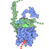

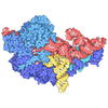














-Map
| File | Download / File: emd_54172.map.gz / Format: CCP4 / Size: 307.5 MB / Type: IMAGE STORED AS FLOATING POINT NUMBER (4 BYTES) | ||||||||||||||||||||||||||||||||||||
|---|---|---|---|---|---|---|---|---|---|---|---|---|---|---|---|---|---|---|---|---|---|---|---|---|---|---|---|---|---|---|---|---|---|---|---|---|---|
| Annotation | Composite map of the ZAK-bound disome complex | ||||||||||||||||||||||||||||||||||||
| Projections & slices | Image control
Images are generated by Spider. | ||||||||||||||||||||||||||||||||||||
| Voxel size | X=Y=Z: 1.454 Å | ||||||||||||||||||||||||||||||||||||
| Density |
| ||||||||||||||||||||||||||||||||||||
| Symmetry | Space group: 1 | ||||||||||||||||||||||||||||||||||||
| Details | EMDB XML:
|
 Z (Sec.)
 Y (Row.)
 X (Col.)


-Supplemental data
- Sample components
+Entire : ZAK-bound human disome
+Supramolecule #1: ZAK-bound human disome
+Macromolecule #1: mRNA of the collided 80S
+Macromolecule #2: mRNA of the stalled 80S
+Macromolecule #3: P/E tRNA on the collided 80S
+Macromolecule #4: P-site tRNA of the stalled 80S
+Macromolecule #5: A/P tRNA on the collided 80S
+Macromolecule #7: E-site tRNA of the stalled 80S
+Macromolecule #8: 28S rRNA of the collided 80S
+Macromolecule #9: 5S rRNA
+Macromolecule #10: 5.8S rRNA of the collided 80S
+Macromolecule #53: 28S rRNA of the stalled 80S
+Macromolecule #54: 5.8S rRNA of the stalled 80S
+Macromolecule #98: 18S rRNA of the collided 80S
+Macromolecule #99: 18S rRNA of the stalled 80S
+Macromolecule #6: Endothelial differentiation-related factor 1
+Macromolecule #11: 60S ribosomal protein L8
+Macromolecule #12: 60S ribosomal protein L3
+Macromolecule #13: 60S ribosomal protein L4
+Macromolecule #14: 60S ribosomal protein L5
+Macromolecule #15: Large ribosomal subunit protein eL6
+Macromolecule #16: Large ribosomal subunit protein uL30
+Macromolecule #17: 60S ribosomal protein L7a
+Macromolecule #18: 60S ribosomal protein L9
+Macromolecule #19: Ribosomal protein uL16-like
+Macromolecule #20: 60S ribosomal protein L11
+Macromolecule #21: Large ribosomal subunit protein eL13
+Macromolecule #22: 60S ribosomal protein L14
+Macromolecule #23: 60S ribosomal protein L15
+Macromolecule #24: 60S ribosomal protein L13a
+Macromolecule #25: 60S ribosomal protein L17
+Macromolecule #26: 60S ribosomal protein L18
+Macromolecule #27: 60S ribosomal protein L19
+Macromolecule #28: 60S ribosomal protein L18a
+Macromolecule #29: 60S ribosomal protein L21
+Macromolecule #30: 60S ribosomal protein L22
+Macromolecule #31: 60S ribosomal protein L23
+Macromolecule #32: 60S ribosomal protein L24
+Macromolecule #33: 60S ribosomal protein L23a
+Macromolecule #34: 60S ribosomal protein L26
+Macromolecule #35: 60S ribosomal protein L27
+Macromolecule #36: 60S ribosomal protein L27a
+Macromolecule #37: 60S ribosomal protein L29
+Macromolecule #38: 60S ribosomal protein L30
+Macromolecule #39: 60S ribosomal protein L31
+Macromolecule #40: 60S ribosomal protein L32
+Macromolecule #41: 60S ribosomal protein L35a
+Macromolecule #42: 60S ribosomal protein L34
+Macromolecule #43: 60S ribosomal protein L35
+Macromolecule #44: 60S ribosomal protein L36
+Macromolecule #45: Large ribosomal subunit protein eL37
+Macromolecule #46: 60S ribosomal protein L38
+Macromolecule #47: 60S ribosomal protein L39
+Macromolecule #48: Ubiquitin-60S ribosomal protein L40
+Macromolecule #49: 60S ribosomal protein L41
+Macromolecule #50: 60S ribosomal protein L36a
+Macromolecule #51: 60S ribosomal protein L37a
+Macromolecule #52: 60S ribosomal protein L28
+Macromolecule #55: Large ribosomal subunit protein uL2
+Macromolecule #56: Large ribosomal subunit protein uL3
+Macromolecule #57: 60S ribosomal protein L10
+Macromolecule #58: Large ribosomal subunit protein uL15
+Macromolecule #59: Large ribosomal subunit protein eL29
+Macromolecule #60: Ubiquitin-ribosomal protein eL40 fusion protein
+Macromolecule #61: Large ribosomal subunit protein eL42
+Macromolecule #62: Large ribosomal subunit protein eL28
+Macromolecule #63: 60S ribosomal protein L10a
+Macromolecule #64: Mitogen-activated protein kinase kinase kinase 20
+Macromolecule #65: Small ribosomal subunit protein uS2
+Macromolecule #66: 40S ribosomal protein S3a
+Macromolecule #67: 40S ribosomal protein S2
+Macromolecule #68: 40S ribosomal protein S3
+Macromolecule #69: Small ribosomal subunit protein eS4, X isoform
+Macromolecule #70: 40S ribosomal protein S5
+Macromolecule #71: 40S ribosomal protein S6
+Macromolecule #72: 40S ribosomal protein S7
+Macromolecule #73: 40S ribosomal protein S8
+Macromolecule #74: 40S ribosomal protein S9
+Macromolecule #75: 40S ribosomal protein S10
+Macromolecule #76: 40S ribosomal protein S11
+Macromolecule #77: 40S ribosomal protein S12
+Macromolecule #78: 40S ribosomal protein S13
+Macromolecule #79: 40S ribosomal protein S14
+Macromolecule #80: 40S ribosomal protein S15
+Macromolecule #81: 40S ribosomal protein S16
+Macromolecule #82: 40S ribosomal protein S17
+Macromolecule #83: 40S ribosomal protein S18
+Macromolecule #84: 40S ribosomal protein S19
+Macromolecule #85: 40S ribosomal protein S20
+Macromolecule #86: Small ribosomal subunit protein eS21
+Macromolecule #87: 40S ribosomal protein S15a
+Macromolecule #88: 40S ribosomal protein S23
+Macromolecule #89: 40S ribosomal protein S24
+Macromolecule #90: 40S ribosomal protein S25
+Macromolecule #91: 40S ribosomal protein S26
+Macromolecule #92: 40S ribosomal protein S27
+Macromolecule #93: 40S ribosomal protein S28
+Macromolecule #94: 40S ribosomal protein S29
+Macromolecule #95: Ubiquitin-like FUBI-ribosomal protein eS30 fusion protein
+Macromolecule #96: Ubiquitin
+Macromolecule #97: Receptor of activated protein C kinase 1
+Macromolecule #100: 40S ribosomal protein SA
+Macromolecule #101: 40S ribosomal protein S21
+Macromolecule #102: MAGNESIUM ION
+Macromolecule #103: ZINC ION
+Macromolecule #104: ANISOMYCIN

-Experimental details
-Structure determination
| Method | cryo EM |
|---|---|
 Processing Processing | single particle reconstruction |
| Aggregation state | particle |
-Sample preparation
| Buffer | pH: 7.5 |
|---|---|
| Vitrification | Cryogen name: ETHANE |
- Electron microscopy
| Microscope | TFS KRIOS |
|---|---|
| Image recording | Film or detector model: FEI FALCON IV (4k x 4k) / Average electron dose: 40.0 e/Å2 |
| Electron beam | Acceleration voltage: 300 kV / Electron source:  FIELD EMISSION GUN FIELD EMISSION GUN |
| Electron optics | Illumination mode: FLOOD BEAM / Imaging mode: BRIGHT FIELD / Nominal defocus max: 3.5 µm / Nominal defocus min: 0.5 µm |
| Experimental equipment |  Model: Titan Krios / Image courtesy: FEI Company |
