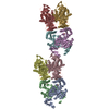
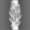
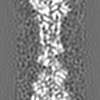
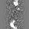
Journal: Proc Natl Acad Sci U S A / Year: 2011 Title: Remodeling of actin filaments by ADF/cofilin proteins. Authors: Vitold E Galkin / Albina Orlova / Dmitri S Kudryashov / Alexander Solodukhin / Emil Reisler / Gunnar F Schröder / Edward H Egelman / Abstract: Cofilin/ADF proteins play key roles in the dynamics of actin, one of the most abundant and highly conserved eukaryotic proteins. We used cryoelectron microscopy to generate a 9-Å resolution three- ...Cofilin/ADF proteins play key roles in the dynamics of actin, one of the most abundant and highly conserved eukaryotic proteins. We used cryoelectron microscopy to generate a 9-Å resolution three-dimensional reconstruction of cofilin-decorated actin filaments, the highest resolution achieved for a complex of F-actin with an actin-binding protein. We show that the cofilin-induced change in the filament twist is due to a unique conformation of the actin molecule unrelated to any previously observed state. The changes between the actin protomer in naked F-actin and in the actin-cofilin filament are greater than the conformational changes between G- and F-actin. Our results show the structural plasticity of actin, suggest that other actin-binding proteins may also induce large but different conformational changes, and show that F-actin cannot be described by a single molecular model.
History
Deposition
Nov 23, 2011
-
Header (metadata) release
Dec 7, 2011
-
Map release
Dec 13, 2011
-
Update
Mar 20, 2013
-
Current status
Mar 20, 2013
Processing site: RCSB / Status: Released
-
Structure visualization
Movie
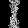
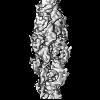
Surface view with section colored by density value
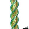
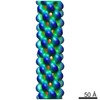
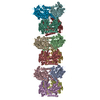
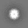
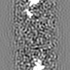
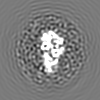
Name: actin decorated with cofilin / type: sample / ID: 1000 Oligomeric state: filament containing one cofilin to one actin Number unique components: 2
In the structure databanks used in Yorodumi, some data are registered as the other names, "COVID-19 virus" and "2019-nCoV". Here are the details of the virus and the list of structure data.
Jan 31, 2019. EMDB accession codes are about to change! (news from PDBe EMDB page)
EMDB accession codes are about to change! (news from PDBe EMDB page)
The allocation of 4 digits for EMDB accession codes will soon come to an end. Whilst these codes will remain in use, new EMDB accession codes will include an additional digit and will expand incrementally as the available range of codes is exhausted. The current 4-digit format prefixed with “EMD-” (i.e. EMD-XXXX) will advance to a 5-digit format (i.e. EMD-XXXXX), and so on. It is currently estimated that the 4-digit codes will be depleted around Spring 2019, at which point the 5-digit format will come into force.
The EM Navigator/Yorodumi systems omit the EMD- prefix.
Related info.:Q: What is EMD? / ID/Accession-code notation in Yorodumi/EM Navigator
Yorodumi is a browser for structure data from EMDB, PDB, SASBDB, etc.
This page is also the successor to EM Navigator detail page, and also detail information page/front-end page for Omokage search.
The word "yorodu" (or yorozu) is an old Japanese word meaning "ten thousand". "mi" (miru) is to see.
Related info.:EMDB / PDB / SASBDB / Comparison of 3 databanks / Yorodumi Search / Aug 31, 2016. New EM Navigator & Yorodumi / Yorodumi Papers / Jmol/JSmol / Function and homology information / Changes in new EM Navigator and Yorodumi
 Movie
Movie Controller


 Open data
 Basic information
 Map data
 Sample
 Keywords
 Function and homology information
 Homo sapiens (human)
 Authors
 Citation
 Structure visualization
 Downloads & links
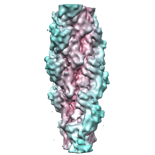 emd_5354_1.png
 http://ftp.pdbj.org/pub/emdb/structures/EMD-5354


 Z (Sec.)
 Y (Row.)
 X (Col.)


 Sample components
 Processing
 Electron microscopy
 FIELD EMISSION GUN
