 Movie
Movie Controller

[English] 日本語
 Yorodumi
- EMDB-53175: Cryo-EM structure of mouse TRPM3 alpha 2 in complex with antagoni... -
+ Open data

- Basic information
| Entry |  | |||||||||
|---|---|---|---|---|---|---|---|---|---|---|
| Title | Cryo-EM structure of mouse TRPM3 alpha 2 in complex with antagonist Primidone | |||||||||
 Map data Map data | Cryo-EM structure of mouse TRPM3 alpha 2 in complex with antagonist Primidone | |||||||||
 Sample Sample |
| |||||||||
 Keywords Keywords | Ca2+ channel Primidone-bound Closed conformation / MEMBRANE PROTEIN | |||||||||
| Function / homology |  Function and homology information Function and homology informationprotein tetramerization / calcium channel activity / calmodulin binding / plasma membrane Similarity search - Function | |||||||||
| Biological species |  | |||||||||
| Method | single particle reconstruction / cryo EM / Resolution: 3.28 Å | |||||||||
 Authors Authors | Shkumatov AV / Schenck S / Brunner JD | |||||||||
| Funding support | 1 items
| |||||||||
 Citation Citation | Journal: To Be Published Title: Cryo-EM structure of mouse TRPM3 alpha 2 in complex with antagonist Primidone Authors: Shkumatov AV / Schenck S / Brunner JD | |||||||||
| History |
|
- Structure visualization
| Supplemental images |
|---|

- Downloads & links
-EMDB archive
| Map data | emd_53175.map.gz | 209.3 MB | EMDB map data format | |
|---|---|---|---|---|
| Header (meta data) | emd-53175-v30.xmlemd-53175.xml | 22 KB 22 KB | Display Display | EMDB header |
| FSC (resolution estimation) | emd_53175_fsc.xml | 18 KB | Display | FSC data file |
| Images |  emd_53175.png emd_53175.png | 131.1 KB | ||
| Masks | emd_53175_msk_1.map | 421.9 MB | Mask map | |
| Filedesc metadata | emd-53175.cif.gz | 7.2 KB | ||
| Others | emd_53175_additional_1.map.gzemd_53175_half_map_1.map.gzemd_53175_half_map_2.map.gz | 398.3 MB 391.2 MB 391.1 MB | ||
| Archive directory |  http://ftp.pdbj.org/pub/emdb/structures/EMD-53175ftp://ftp.pdbj.org/pub/emdb/structures/EMD-53175 http://ftp.pdbj.org/pub/emdb/structures/EMD-53175ftp://ftp.pdbj.org/pub/emdb/structures/EMD-53175 | HTTPS FTP |
-Related structure data
| Related structure data |  9qhoMC M: atomic model generated by this map C: citing same article ( |
|---|---|
| Similar structure data |


-Links
| EMDB pages | EMDB (EBI/PDBe) / EMDataResource |
|---|
-Map
| File | Download / File: emd_53175.map.gz / Format: CCP4 / Size: 421.9 MB / Type: IMAGE STORED AS FLOATING POINT NUMBER (4 BYTES) | ||||||||||||||||||||||||||||||||||||
|---|---|---|---|---|---|---|---|---|---|---|---|---|---|---|---|---|---|---|---|---|---|---|---|---|---|---|---|---|---|---|---|---|---|---|---|---|---|
| Annotation | Cryo-EM structure of mouse TRPM3 alpha 2 in complex with antagonist Primidone | ||||||||||||||||||||||||||||||||||||
| Projections & slices | Image control
Images are generated by Spider. | ||||||||||||||||||||||||||||||||||||
| Voxel size | X=Y=Z: 0.7 Å | ||||||||||||||||||||||||||||||||||||
| Density |
| ||||||||||||||||||||||||||||||||||||
| Symmetry | Space group: 1 | ||||||||||||||||||||||||||||||||||||
| Details | EMDB XML:
|
 Z (Sec.)
 Y (Row.)
 X (Col.)


-Supplemental data
-Mask #1
| File | emd_53175_msk_1.map | ||||||||||||
|---|---|---|---|---|---|---|---|---|---|---|---|---|---|
| Projections & Slices |
| ||||||||||||
| Density Histograms |


-Additional map: Sharpened map
| File | emd_53175_additional_1.map | ||||||||||||
|---|---|---|---|---|---|---|---|---|---|---|---|---|---|
| Annotation | Sharpened map | ||||||||||||
| Projections & Slices |
| ||||||||||||
| Density Histograms |


-Half map: halfB
| File | emd_53175_half_map_1.map | ||||||||||||
|---|---|---|---|---|---|---|---|---|---|---|---|---|---|
| Annotation | halfB | ||||||||||||
| Projections & Slices |
| ||||||||||||
| Density Histograms |


-Half map: halfA
| File | emd_53175_half_map_2.map | ||||||||||||
|---|---|---|---|---|---|---|---|---|---|---|---|---|---|
| Annotation | halfA | ||||||||||||
| Projections & Slices |
| ||||||||||||
| Density Histograms |


- Sample components
-Entire : Tetrameric assembly of mouse TRPM3 alpha 2 in complex with antago...
| Entire | Name: Tetrameric assembly of mouse TRPM3 alpha 2 in complex with antagonist Primidone |
|---|---|
| Components |
|
-Supramolecule #1: Tetrameric assembly of mouse TRPM3 alpha 2 in complex with antago...
| Supramolecule | Name: Tetrameric assembly of mouse TRPM3 alpha 2 in complex with antagonist Primidone type: complex / ID: 1 / Parent: 0 / Macromolecule list: #1 |
|---|---|
| Source (natural) | Organism: |
| Molecular weight | Theoretical: 161 KDa |
-Macromolecule #1: MKIAA1616 protein
| Macromolecule | Name: MKIAA1616 protein / type: protein_or_peptide / ID: 1 / Number of copies: 4 / Enantiomer: LEVO |
|---|---|
| Source (natural) | Organism: |
| Molecular weight | Theoretical: 161.96325 KDa |
| Recombinant expression | Organism:  Homo sapiens (human) Homo sapiens (human) |
| Sequence | String: MSGKKWRDAG ELERGCSDRE DSAESRRRSR SASRGRFAES WKRLSSKQGS TKRSGLPAQQ TPAQKSWIER AFYKRECVHI IPSTKDPHR CCCGRLIGQH VGLTPSISVL QNEKNESRLS RNDIQSEKWS ISKHTQLSPT DAFGTIEFQG GGHSNKAMYV R VSFDTKPD ...String: MSGKKWRDAG ELERGCSDRE DSAESRRRSR SASRGRFAES WKRLSSKQGS TKRSGLPAQQ TPAQKSWIER AFYKRECVHI IPSTKDPHR CCCGRLIGQH VGLTPSISVL QNEKNESRLS RNDIQSEKWS ISKHTQLSPT DAFGTIEFQG GGHSNKAMYV R VSFDTKPD LLLHLMTKEW QLELPKLLIS VHGGLQNFEL QPKLKQVFGK GLIKAAMTTG AWIFTGGVNT GVIRHVGDAL KD HASKSRG KICTIGIAPW GIVENQEDLI GRDVVRPYQT MSNPMSKLTV LNSMHSHFIL ADNGTTGKYG AEVKLRRQLE KHI SLQKIN TRIGQGVPVV ALIVEGGPNV ISIVLEYLRD TPPVPVVVCD GSGRASDILA FGHKYSEEGG LINESLRDQL LVTI QKTFT YTRTQAQHLF IILMECMKKK ELITVFRMGS EGHQDIDLAI LTALLKGANA SAPDQLSLAL AWNRVDIARS QIFIY GQQW PVGSLEQAML DALVLDRVDF VKLLIENGVS MHRFLTISRL EELYNTRHGP SNTLYHLVRD VKKGNLPPDY RISLID IGL VIEYLMGGAY RCNYTRKRFR TLYHNLFGPK RPKALKLLGM EDDIPLRRGR KTTKKREEEV DIDLDDPEIN HFPFPFH EL MVWAVLMKRQ KMALFFWQHG EEAMAKALVA CKLCKAMAHE ASENDMVDDI SQELNHNSRD FGQLAVELLD QSYKQDEQ L AMKLLTYELK NWSNATCLQL AVAAKHRDFI AHTCSQMLLT DMWMGRLRMR KNSGLKVILG ILLPPSILSL EFKNKDDMP YMTQAQEIHL QEKEPEEPEK PTKEKDEEDM ELTAMLGRSN GESSRKKDEE EVQSRHRLIP VGRKIYEFYN APIVKFWFYT LAYIGYLML FNYIVLVKME RWPSTQEWIV ISYIFTLGIE KMREILMSEP GKLLQKVKVW LQEYWNVTDL IAILLFSVGM I LRLQDQPF RSDGRVIYCV NIIYWYIRLL DIFGVNKYLG PYVMMIGKMM IDMMYFVIIM LVVLMSFGVA RQAILFPNEE PS WKLAKNI FYMPYWMIYG EVFADQIDPP CGQNETREDG KTIQLPPCKT GAWIVPAIMA CYLLVANILL VNLLIAVFNN TFF EVKSIS NQVWKFQRYQ LIMTFHERPV LPPPLIIFSH MTMIFQHVCC RWRKHESDQD ERDYGLKLFI TDDELKKVHD FEEQ CIEEY FREKDDRFNS SNDERIRVTS ERVENMSMRL EEVNEREHSM KASLQTVDIR LAQLEDLIGR MATALERLTG LERAE SNKI RSRTSSDCTD AAYIVRQSSF NSQEGNTFKL QESIDPAGEE TISPTSPTLM PRMRSHSFYS VALEVLFQGP QGTEQK LIS EEDLRGASMD EKTTGWRGGH VVEGLAGELE QLRARLEHHP QGQREP UniProtKB: Transient receptor potential cation channel subfamily M member 3 |
-Macromolecule #2: (3beta,14beta,17beta,25R)-3-[4-methoxy-3-(methoxymethyl)butoxy]sp...
| Macromolecule | Name: (3beta,14beta,17beta,25R)-3-[4-methoxy-3-(methoxymethyl)butoxy]spirost-5-en type: ligand / ID: 2 / Number of copies: 4 / Formula: 9Z9 |
|---|---|
| Molecular weight | Theoretical: 544.805 Da |
| Chemical component information | 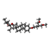 ChemComp-9Z9: |
-Macromolecule #3: primidone
| Macromolecule | Name: primidone / type: ligand / ID: 3 / Number of copies: 4 / Formula: A1AIA |
|---|---|
| Molecular weight | Theoretical: 218.252 Da |
-Macromolecule #4: 1,2-DISTEAROYL-SN-GLYCERO-3-PHOSPHATE
| Macromolecule | Name: 1,2-DISTEAROYL-SN-GLYCERO-3-PHOSPHATE / type: ligand / ID: 4 / Number of copies: 4 / Formula: PX8 |
|---|---|
| Molecular weight | Theoretical: 703.99 Da |
| Chemical component information | 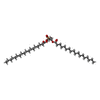 ChemComp-PX8: |
-Experimental details
-Structure determination
| Method | cryo EM |
|---|---|
 Processing Processing | single particle reconstruction |
| Aggregation state | particle |
-Sample preparation
| Concentration | 0.1 mg/mL | |||||||||||||||
|---|---|---|---|---|---|---|---|---|---|---|---|---|---|---|---|---|
| Buffer | pH: 7.5 Component:
Details: 10 mM Hepes pH 7.5, 150 mM NaCl, 0.063% Glycodiosgenin, 100 uM Primidone | |||||||||||||||
| Grid | Model: Quantifoil R1.2/1.3 / Material: GOLD / Mesh: 300 / Support film - Material: GRAPHENE / Support film - topology: CONTINUOUS | |||||||||||||||
| Vitrification | Cryogen name: ETHANE / Chamber humidity: 90 % / Chamber temperature: 298.15 K / Instrument: GATAN CRYOPLUNGE 3 |
- Electron microscopy
| Microscope | JEOL CRYO ARM 300 |
|---|---|
| Image recording | Film or detector model: GATAN K3 (6k x 4k) / Average electron dose: 62.0 e/Å2 |
| Electron beam | Acceleration voltage: 300 kV / Electron source:  FIELD EMISSION GUN FIELD EMISSION GUN |
| Electron optics | Illumination mode: FLOOD BEAM / Imaging mode: BRIGHT FIELD / Nominal defocus max: 1.2 µm / Nominal defocus min: 0.7000000000000001 µm |
+Image processing

-Atomic model buiding 1
| Initial model | Chain - Source name: PDB / Chain - Initial model type: experimental model |
|---|---|
| Output model | PDB-9qho: |
