 Movie
Movie Controller

[English] 日本語
 Yorodumi
- EMDB-51916: Structure of the outer membrane exopolysaccharide transporter PelBC -
+ Open data

- Basic information
| Entry |  | |||||||||
|---|---|---|---|---|---|---|---|---|---|---|
| Title | Structure of the outer membrane exopolysaccharide transporter PelBC | |||||||||
 Map data Map data | ||||||||||
 Sample Sample |
| |||||||||
 Keywords Keywords | Exopolysaccharide / Complex / Acyl-chain / TRANSPORT PROTEIN | |||||||||
| Function / homology |  Function and homology information Function and homology information | |||||||||
| Biological species |   Pseudomonas aeruginosa (bacteria) Pseudomonas aeruginosa (bacteria) | |||||||||
| Method | single particle reconstruction / cryo EM / Resolution: 2.5 Å | |||||||||
 Authors Authors | Benedens M / Rosales C / Beckmann R / Kedrov A | |||||||||
| Funding support |  Germany, 1 items Germany, 1 items
| |||||||||
 Citation Citation | Journal: Nat Commun / Year: 2025 Title: Assembly and the gating mechanism of the Pel exopolysaccharide export complex PelBC of Pseudomonas aeruginosa. Authors: Marius Benedens / Cristian Rosales-Hernandez / Sabine A P Straathof / Jennifer Loschwitz / Otto Berninghausen / Giovanni Maglia / Roland Beckmann / Alexej Kedrov /  Abstract: The pathogen Pseudomonas aeruginosa enhances its virulence and antibiotic resistance upon formation of durable biofilms. The exopolysaccharides Pel, Psl and alginate essentially contribute to the ...The pathogen Pseudomonas aeruginosa enhances its virulence and antibiotic resistance upon formation of durable biofilms. The exopolysaccharides Pel, Psl and alginate essentially contribute to the biofilm matrix, but their secretion mechanisms are barely understood. Here, we reveal the architecture of the outer membrane complex PelBC for Pel export, where the essential periplasmic ring of twelve lipoproteins PelC is mounted on top of the nanodisc-embedded β-barrel PelB. The PelC assembly is stabilized by electrostatic contacts with the periplasmic rim of PelB and via the membrane-anchored acyl chains. The negatively charged interior of the PelB β-barrel forms a route for the cationic Pel exopolysaccharide. The β-barrel is sealed at the extracellular side, but molecular dynamic simulations suggest that the short loop Plug-S is sufficiently flexible to open a tunnel for the exopolysaccharide transport. This gating model is corroborated by single-channel conductivity measurements, where a deletion of Plug-S renders a constitutively open β-barrel. Our structural and functional analysis offers a comprehensive view on this pathogenicity-relevant complex and suggests the route taken by the exopolysaccharide at the final secretion step. | |||||||||
| History |
|
- Structure visualization
| Supplemental images |
|---|

- Downloads & links
-EMDB archive
| Map data | emd_51916.map.gz | 88.9 MB | EMDB map data format | |
|---|---|---|---|---|
| Header (meta data) | emd-51916-v30.xmlemd-51916.xml | 17.3 KB 17.3 KB | Display Display | EMDB header |
| Images |  emd_51916.png emd_51916.png | 109.1 KB | ||
| Filedesc metadata | emd-51916.cif.gz | 6.4 KB | ||
| Others | emd_51916_half_map_1.map.gzemd_51916_half_map_2.map.gz | 165 MB 165 MB | ||
| Archive directory |  http://ftp.pdbj.org/pub/emdb/structures/EMD-51916ftp://ftp.pdbj.org/pub/emdb/structures/EMD-51916 http://ftp.pdbj.org/pub/emdb/structures/EMD-51916ftp://ftp.pdbj.org/pub/emdb/structures/EMD-51916 | HTTPS FTP |
-Related structure data
| Related structure data |  9h80MC M: atomic model generated by this map C: citing same article ( |
|---|---|
| Similar structure data |
-Links
| EMDB pages | EMDB (EBI/PDBe) / EMDataResource |
|---|---|
| Related items in Molecule of the Month |


-Map
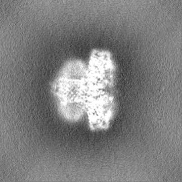
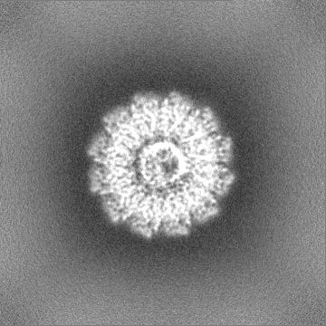
| File | Download / File: emd_51916.map.gz / Format: CCP4 / Size: 178 MB / Type: IMAGE STORED AS FLOATING POINT NUMBER (4 BYTES) | ||||||||||||||||||||||||||||||||||||
|---|---|---|---|---|---|---|---|---|---|---|---|---|---|---|---|---|---|---|---|---|---|---|---|---|---|---|---|---|---|---|---|---|---|---|---|---|---|
| Projections & slices | Image control
Images are generated by Spider. | ||||||||||||||||||||||||||||||||||||
| Voxel size | X=Y=Z: 0.727 Å | ||||||||||||||||||||||||||||||||||||
| Density |
| ||||||||||||||||||||||||||||||||||||
| Symmetry | Space group: 1 | ||||||||||||||||||||||||||||||||||||
| Details | EMDB XML:
|
 Z (Sec.)
 Y (Row.)
 X (Col.)


-Supplemental data
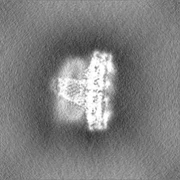
-Half map: #1
| File | emd_51916_half_map_1.map | ||||||||||||
|---|---|---|---|---|---|---|---|---|---|---|---|---|---|
| Projections & Slices |
| ||||||||||||
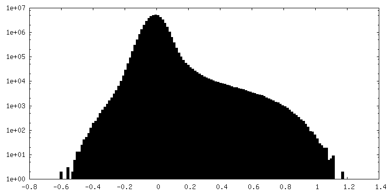
| Density Histograms |


-Half map: #2
| File | emd_51916_half_map_2.map | ||||||||||||
|---|---|---|---|---|---|---|---|---|---|---|---|---|---|
| Projections & Slices |
| ||||||||||||
| Density Histograms |


- Sample components
-Entire : Exopolysaccharide transporter PelBC
| Entire | Name: Exopolysaccharide transporter PelBC |
|---|---|
| Components |
|
-Supramolecule #1: Exopolysaccharide transporter PelBC
| Supramolecule | Name: Exopolysaccharide transporter PelBC / type: complex / ID: 1 / Parent: 0 / Macromolecule list: #1-#2 |
|---|---|
| Source (natural) | Organism: Pseudomonas aeruginosa (bacteria) |
-Macromolecule #1: PelB
| Macromolecule | Name: PelB / type: protein_or_peptide / ID: 1 / Number of copies: 1 / Enantiomer: LEVO |
|---|---|
| Source (natural) | Organism: Pseudomonas aeruginosa (bacteria) |
| Molecular weight | Theoretical: 135.273703 KDa |
| Recombinant expression | Organism: |
| Sequence | String: MANSSAADKH PQARLLNPWA LLPVALGVAL VLWLTFNSEE VFMPSGDGEP DAVSVNYAEL LLQAHPENDA LRLTLIDLLV KLGDFEQAR HHLARLRGKD RLATPFYEVE LDILGALARP EGMDEEQTRR LLERLRKIEH VSLNDAMLER LARHALALDA P DLAARTFA ...String: MANSSAADKH PQARLLNPWA LLPVALGVAL VLWLTFNSEE VFMPSGDGEP DAVSVNYAEL LLQAHPENDA LRLTLIDLLV KLGDFEQAR HHLARLRGKD RLATPFYEVE LDILGALARP EGMDEEQTRR LLERLRKIEH VSLNDAMLER LARHALALDA P DLAARTFA ELAGRDPQGR QRWLDEAARW YLASGEPLPA ADIQRQLAEA QTEPAKRLAY LRQAFASLLA GERGEQAALL LD ERLDALP EDESTLAWLA QGVRAAEGSQ RYDLAERFIR RWRELRPEDH EALAADLRLN MAAGRVERAW EVGQELLALR PED RTLLAD LARLGEWTGN GPRALGFWKQ LLAGADDPAL REHAWRLSLQ MFDFDSAIEL LAPIGAQRQM TDEELDALVY SHET RGTPE EGEAWLRGYV QRYPKQRLAW QRLQQILEHT QQLQEETGVW ARMARHFPLS VKERMQWAET HWNLFDPRQA WKVLA GVDT RAIREPEFWR LRAALAWALE QDDDARAAYE RMLALDIRLN SRDEDQLIAL YRDSNPKQAL QVLIGSWQRS RDPRRL ASA LQLAENLHDW PALKSLLAEA EGLPEAQGSP YYWVARARLA EQEGHGDVAE RLYREALVRF PGENLVRERL LWFYIDR GR RDSLAPLLAQ WHGLALRDST LWLPFASASL LLERNDQALA WFRLYLKSNP NDWLVQAAYA DALDASGYQD KALRLRRL L LRRLDREAVR ATPDSFATYL RLLAVAQGPL LAQGEARRAW NGEPAMLQLW FEQFLDQLAA TNQEPLKDNW LAWARGRGL KIGRNEEIQA ALRSQNRAAL QRLLERGELD PAQRVEALVR LGHGGEALGE ALGALGDGHS RDNREQLRRQ AAEILERTPQ GLQLGWNKR DFGGLDFKGP TLRAARHLGD DWYADLELGS GRYHGDALDS SLLGSERNAR LTLRRELADG FAAATLDGSW R DDEDRHGL GVLRNWRLSS RDELEAGLDW HRETDETGLM RALGMRDSLR LGGRHTLSGR DQLSWSLAHN RFSTRQGDDL GN GEALSLE WAHTLFFDGP AWQLRGGIDY QRNRLENRVP DDLLAAHGGA LALDGARSQD LLQDRYGQVY LGSTWRRGFP GAL NRSRPQ YTWIVDTLAG WQWTEKEFNY GIDLGIGMEL LGDDELAFTF GYQSAPQGGG GDAGGTLGVT YSTRFGR UniProtKB: PelB |
-Macromolecule #2: PelC
| Macromolecule | Name: PelC / type: protein_or_peptide / ID: 2 / Number of copies: 12 / Enantiomer: LEVO |
|---|---|
| Source (natural) | Organism: Pseudomonas aeruginosa (bacteria) |
| Molecular weight | Theoretical: 18.693111 KDa |
| Recombinant expression | Organism: |
| Sequence | String: MQSIRCLALA AVALFMAGCS SFTSESATPL ARGAQWGLVP LLNYSQAPQA GERAEQILLS VLAEEGVRPR LYPAQPQGDL QLVDDRERQ QRALDWARQQ KLAYVVTGSV EEWQYKNGLD GEPAVGVSLQ VLEPASGRVL WSTSGARAGW SRESLAGAAQ K VLRELVGD LRLE UniProtKB: PelC |
-Macromolecule #3: PHOSPHATIDYLETHANOLAMINE
| Macromolecule | Name: PHOSPHATIDYLETHANOLAMINE / type: ligand / ID: 3 / Number of copies: 27 / Formula: PTY |
|---|---|
| Molecular weight | Theoretical: 734.039 Da |
| Chemical component information |  ChemComp-PTY: |
-Experimental details
-Structure determination
| Method | cryo EM |
|---|---|
 Processing Processing | single particle reconstruction |
| Aggregation state | particle |
-Sample preparation
| Buffer | pH: 7.5 |
|---|---|
| Vitrification | Cryogen name: ETHANE |
- Electron microscopy
| Microscope | TFS KRIOS |
|---|---|
| Image recording | Film or detector model: FEI FALCON IV (4k x 4k) / Average electron dose: 40.0 e/Å2 |
| Electron beam | Acceleration voltage: 300 kV / Electron source:  FIELD EMISSION GUN FIELD EMISSION GUN |
| Electron optics | Illumination mode: FLOOD BEAM / Imaging mode: BRIGHT FIELD / Nominal defocus max: 3.5 µm / Nominal defocus min: 0.5 µm |
| Experimental equipment |  Model: Titan Krios / Image courtesy: FEI Company |
