 Movie
Movie Controller


+ Open data

- Basic information
| Entry |  | |||||||||
|---|---|---|---|---|---|---|---|---|---|---|
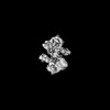
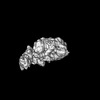
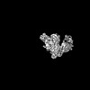
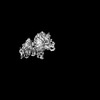
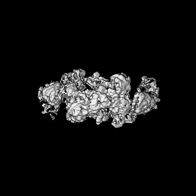
| Title | Composite map of MtUvrA2UvrB2-DNA | |||||||||
 Map data Map data | ||||||||||
 Sample Sample |
| |||||||||
 Keywords Keywords | DNA repair / NER / UVRA / UVRB / UVR / MTB / DNA BINDING PROTEIN | |||||||||
| Function / homology |  Function and homology information Function and homology informationnegative regulation of strand invasion / symbiont-mediated perturbation of host innate immune response / nucleotide-excision repair, DNA damage recognition / response to nitrosative stress / excinuclease ABC activity / excinuclease repair complex / SOS response / peptidoglycan-based cell wall / nucleotide-excision repair / DNA damage response ...negative regulation of strand invasion / symbiont-mediated perturbation of host innate immune response / nucleotide-excision repair, DNA damage recognition / response to nitrosative stress / excinuclease ABC activity / excinuclease repair complex / SOS response / peptidoglycan-based cell wall / nucleotide-excision repair / DNA damage response / ATP hydrolysis activity / DNA binding / zinc ion binding / ATP binding / plasma membrane / cytosol / cytoplasm Similarity search - Function | |||||||||
| Biological species |   Mycobacterium tuberculosis (bacteria) / Mycobacterium tuberculosis (bacteria) / | |||||||||
| Method | single particle reconstruction / cryo EM / Resolution: 3.7 Å | |||||||||
 Authors Authors | Genta M / Capelli R / Ferrara G / Rizzi M / Rossi F / Jeruzalmi D / Bolognesi M / Chaves-Sanjuan A / Miggiano R | |||||||||
| Funding support |  Italy, 1 items Italy, 1 items
| |||||||||
 Citation Citation | Journal: Nat Commun / Year: 2025 Title: Mechanistic understanding of UvrA damage detection and lesion hand-off to UvrB in Nucleotide Excision Repair. Authors: Marianna Genta / Giulia Ferrara / Riccardo Capelli / Diego Rondelli / Sarah Sertic / Martino Bolognesi / Menico Rizzi / Franca Rossi / David Jeruzalmi / Antonio Chaves-Sanjuan / Riccardo Miggiano /  Abstract: Nucleotide excision repair (NER) represents one of the major molecular machineries that control chromosome stability in all living species. In Eubacteria, the initial stages of the repair process are ...Nucleotide excision repair (NER) represents one of the major molecular machineries that control chromosome stability in all living species. In Eubacteria, the initial stages of the repair process are carried out by the UvrABC excinuclease complex. Despite the wealth of structural data available, some crucial details of the pathway remain elusive. In this study, we present a structural investigation of the Mycobacterium tuberculosis UvrAUvrB complex and of the UvrA dimer, both in complex with damaged DNA. Our analyses yield insights into the DNA binding mode of UvrA, showing an unexplored conformation of Insertion Domains (IDs), underlying the essential role of these domains in DNA coordination. Furthermore, we observe an interplay between the ID and the UvrB Binding Domain (UBD): after the recognition of the damage, the IDs repositions with the concomitant reorganization of UBD, allowing the formation of the complex between UvrA and UvrB. These events are detected along the formation of the uncharacterized UvrAUvrB-DNA and the UvrAUvrB-DNA complexes which we interpret as hierarchical steps initiating the DNA repair cascade in the NER pathway, resulting in the formation of the pre-incision complex. | |||||||||
| History |
|
- Structure visualization
| Supplemental images |
|---|

- Downloads & links
-EMDB archive
| Map data | emd_51173.map.gz | 116 MB | EMDB map data format | |
|---|---|---|---|---|
| Header (meta data) | emd-51173-v30.xmlemd-51173.xml | 21.3 KB 21.3 KB | Display Display | EMDB header |
| Images |  emd_51173.png emd_51173.png | 102 KB | ||
| Filedesc metadata | emd-51173.cif.gz | 8 KB | ||
| Archive directory |  http://ftp.pdbj.org/pub/emdb/structures/EMD-51173ftp://ftp.pdbj.org/pub/emdb/structures/EMD-51173 http://ftp.pdbj.org/pub/emdb/structures/EMD-51173ftp://ftp.pdbj.org/pub/emdb/structures/EMD-51173 | HTTPS FTP |
-Related structure data
| Related structure data | 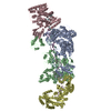 9ga4MC 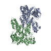 9ga2C 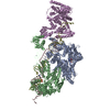 9ga3C 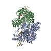 9ga5C C: citing same article ( M: atomic model generated by this map |
|---|---|
| Similar structure data |

-Links
| EMDB pages | EMDB (EBI/PDBe) / EMDataResource |
|---|---|
| Related items in Molecule of the Month |


-Map
| File | Download / File: emd_51173.map.gz / Format: CCP4 / Size: 244.1 MB / Type: IMAGE STORED AS FLOATING POINT NUMBER (4 BYTES) | ||||||||||||||||||||||||||||||||||||
|---|---|---|---|---|---|---|---|---|---|---|---|---|---|---|---|---|---|---|---|---|---|---|---|---|---|---|---|---|---|---|---|---|---|---|---|---|---|
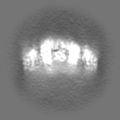
| Projections & slices | Image control
Images are generated by Spider. | ||||||||||||||||||||||||||||||||||||
| Voxel size | X=Y=Z: 0.84 Å | ||||||||||||||||||||||||||||||||||||
| Density |
| ||||||||||||||||||||||||||||||||||||
| Symmetry | Space group: 1 | ||||||||||||||||||||||||||||||||||||
| Details | EMDB XML:
|
 Z (Sec.)
 Y (Row.)
 X (Col.)


-Supplemental data
- Sample components
-Entire : MtUvrA2UvrB2-DNA complex
| Entire | Name: MtUvrA2UvrB2-DNA complex |
|---|---|
| Components |
|
-Supramolecule #1: MtUvrA2UvrB2-DNA complex
| Supramolecule | Name: MtUvrA2UvrB2-DNA complex / type: complex / ID: 1 / Parent: 0 / Macromolecule list: #1-#3 / Details: Heterotetrameric MtUvrA2UvrB2 in complex with DNA |
|---|---|
| Molecular weight | Theoretical: 400 KDa |
-Supramolecule #2: MtUvrA2UvrB2
| Supramolecule | Name: MtUvrA2UvrB2 / type: complex / ID: 2 / Parent: 1 / Macromolecule list: #1-#2 |
|---|---|
| Source (natural) | Organism: Mycobacterium tuberculosis (bacteria) |
-Supramolecule #3: DNA
| Supramolecule | Name: DNA / type: complex / ID: 3 / Parent: 1 / Macromolecule list: #3 |
|---|---|
| Source (natural) | Organism: |
-Macromolecule #1: UvrABC system protein A
| Macromolecule | Name: UvrABC system protein A / type: protein_or_peptide / ID: 1 / Number of copies: 2 / Enantiomer: LEVO |
|---|---|
| Source (natural) | Organism: Mycobacterium tuberculosis (bacteria) |
| Molecular weight | Theoretical: 108.806398 KDa |
| Recombinant expression | Organism: |
| Sequence | String: MGHHHHHHHH HHSSGHIEGR HMADRLIVKG AREHNLRSVD LDLPRDALIV FTGLSGSGKS SLAFDTIFAE GQRRYVESLS AYARQFLGQ MDKPDVDFIE GLSPAVSIDQ KSTNRNPRST VGTITEVYDY LRLLYARAGT PHCPTCGERV ARQTPQQIVD Q VLAMPEGT ...String: MGHHHHHHHH HHSSGHIEGR HMADRLIVKG AREHNLRSVD LDLPRDALIV FTGLSGSGKS SLAFDTIFAE GQRRYVESLS AYARQFLGQ MDKPDVDFIE GLSPAVSIDQ KSTNRNPRST VGTITEVYDY LRLLYARAGT PHCPTCGERV ARQTPQQIVD Q VLAMPEGT RFLVLAPVVR TRKGEFADLF DKLNAQGYSR VRVDGVVHPL TDPPKLKKQE KHDIEVVVDR LTVKAAAKRR LT DSVETAL NLADGIVVLE FVDHELGAPH REQRFSEKLA CPNGHALAVD DLEPRSFSFN SPYGACPECS GLGIRKEVDP ELV VPDPDR TLAQGAVAPW SNGHTAEYFT RMMAGLGEAL GFDVDTPWRK LPAKARKAIL EGADEQVHVR YRNRYGRTRS YYAD FEGVL AFLQRKMSQT ESEQMKERYE GFMRDVPCPV CAGTRLKPEI LAVTLAGESK GEHGAKSIAE VCELSIADCA DFLNA LTLG PREQAIAGQV LKEIRSRLGF LLDVGLEYLS LSRAAATLSG GEAQRIRLAT QIGSGLVGVL YVLDEPSIGL HQRDNR RLI ETLTRLRDLG NTLIVVEHDE DTIEHADWIV DIGPGAGEHG GRIVHSGPYD ELLRNKDSIT GAYLSGRESI EIPAIRR SV DPRRQLTVVG AREHNLRGID VSFPLGVLTS VTGVSGSGKS TLVNDILAAV LANRLNGARQ VPGRHTRVTG LDYLDKLV R VDQSPIGRTP RSNPATYTGV FDKIRTLFAA TTEAKVRGYQ PGRFSFNVKG GRCEACTGDG TIKIEMNFLP DVYVPCEVC QGARYNRETL EVHYKGKTVS EVLDMSIEEA AEFFEPIAGV HRYLRTLVDV GLGYVRLGQP APTLSGGEAQ RVKLASELQK RSTGRTVYI LDEPTTGLHF DDIRKLLNVI NGLVDKGNTV IVIEHNLDVI KTSDWIIDLG PEGGAGGGTV VAQGTPEDVA A VPASYTGK FLAEVVGGGA SAATSRSNRR RNVSA UniProtKB: UvrABC system protein A |
-Macromolecule #2: UvrABC system protein B
| Macromolecule | Name: UvrABC system protein B / type: protein_or_peptide / ID: 2 / Number of copies: 2 / Enantiomer: LEVO |
|---|---|
| Source (natural) | Organism: Mycobacterium tuberculosis (bacteria) |
| Molecular weight | Theoretical: 80.800156 KDa |
| Recombinant expression | Organism: |
| Sequence | String: MGHHHHHHHH HHSSGHIEGR HMVRAGGHFE VVSPHAPAGD QPAAIDELER RINAGERDVV LLGATGTGKS ATTAWLIERL QRPTLVMAP NKTLAAQLAN ELREMLPHNA VEYFVSYYDY YQPEAYIAQT DTYIEKDSSI NDDVERLRHS ATSALLSRRD V VVVASVSC ...String: MGHHHHHHHH HHSSGHIEGR HMVRAGGHFE VVSPHAPAGD QPAAIDELER RINAGERDVV LLGATGTGKS ATTAWLIERL QRPTLVMAP NKTLAAQLAN ELREMLPHNA VEYFVSYYDY YQPEAYIAQT DTYIEKDSSI NDDVERLRHS ATSALLSRRD V VVVASVSC IYGLGTPQSY LDRSVELKVG EEVPRDGLLR LLVDVQYTRN DMSFTRGSFR VRGDTVEIIP SYEELAVRIE FF GDEIEAL YYLHPLTGEV IRQVDSLRIF PATHYVAGPE RMAHAVSAIE EELAERLAEL ESQGKLLEAQ RLRMRTNYDI EMM RQVGFC SGIENYSRHI DGRGPGTPPA TLLDYFPEDF LLVIDESHVT VPQIGGMYEG DISRKRNLVE YGFRLPSACD NRPL TWEEF ADRIGQTVYL SATPGPYELS QTGGEFVEQV IRPTGLVDPK VVVKPTKGQI DDLIGEIRTR ADADQRVLVT TLTKK MAED LTDYLLEMGI RVRYLHSEVD TLRRVELLRQ LRLGDYDVLV GINLLREGLD LPEVSLVAIL DADKEGFLRS SRSLIQ TIG RAARNVSGEV HMYADKITDS MREAIDETER RRAKQIAYNE ANGIDPQPLR KKIADILDQV YREADDTAVV EVGGSGR NA SRGRRAQGEP GRAVSAGVFE GRDTSAMPRA ELADLIKDLT AQMMAAARDL QFELAARFRD EIADLKRELR GMDAAGLK UniProtKB: UvrABC system protein B |
-Macromolecule #3: DNA
| Macromolecule | Name: DNA / type: dna / ID: 3 / Number of copies: 2 / Classification: DNA |
|---|---|
| Source (natural) | Organism: |
| Molecular weight | Theoretical: 12.89029 KDa |
| Sequence | String: (DT)(DA)(DG)(DT)(DC)(DA)(DC)(DA)(DT)(DC) (DA)(DG)(DT)(DG)(DA)(DT)(DC)(DA)(DG)(DT) (DG)(DG)(DT)(DT)(DC)(DC)(DG)(DG)(DA) (DA)(DC)(DC)(DA)(DC)(DT)(DG)(DA)(DT)(DC) (DA) (DC)(DT) |
-Macromolecule #4: ZINC ION
| Macromolecule | Name: ZINC ION / type: ligand / ID: 4 / Number of copies: 4 / Formula: ZN |
|---|---|
| Molecular weight | Theoretical: 65.409 Da |
-Experimental details
-Structure determination
| Method | cryo EM |
|---|---|
 Processing Processing | single particle reconstruction |
| Aggregation state | particle |
-Sample preparation
| Concentration | 0.5 mg/mL | |||||||||
|---|---|---|---|---|---|---|---|---|---|---|
| Buffer | pH: 8 Component:
| |||||||||
| Grid | Model: Quantifoil R1.2/1.3 / Material: COPPER / Mesh: 300 / Support film - Material: CARBON / Support film - topology: HOLEY / Pretreatment - Type: GLOW DISCHARGE / Pretreatment - Time: 30 sec. / Pretreatment - Atmosphere: AIR / Details: 30mA | |||||||||
| Vitrification | Cryogen name: ETHANE / Chamber humidity: 100 % / Chamber temperature: 277 K / Instrument: FEI VITROBOT MARK IV |
- Electron microscopy
| Microscope | FEI TALOS ARCTICA |
|---|---|
| Image recording | Film or detector model: GATAN K3 (6k x 4k) / Number grids imaged: 1 / Number real images: 21232 / Average electron dose: 40.0 e/Å2 |
| Electron beam | Acceleration voltage: 300 kV / Electron source:  FIELD EMISSION GUN FIELD EMISSION GUN |
| Electron optics | Illumination mode: FLOOD BEAM / Imaging mode: BRIGHT FIELD / Cs: 2.7 mm / Nominal defocus max: 2.5 µm / Nominal defocus min: 0.8 µm / Nominal magnification: 105000 |
| Sample stage | Specimen holder model: FEI TITAN KRIOS AUTOGRID HOLDER / Cooling holder cryogen: NITROGEN |
| Experimental equipment |  Model: Talos Arctica / Image courtesy: FEI Company |
