 Movie
Movie Controller

[English] 日本語
 Yorodumi
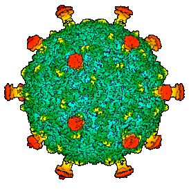
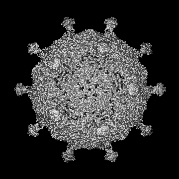
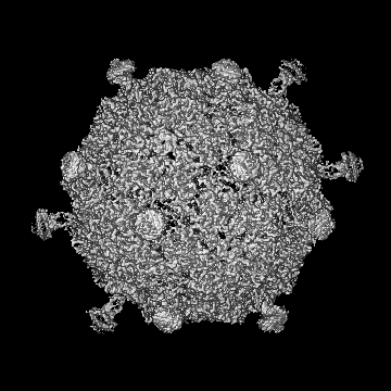
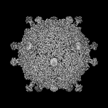
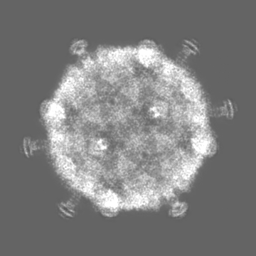
- EMDB-50357: Native capsid of Rhodobacter microvirus Ebor computed with I4 symmetry -
+ Open data

- Basic information
| Entry |  | ||||||||||||
|---|---|---|---|---|---|---|---|---|---|---|---|---|---|
| Title | Native capsid of Rhodobacter microvirus Ebor computed with I4 symmetry | ||||||||||||
 Map data Map data | |||||||||||||
 Sample Sample |
| ||||||||||||
 Keywords Keywords | ssDNA virus / Microviridae / Tainavirinae / Alphaproteobacteria / Rhodobacter capsulatus / aquatic virus / jelly-roll fold / VIRUS | ||||||||||||
| Biological species |  Rhodobacter capsulatus SB 1003 (bacteria) / Rhodobacter capsulatus SB 1003 (bacteria) /  Rhodobacter phage Ebor (virus) Rhodobacter phage Ebor (virus) | ||||||||||||
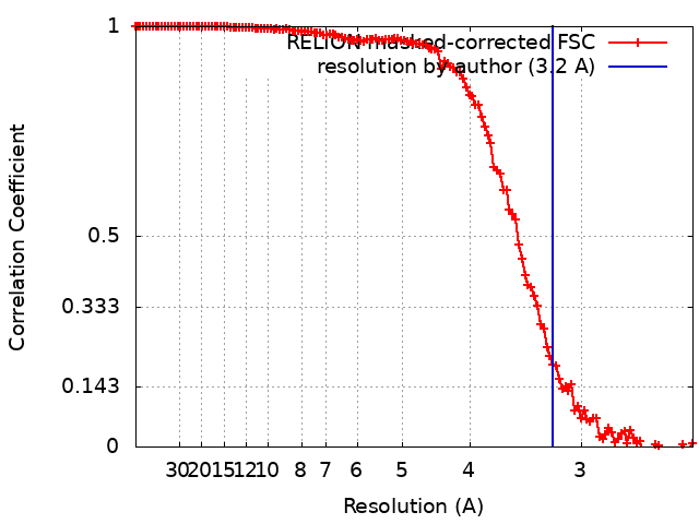
| Method | single particle reconstruction / cryo EM / Resolution: 3.2 Å | ||||||||||||
 Authors Authors | Bardy P / MacDonald CIW / Jenkins HT / Chechik M / Hart SJ / Turkenburg JP / Blaza JN / Fogg PCM / Antson AA | ||||||||||||
| Funding support |  United Kingdom, 3 items United Kingdom, 3 items
| ||||||||||||
 Citation Citation | Journal: bioRxiv / Year: 2024 Title: A stargate mechanism of genome delivery unveiled by cryogenic electron tomography. Authors: Pavol Bardy / Conor I W MacDonald / Paul C Kirchberger / Huw T Jenkins / Tibor Botka / Lewis Byrom / Nawshin T B Alim / Daouda A K Traore / Hannah C König / Tristan R Nicholas / Maria ...Authors: Pavol Bardy / Conor I W MacDonald / Paul C Kirchberger / Huw T Jenkins / Tibor Botka / Lewis Byrom / Nawshin T B Alim / Daouda A K Traore / Hannah C König / Tristan R Nicholas / Maria Chechik / Samuel J Hart / Johan P Turkenburg / James N Blaza / J Thomas Beatty / Paul C M Fogg / Alfred A Antson /    Abstract: Single-stranded DNA bacteriophages of the family are major components of the global virosphere. Microviruses are highly abundant in aquatic ecosystems and are prominent members of the mammalian gut ...Single-stranded DNA bacteriophages of the family are major components of the global virosphere. Microviruses are highly abundant in aquatic ecosystems and are prominent members of the mammalian gut microbiome, where their diversity has been linked to various chronic health disorders. Despite the clear importance of microviruses, little is known about the molecular mechanism of host infection. Here, we have characterized an exceptionally large microvirus, Ebor, and provide crucial insights into long-standing mechanistic questions. Cryogenic electron microscopy of Ebor revealed a capsid with trimeric protrusions that recognise lipopolysaccharides on the host surface. Cryogenic electron tomography of the host cell colonized with virus particles demonstrated that the virus initially attaches to the cell via five such protrusions, located at the corners of a single pentamer. This interaction triggers a stargate mechanism of capsid opening along the 5-fold symmetry axis, enabling delivery of the virus genome. Despite variations in specific virus-host interactions among different family viruses, structural data indicate that the stargate mechanism of infection is universally employed by all members of the family. Startlingly, our data reveal a mechanistic link for the opening of relatively small capsids made out of a single jelly-roll fold with the structurally unrelated giant viruses. | ||||||||||||
| History |
|
- Structure visualization
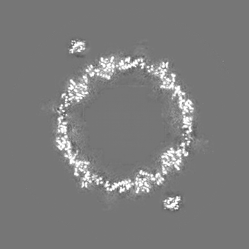
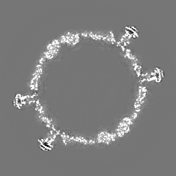
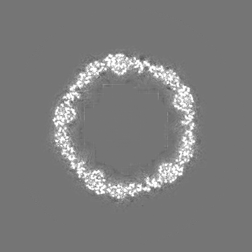
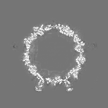
| Supplemental images |
|---|
- Downloads & links
-EMDB archive
| Map data | emd_50357.map.gz | 89.4 MB |  EMDB map data format EMDB map data format | |
|---|---|---|---|---|
| Header (meta data) | emd-50357-v30.xmlemd-50357.xml | 20.7 KB 20.7 KB | Display Display | EMDB header |
| FSC (resolution estimation) | emd_50357_fsc.xml | 12.7 KB | Display | FSC data file |
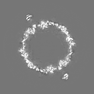
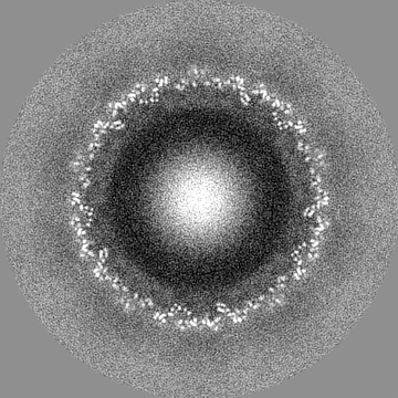
| Images |  emd_50357.png emd_50357.png | 89.6 KB | ||
| Filedesc metadata | emd-50357.cif.gz | 6.5 KB | ||
| Others | emd_50357_half_map_1.map.gzemd_50357_half_map_2.map.gz | 140.6 MB 140.5 MB | ||
| Archive directory |  http://ftp.pdbj.org/pub/emdb/structures/EMD-50357ftp://ftp.pdbj.org/pub/emdb/structures/EMD-50357 http://ftp.pdbj.org/pub/emdb/structures/EMD-50357ftp://ftp.pdbj.org/pub/emdb/structures/EMD-50357 | HTTPS FTP |
-Related structure data


-Links
| EMDB pages | EMDB (EBI/PDBe) / EMDataResource |
|---|
-Map
| File | Download / File: emd_50357.map.gz / Format: CCP4 / Size: 178 MB / Type: IMAGE STORED AS FLOATING POINT NUMBER (4 BYTES) | ||||||||||||||||||||||||||||||||||||
|---|---|---|---|---|---|---|---|---|---|---|---|---|---|---|---|---|---|---|---|---|---|---|---|---|---|---|---|---|---|---|---|---|---|---|---|---|---|
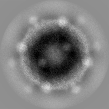
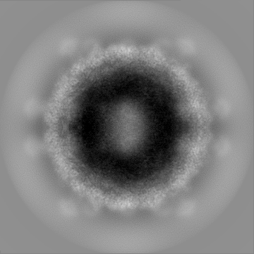
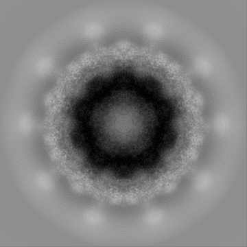
| Projections & slices | Image control
Images are generated by Spider. | ||||||||||||||||||||||||||||||||||||
| Voxel size | X=Y=Z: 1.2 Å | ||||||||||||||||||||||||||||||||||||
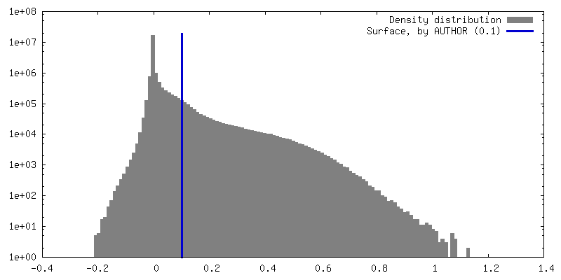
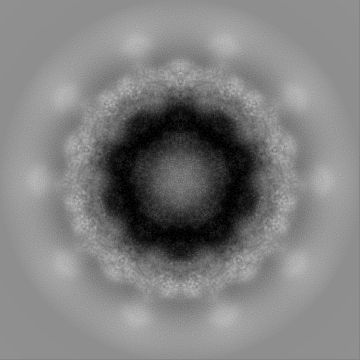
| Density |
| ||||||||||||||||||||||||||||||||||||
| Symmetry | Space group: 1 | ||||||||||||||||||||||||||||||||||||
| Details | EMDB XML:
|
 Z (Sec.)
 Y (Row.)
 X (Col.)


-Supplemental data
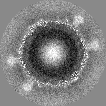
-Half map: #1
| File | emd_50357_half_map_1.map | ||||||||||||
|---|---|---|---|---|---|---|---|---|---|---|---|---|---|
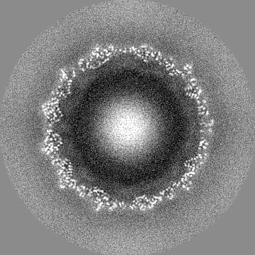
| Projections & Slices |
| ||||||||||||
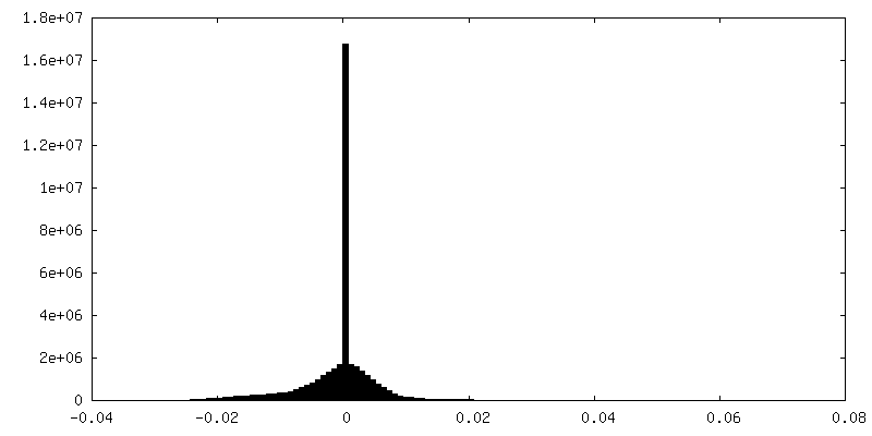
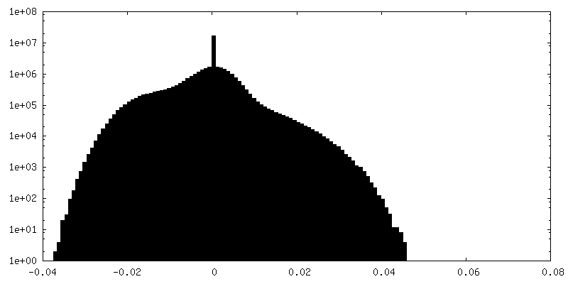
| Density Histograms |


-Half map: #2
| File | emd_50357_half_map_2.map | ||||||||||||
|---|---|---|---|---|---|---|---|---|---|---|---|---|---|
| Projections & Slices |
| ||||||||||||
| Density Histograms |


- Sample components
-Entire : Rhodobacter phage Ebor
| Entire | Name: Rhodobacter phage Ebor (virus) |
|---|---|
| Components |
|
-Supramolecule #1: Rhodobacter phage Ebor
| Supramolecule | Name: Rhodobacter phage Ebor / type: virus / ID: 1 / Parent: 0 / Macromolecule list: all Details: purified from Rhodobacter capsulatus strain SB1003 by CsCl ultracentrifugation NCBI-ID: 3144506 / Sci species name: Rhodobacter phage Ebor / Sci species strain: variant R120 / Virus type: VIRION / Virus isolate: OTHER / Virus enveloped: No / Virus empty: No |
|---|---|
| Host (natural) | Organism: Rhodobacter capsulatus (bacteria) / Strain: SB1003 |
| Molecular weight | Theoretical: 5.7 MDa |
| Virus shell | Shell ID: 1 / Name: Native capsid / Diameter: 460.0 Å / T number (triangulation number): 1 |
-Macromolecule #1: Major capsid protein
| Macromolecule | Name: Major capsid protein / type: protein_or_peptide / ID: 1 / Details: Ebor variant R120 / Number of copies: 1 / Enantiomer: LEVO |
|---|---|
| Source (natural) | Organism: Rhodobacter capsulatus SB 1003 (bacteria) / Strain: SB1003 |
| Molecular weight | Theoretical: 55.885539 KDa |
| Sequence | String: MSYQTKRNVR REARTLMGRF KAGKLAPVMA VPVKGSEGGM LSQSVSFELD PIAGRMATPI TAEMCAVFVP VQACDALKNP EADYAGMTE IVREKLLSGN PLFVLEPETD VSKRCGVNPR RNNGLMRVNE IVRLAHNCAV NFLRRRRYVD AVQLTAANHS T TPAILSQT ...String: MSYQTKRNVR REARTLMGRF KAGKLAPVMA VPVKGSEGGM LSQSVSFELD PIAGRMATPI TAEMCAVFVP VQACDALKNP EADYAGMTE IVREKLLSGN PLFVLEPETD VSKRCGVNPR RNNGLMRVNE IVRLAHNCAV NFLRRRRYVD AVQLTAANHS T TPAILSQT VLDRFNGALD PDPNVNGAVQ LSMPDMRLPV ASDAEKAVTA PLTVKRGNEN RRLDAGISRL AAAEVAAATD AA LYAVFGG REAGNVSLTD FYNAQKMDEL TRVMRKICDD NPEYGEEMVL RWAHGLSVDP GRVPFLLAEK SVVLGRQIIG ATD TAGVED GVKRSDMAAQ LSFTVPIPTT ELGGIIVTFA CIKPDETLSS QPHPILADHW RLDNFVADEL ALDPQPVMAR ELDY KVAQA NETTVVFYTG LNELKKTYVS YGLCRALDPN TVESKNAVWQ LEVPLSVTPE TVLYPADLPQ YPFADQQAEV CTYVV QSTA VMPTPMIFGP SPVEQLAVIE TEDLFED |
-Experimental details
-Structure determination
| Method | cryo EM |
|---|---|
 Processing Processing | single particle reconstruction |
| Aggregation state | particle |
-Sample preparation
| Buffer | pH: 7.75 Component:
| ||||||||||||
|---|---|---|---|---|---|---|---|---|---|---|---|---|---|
| Grid | Model: Quantifoil R2/1 / Material: COPPER / Mesh: 300 / Support film - Material: CARBON / Support film - topology: HOLEY / Pretreatment - Type: GLOW DISCHARGE / Pretreatment - Time: 30 sec. / Pretreatment - Atmosphere: AIR | ||||||||||||
| Vitrification | Cryogen name: ETHANE / Chamber humidity: 100 % / Chamber temperature: 295.15 K / Instrument: FEI VITROBOT MARK IV / Details: blot time 2 sec, blot force 0, wait time 8 sec. | ||||||||||||
| Details | concentration between 10^10-11 PFU/ml |
- Electron microscopy
| Microscope | TFS GLACIOS |
|---|---|
| Image recording | Film or detector model: FEI FALCON IV (4k x 4k) / Number grids imaged: 1 / Number real images: 996 / Average electron dose: 50.0 e/Å2 |
| Electron beam | Acceleration voltage: 200 kV / Electron source:  FIELD EMISSION GUN FIELD EMISSION GUN |
| Electron optics | Illumination mode: FLOOD BEAM / Imaging mode: BRIGHT FIELD / Nominal defocus max: 2.5 µm / Nominal defocus min: 0.5 µm |
| Sample stage | Specimen holder model: FEI TITAN KRIOS AUTOGRID HOLDER / Cooling holder cryogen: NITROGEN |
+Image processing
-Atomic model buiding 1
| Initial model | Chain - Chain ID: A / Chain - Source name: Other / Chain - Initial model type: experimental model / Details: Related entry - empty capsid |
|---|---|
| Refinement | Space: REAL / Protocol: AB INITIO MODEL |
| Output model |  PDB-9ffh: |
