 Movie
Movie Controller

[English] 日本語
 Yorodumi
- EMDB-48929: Structure of VcINDY-alpha ketoglutarate complex in Ci-Ci conformation -
+ Open data

- Basic information
| Entry |  | |||||||||||||||||||||
|---|---|---|---|---|---|---|---|---|---|---|---|---|---|---|---|---|---|---|---|---|---|---|
| Title | Structure of VcINDY-alpha ketoglutarate complex in Ci-Ci conformation | |||||||||||||||||||||
 Map data Map data | sharpened map of VcINDY in alpha ketoglutarate | |||||||||||||||||||||
 Sample Sample |
| |||||||||||||||||||||
 Keywords Keywords | Na(+)/dicarboxylate cotransporter(VcINDY) / Solute carries / Elevator type alternating access / membrane protein / TRANSPORT PROTEIN | |||||||||||||||||||||
| Function / homology |  Function and homology information Function and homology informationsuccinate transmembrane transporter activity / transmembrane transporter activity / transmembrane transport / identical protein binding / plasma membrane Similarity search - Function | |||||||||||||||||||||
| Biological species |  Vibrio cholerae O1 biovar El Tor str. N16961 (bacteria) Vibrio cholerae O1 biovar El Tor str. N16961 (bacteria) | |||||||||||||||||||||
| Method | single particle reconstruction / cryo EM / Resolution: 2.35 Å | |||||||||||||||||||||
 Authors Authors | Li Y / Daab A / Song JM / Marden JJ / Mulligan C / Wang DN | |||||||||||||||||||||
| Funding support |  United States, United States,  United Kingdom, 6 items United Kingdom, 6 items
| |||||||||||||||||||||
 Citation Citation | Journal: To Be Published Title: Structure of VcINDY-alpha ketoglutarate complex in Ci-Ci conformation Authors: Li Y / Daab A / Song JM / Marden JJ / Mulligan C / Wang DN | |||||||||||||||||||||
| History |
|
- Structure visualization
| Supplemental images |
|---|

- Downloads & links
-EMDB archive
| Map data | emd_48929.map.gz | 50.3 MB | EMDB map data format | |
|---|---|---|---|---|
| Header (meta data) | emd-48929-v30.xmlemd-48929.xml | 28.1 KB 28.1 KB | Display Display | EMDB header |
| FSC (resolution estimation) | emd_48929_fsc.xml | 8.4 KB | Display | FSC data file |
| Images |  emd_48929.png emd_48929.png | 57.9 KB | ||
| Masks | emd_48929_msk_1.map | 64 MB | Mask map | |
| Filedesc metadata | emd-48929.cif.gz | 7.7 KB | ||
| Others | emd_48929_additional_1.map.gzemd_48929_half_map_1.map.gzemd_48929_half_map_2.map.gz | 30.8 MB 49.6 MB 49.6 MB | ||
| Archive directory |  http://ftp.pdbj.org/pub/emdb/structures/EMD-48929ftp://ftp.pdbj.org/pub/emdb/structures/EMD-48929 http://ftp.pdbj.org/pub/emdb/structures/EMD-48929ftp://ftp.pdbj.org/pub/emdb/structures/EMD-48929 | HTTPS FTP |
-Related structure data
| Related structure data |  9n5nMC M: atomic model generated by this map C: citing same article ( |
|---|---|
| Similar structure data |
-Links
| EMDB pages | EMDB (EBI/PDBe) / EMDataResource |
|---|
-Map
| File | Download / File: emd_48929.map.gz / Format: CCP4 / Size: 64 MB / Type: IMAGE STORED AS FLOATING POINT NUMBER (4 BYTES) | ||||||||||||||||||||||||||||||||||||
|---|---|---|---|---|---|---|---|---|---|---|---|---|---|---|---|---|---|---|---|---|---|---|---|---|---|---|---|---|---|---|---|---|---|---|---|---|---|
| Annotation | sharpened map of VcINDY in alpha ketoglutarate | ||||||||||||||||||||||||||||||||||||
| Projections & slices | Image control
Images are generated by Spider. | ||||||||||||||||||||||||||||||||||||
| Voxel size | X=Y=Z: 0.825 Å | ||||||||||||||||||||||||||||||||||||
| Density |
| ||||||||||||||||||||||||||||||||||||
| Symmetry | Space group: 1 | ||||||||||||||||||||||||||||||||||||
| Details | EMDB XML:
|
 Z (Sec.)
 Y (Row.)
 X (Col.)


-Supplemental data
-Mask #1
| File | emd_48929_msk_1.map | ||||||||||||
|---|---|---|---|---|---|---|---|---|---|---|---|---|---|
| Projections & Slices |
| ||||||||||||
| Density Histograms |


-Additional map: unsharpened map of VcINDY in alpha ketoglutarate
| File | emd_48929_additional_1.map | ||||||||||||
|---|---|---|---|---|---|---|---|---|---|---|---|---|---|
| Annotation | unsharpened map of VcINDY in alpha ketoglutarate | ||||||||||||
| Projections & Slices |
| ||||||||||||
| Density Histograms |


-Half map: half map B of VcINDY in alpha ketoglutarate
| File | emd_48929_half_map_1.map | ||||||||||||
|---|---|---|---|---|---|---|---|---|---|---|---|---|---|
| Annotation | half map B of VcINDY in alpha ketoglutarate | ||||||||||||
| Projections & Slices |
| ||||||||||||
| Density Histograms |


-Half map: half map A of VcINDY in alpha ketoglutarate
| File | emd_48929_half_map_2.map | ||||||||||||
|---|---|---|---|---|---|---|---|---|---|---|---|---|---|
| Annotation | half map A of VcINDY in alpha ketoglutarate | ||||||||||||
| Projections & Slices |
| ||||||||||||
| Density Histograms |


- Sample components
-Entire : Dimer of VcINDY in complex with alpha ketoglutarate
| Entire | Name: Dimer of VcINDY in complex with alpha ketoglutarate |
|---|---|
| Components |
|
-Supramolecule #1: Dimer of VcINDY in complex with alpha ketoglutarate
| Supramolecule | Name: Dimer of VcINDY in complex with alpha ketoglutarate / type: complex / ID: 1 / Parent: 0 / Macromolecule list: #1 |
|---|---|
| Source (natural) | Organism: Vibrio cholerae O1 biovar El Tor str. N16961 (bacteria) |
| Molecular weight | Theoretical: 192 KDa |
-Macromolecule #1: Transporter, NadC family
| Macromolecule | Name: Transporter, NadC family / type: protein_or_peptide / ID: 1 / Details: Indy_Vibrio, wild type / Number of copies: 2 / Enantiomer: LEVO |
|---|---|
| Source (natural) | Organism: Vibrio cholerae O1 biovar El Tor str. N16961 (bacteria) |
| Molecular weight | Theoretical: 49.59893 KDa |
| Recombinant expression | Organism:  Escherichia coli (E. coli) Escherichia coli (E. coli) |
| Sequence | String: MNRNDSVPLP TNTREWFLHR NSLIVLADVA LFLALYHFLP FEHNVVLGIS MLAFIAVLWL TEALHVTVTA ILVPVMAVFF GIFETQAAL NNFANSIIFL FLGGFALAAA MHHQGLDKVI ADKVLAMAQG KMSVAVFMLF GVTALLSMWI SNTATAAMML P LVLGVLSK ...String: MNRNDSVPLP TNTREWFLHR NSLIVLADVA LFLALYHFLP FEHNVVLGIS MLAFIAVLWL TEALHVTVTA ILVPVMAVFF GIFETQAAL NNFANSIIFL FLGGFALAAA MHHQGLDKVI ADKVLAMAQG KMSVAVFMLF GVTALLSMWI SNTATAAMML P LVLGVLSK VDADKQRSTY VFVLLGVAYS ASIGGIATLV GSPPNAIAAA EVGLSFTDWM KFGLPTAMMM LPMAIAILYF LL KPTLNGM FELDRAPVNW DKGKVVTLGI FGLTVFLWIF SSPINAALGG FKSFDTLVAL GAILMLSFAR VVHWKEIQKT ADW GVLLLF GGGLCLSNVL KQTGTSVFLA NALSDMVSHM GIFVVILVVA TFVVFLTEFA SNTASAALLI PVFATVAEAF GMSP VLLSV LIAVAASCAF MLPVATPPNA IVFASGHIKQ SEMMRVGLYL NIACIGLLTA IAMLFWQ UniProtKB: Transporter, NadC family |
-Macromolecule #2: SODIUM ION
| Macromolecule | Name: SODIUM ION / type: ligand / ID: 2 / Number of copies: 4 |
|---|---|
| Molecular weight | Theoretical: 22.99 Da |
-Macromolecule #3: 2-OXOGLUTARIC ACID
| Macromolecule | Name: 2-OXOGLUTARIC ACID / type: ligand / ID: 3 / Number of copies: 2 / Formula: AKG |
|---|---|
| Molecular weight | Theoretical: 146.098 Da |
| Chemical component information | 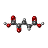 ChemComp-AKG: |
-Macromolecule #4: water
| Macromolecule | Name: water / type: ligand / ID: 4 / Number of copies: 2 / Formula: HOH |
|---|---|
| Molecular weight | Theoretical: 18.015 Da |
| Chemical component information |  ChemComp-HOH: |
-Experimental details
-Structure determination
| Method | cryo EM |
|---|---|
 Processing Processing | single particle reconstruction |
| Aggregation state | particle |
-Sample preparation
| Concentration | 4 mg/mL | |||||||||||||||
|---|---|---|---|---|---|---|---|---|---|---|---|---|---|---|---|---|
| Buffer | pH: 8 Component:
| |||||||||||||||
| Grid | Model: UltrAuFoil R1.2/1.3 / Material: GOLD / Mesh: 300 / Support film - Material: GOLD / Support film - topology: HOLEY / Support film - Film thickness: 50 / Pretreatment - Type: GLOW DISCHARGE / Pretreatment - Time: 2 sec. / Pretreatment - Atmosphere: AIR / Pretreatment - Pressure: 0.026000000000000002 kPa / Details: Hold 10s before glow discharge | |||||||||||||||
| Vitrification | Cryogen name: ETHANE / Chamber humidity: 100 % / Chamber temperature: 281.15 K / Instrument: FEI VITROBOT MARK IV |
- Electron microscopy
| Microscope | TFS KRIOS |
|---|---|
| Temperature | Min: 80.0 K / Max: 80.0 K |
| Alignment procedure | Coma free - Residual tilt: 0.05 mrad |
| Specialist optics | Energy filter - Name: GIF Bioquantum / Energy filter - Slit width: 20 eV |
| Image recording | Film or detector model: GATAN K3 BIOCONTINUUM (6k x 4k) / Digitization - Dimensions - Width: 5760 pixel / Digitization - Dimensions - Height: 4092 pixel / Number grids imaged: 1 / Number real images: 3948 / Average exposure time: 1.8 sec. / Average electron dose: 53.11 e/Å2 Details: 3948 untilted images were collected in super resolution mode at 40 frames per micrograph |
| Electron beam | Acceleration voltage: 300 kV / Electron source:  FIELD EMISSION GUN FIELD EMISSION GUN |
| Electron optics | C2 aperture diameter: 70.0 µm / Calibrated defocus max: 3.0 µm / Calibrated defocus min: 0.8 µm / Calibrated magnification: 105000 / Illumination mode: FLOOD BEAM / Imaging mode: BRIGHT FIELD / Cs: 2.7 mm / Nominal defocus max: 1.6 µm / Nominal defocus min: 1.2 µm / Nominal magnification: 105000 |
| Sample stage | Specimen holder model: FEI TITAN KRIOS AUTOGRID HOLDER / Cooling holder cryogen: NITROGEN |
| Experimental equipment |  Model: Titan Krios / Image courtesy: FEI Company |
+Image processing

-Atomic model buiding 1
| Initial model | PDB ID: Chain - Chain ID: AB / Chain - Residue range: 1-462 / Chain - Source name: PDB / Chain - Initial model type: experimental model / Details: the whole model was used |
|---|---|
| Details | Initial local fitting was done using Chimera and then coot was used for ajustment. |
| Refinement | Space: REAL / Protocol: OTHER / Overall B value: 52.28 / Target criteria: cross-correlation coefficient |
| Output model | PDB-9n5n: |

