[English] 日本語
Yorodumi
- EMDB-48096: Human FANCJ helicase bound to a parallel G4 DNA -

+
Open data


ID or keywords:

Loading...

-
Basic information

Entry
Database: EMDB / ID: EMD-48096
TitleHuman FANCJ helicase bound to a parallel G4 DNA
Map dataThe 3D EM map of human FANCJ with G4 DNA
Sample
  • Complex: Complex of Human FANCJ with a parallel G4 DNA with 5' ssDNA overhang
    • Protein or peptide: Fanconi anemia group J protein
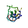
    • DNA: DNA (31-MER)
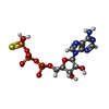
  • Ligand: PHOSPHOTHIOPHOSPHORIC ACID-ADENYLATE ESTER
  • Ligand: MAGNESIUM ION
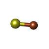
  • Ligand: IRON/SULFUR CLUSTER
  • Ligand: POTASSIUM ION
KeywordsHelicase / G4 / ISOMERASE-DNA complex
Function / homology
Function and homology information


G-quadruplex unwinding activity / Cytosolic iron-sulfur cluster assembly / double-strand break repair involved in meiotic recombination / homologous recombination / protein-DNA covalent cross-linking repair / Impaired BRCA2 binding to PALB2 / DNA 5'-3' helicase / HDR through Single Strand Annealing (SSA) / Homologous DNA Pairing and Strand Exchange / Defective homologous recombination repair (HRR) due to BRCA1 loss of function ...G-quadruplex unwinding activity / Cytosolic iron-sulfur cluster assembly / double-strand break repair involved in meiotic recombination / homologous recombination / protein-DNA covalent cross-linking repair / Impaired BRCA2 binding to PALB2 / DNA 5'-3' helicase / HDR through Single Strand Annealing (SSA) / Homologous DNA Pairing and Strand Exchange / Defective homologous recombination repair (HRR) due to BRCA1 loss of function / Defective HDR through Homologous Recombination Repair (HRR) due to PALB2 loss of BRCA1 binding function / Defective HDR through Homologous Recombination Repair (HRR) due to PALB2 loss of BRCA2/RAD51/RAD51C binding function / Resolution of D-loop Structures through Synthesis-Dependent Strand Annealing (SDSA) / Resolution of D-loop Structures through Holliday Junction Intermediates / Impaired BRCA2 binding to RAD51 / Presynaptic phase of homologous DNA pairing and strand exchange / DNA damage checkpoint signaling / DNA helicase activity / replication fork / nucleotide-excision repair / G2/M DNA damage checkpoint / HDR through Homologous Recombination (HRR) / double-strand break repair / 4 iron, 4 sulfur cluster binding / nuclear membrane / Processing of DNA double-strand break ends / 5'-3' DNA helicase activity / Regulation of TP53 Activity through Phosphorylation / DNA repair / regulation of transcription by RNA polymerase II / ATP hydrolysis activity / DNA binding / nucleoplasm / ATP binding / metal ion binding / nucleus / cytoplasm
Similarity search - Function
ATP-dependent helicase Rad3/Chl1-like / Helicase-like, DEXD box c2 type / DEAD2 / DEAD_2 / DEXDc2 / Helicase superfamily 1/2, DinG/Rad3-like / Helicase superfamily 1/2, ATP-binding domain, DinG/Rad3-type / Superfamilies 1 and 2 helicase ATP-binding type-2 domain profile. / HELICc2 / ATP-dependent helicase, C-terminal ...ATP-dependent helicase Rad3/Chl1-like / Helicase-like, DEXD box c2 type / DEAD2 / DEAD_2 / DEXDc2 / Helicase superfamily 1/2, DinG/Rad3-like / Helicase superfamily 1/2, ATP-binding domain, DinG/Rad3-type / Superfamilies 1 and 2 helicase ATP-binding type-2 domain profile. / HELICc2 / ATP-dependent helicase, C-terminal / Helicase C-terminal domain / DEAD-like helicases superfamily / Helicase superfamily 1/2, ATP-binding domain / P-loop containing nucleoside triphosphate hydrolase
Similarity search - Domain/homology
Fanconi anemia group J protein
Similarity search - Component
Biological speciesHomo sapiens (human) / synthetic construct (others)
Methodsingle particle reconstruction / cryo EM / Resolution: 2.8 Å
AuthorsYou Q / Li H
Funding support United States, 1 items
OrganizationGrant numberCountry
National Institutes of Health/National Cancer Institute (NIH/NCI)GM131754 United States
CitationJournal: Nat Commun / Year: 2026
Title: Cryo-EM structures of human FANCJ reveal the mechanism of G-quadruplex unwinding and disease-associated mutations.
Authors: Qinglong You / Naoko Kakusho / Hiroyuki Sasanuma / Hisao Masai / Huilin Li /
Abstract: Guanine-rich nucleic acid sequences can fold into G-quadruplex (G4) structures that regulate DNA replication, transcription, and translation. Fanconi anemia group J helicase (FANCJ) resolves G4 ...Guanine-rich nucleic acid sequences can fold into G-quadruplex (G4) structures that regulate DNA replication, transcription, and translation. Fanconi anemia group J helicase (FANCJ) resolves G4 structures at stalled replication forks. Despite its central role in genome maintenance, the molecular basis of G4 recognition and unwinding by FANCJ has remained unclear. Here, we report cryo-EM structures of human FANCJ bound to a G4-containing DNA substrate and ATPγS. The structures reveal direct engagement of the G4 by the Fe-S domain. Structure-guided mutagenesis demonstrates that this interface is essential for G4 binding and unwinding. The structures further capture open and closed conformational states linked to ATP hydrolysis, providing a mechanism for directional translocation along 5' ssDNA and progressive G4 unwinding. Together, these findings establish the structural basis of G4 recognition by FANCJ and provide mechanistic insights into how disease-associated mutations linked to Fanconi anemia and breast cancer impair helicase function.
History
DepositionNov 27, 2024-
Header (metadata) releaseJul 29, 2026-
Map releaseJul 29, 2026-
UpdateJul 29, 2026-
Current statusJul 29, 2026Processing site: RCSB / Status: Released

-
Structure visualization

Supplemental images

Downloads & links

-
Map

FileDownload / File: emd_48096.map.gz / Format: CCP4 / Size: 64 MB / Type: IMAGE STORED AS FLOATING POINT NUMBER (4 BYTES)
AnnotationThe 3D EM map of human FANCJ with G4 DNA
Projections & slices

Image control

Size
Brightness
Contrast
Others
AxesZ (Sec.)Y (Row.)X (Col.)
0.83 Å/pix.
x 256 pix.
= 211.968 Å
0.83 Å/pix.
x 256 pix.
= 211.968 Å
0.83 Å/pix.
x 256 pix.
= 211.968 Å

Surface

Projections

Slices (1/3)

Slices (1/2)

Slices (2/3)

Images are generated by Spider.

Voxel sizeX=Y=Z: 0.828 Å
Density
Contour LevelBy AUTHOR: 0.245
Minimum - Maximum-1.7445225 - 2.93799
Average (Standard dev.)-0.0002241854 (±0.047273833)
SymmetrySpace group: 1
Details

EMDB XML:

Map geometry
Axis orderXYZ
Origin000
Dimensions256256256
Spacing256256256
CellA=B=C: 211.968 Å
α=β=γ: 90.0 °

-
Supplemental data

-
Half map: The half map B of human FANCJ with G4 DNA

Fileemd_48096_half_map_1.map
AnnotationThe half map B of human FANCJ with G4 DNA
Projections & Slices
AxesZYX

Projections

Slices (1/2)
Density Histograms

-
Half map: The half map A of human FANCJ with G4 DNA

Fileemd_48096_half_map_2.map
AnnotationThe half map A of human FANCJ with G4 DNA
Projections & Slices
AxesZYX

Projections

Slices (1/2)
Density Histograms

-
Sample components

-
Entire : Complex of Human FANCJ with a parallel G4 DNA with 5' ssDNA overhang

EntireName: Complex of Human FANCJ with a parallel G4 DNA with 5' ssDNA overhang
Components
  • Complex: Complex of Human FANCJ with a parallel G4 DNA with 5' ssDNA overhang
    • Protein or peptide: Fanconi anemia group J protein
    • DNA: DNA (31-MER)
  • Ligand: PHOSPHOTHIOPHOSPHORIC ACID-ADENYLATE ESTER
  • Ligand: MAGNESIUM ION
  • Ligand: IRON/SULFUR CLUSTER
  • Ligand: POTASSIUM ION

-
Supramolecule #1: Complex of Human FANCJ with a parallel G4 DNA with 5' ssDNA overhang

SupramoleculeName: Complex of Human FANCJ with a parallel G4 DNA with 5' ssDNA overhang
type: complex / ID: 1 / Parent: 0 / Macromolecule list: #1-#2
Source (natural)Organism: Homo sapiens (human)

-
Macromolecule #1: Fanconi anemia group J protein

MacromoleculeName: Fanconi anemia group J protein / type: protein_or_peptide / ID: 1 / Number of copies: 1 / Enantiomer: LEVO / EC number: DNA 5'-3' helicase
Source (natural)Organism: Homo sapiens (human)
Molecular weightTheoretical: 143.773 KDa
Recombinant expressionOrganism: Spodoptera frugiperda (fall armyworm)
SequenceString: MSSMWSEYTI GGVKIYFPYK AYPSQLAMMN SILRGLNSKQ HCLLESPTGS GKSLALLCSA LAWQQSLSGK PADEGVSEKA EVQLSCCCA CHSKDFTNND MNQGTSRHFN YPSTPPSERN GTSSTCQDSP EKTTLAAKLS AKKQASIYRD ENDDFQVEKK R IRPLETTQ ...String:
MSSMWSEYTI GGVKIYFPYK AYPSQLAMMN SILRGLNSKQ HCLLESPTGS GKSLALLCSA LAWQQSLSGK PADEGVSEKA EVQLSCCCA CHSKDFTNND MNQGTSRHFN YPSTPPSERN GTSSTCQDSP EKTTLAAKLS AKKQASIYRD ENDDFQVEKK R IRPLETTQ QIRKRHCFGT EVHNLDAKVD SGKTVKLNSP LEKINSFSPQ KPPGHCSRCC CSTKQGNSQE SSNTIKKDHT GK SKIPKIY FGTRTHKQIA QITRELRRTA YSGVPMTILS SRDHTCVHPE VVGNFNRNEK CMELLDGKNG KSCYFYHGVH KIS DQHTLQ TFQGMCKAWD IEELVSLGKK LKACPYYTAR ELIQDADIIF CPYNYLLDAQ IRESMDLNLK EQVVILDEAH NIED CARES ASYSVTEVQL RFARDELDSM VNNNIRKKDH EPLRAVCCSL INWLEANAEY LVERDYESAC KIWSGNEMLL TLHKM GITT ATFPILQGHF SAVLQKEEKI SPIYGKEEAR EVPVISASTQ IMLKGLFMVL DYLFRQNSRF ADDYKIAIQQ TYSWTN QID ISDKNGLLVL PKNKKRSRQK TAVHVLNFWC LNPAVAFSDI NGKVQTIVLT SGTLSPMKSF SSELGVTFTI QLEANHI IK NSQVWVGTIG SGPKGRNLCA TFQNTETFEF QDEVGALLLS VCQTVSQGIL CFLPSYKLLE KLKERWLSTG LWHNLELV K TVIVEPQGGE KTNFDELLQV YYDAIKYKGE KDGALLVAVC RGKVSEGLDF SDDNARAVIT IGIPFPNVKD LQVELKRQY NDHHSKLRGL LPGRQWYEIQ AYRALNQALG RCIRHRNDWG ALILVDDRFR NNPSRYISGL SKWVRQQIQH HSTFESALES LAEFSKKHQ KVLNVSIKDR TNIQDNESTL EVTSLKYSTS PYLLEAASHL SPENFVEDEA KICVQELQCP KIITKNSPLP S SIISRKEK NDPVFLEEAG KAEKIVISRS TSPTFNKQTK RVSWSSFNSL GQYFTGKIPK ATPELGSSEN SASSPPRFKT EK MESKTVL PFTDKCESSN LTVNTSFGSC PQSETIISSL KIDATLTRKN HSEHPLCSEE ALDPDIELSL VSEEDKQSTS NRD FETEAE DESIYFTPEL YDPEDTDEEK NDLAETDRGN RLANNSDCIL AKDLFEIRTI KEVDSAREVK AEDCIDTKLN GILH IEESK IDDIDGNVKT TWINELELGK THEIEIKNFK PSPSKNKGMF PGFKDYKDHD GDYKDHDIDY KDDDDK

UniProtKB: Fanconi anemia group J protein

-
Macromolecule #2: DNA (31-MER)

MacromoleculeName: DNA (31-MER) / type: dna / ID: 2 / Number of copies: 1 / Classification: DNA
Source (natural)Organism: synthetic construct (others)
Molecular weightTheoretical: 10.658822 KDa
SequenceString:
(DT)(DT)(DT)(DT)(DT)(DT)(DT)(DT)(DT)(DT) (DT)(DT)(DT)(DG)(DA)(DG)(DG)(DG)(DT)(DG) (DG)(DG)(DT)(DA)(DG)(DG)(DG)(DT)(DG) (DG)(DG)(DT)(DA)(DA)

-
Macromolecule #3: PHOSPHOTHIOPHOSPHORIC ACID-ADENYLATE ESTER

MacromoleculeName: PHOSPHOTHIOPHOSPHORIC ACID-ADENYLATE ESTER / type: ligand / ID: 3 / Number of copies: 1 / Formula: AGS
Molecular weightTheoretical: 523.247 Da
Chemical component information

ChemComp-AGS:
PHOSPHOTHIOPHOSPHORIC ACID-ADENYLATE ESTER / ATP-gamma-S, energy-carrying molecule analogue*YM

-
Macromolecule #4: MAGNESIUM ION

MacromoleculeName: MAGNESIUM ION / type: ligand / ID: 4 / Number of copies: 1 / Formula: MG
Molecular weightTheoretical: 24.305 Da

-
Macromolecule #5: IRON/SULFUR CLUSTER

MacromoleculeName: IRON/SULFUR CLUSTER / type: ligand / ID: 5 / Number of copies: 1 / Formula: SF4
Molecular weightTheoretical: 351.64 Da
Chemical component information

ChemComp-FS1:
IRON/SULFUR CLUSTER

-
Macromolecule #6: POTASSIUM ION

MacromoleculeName: POTASSIUM ION / type: ligand / ID: 6 / Number of copies: 1 / Formula: K
Molecular weightTheoretical: 39.098 Da

-
Experimental details

-
Structure determination

Methodcryo EM
Processingsingle particle reconstruction
Aggregation stateparticle

-
Sample preparation

BufferpH: 7.4
VitrificationCryogen name: ETHANE

-
Electron microscopy

MicroscopeTFS KRIOS
Image recordingFilm or detector model: GATAN K3 BIOQUANTUM (6k x 4k) / Average electron dose: 60.0 e/Å2
Electron beamAcceleration voltage: 300 kV / Electron source: FIELD EMISSION GUN
Electron opticsC2 aperture diameter: 100.0 µm / Illumination mode: FLOOD BEAM / Imaging mode: BRIGHT FIELD / Cs: 2.7 mm / Nominal defocus max: 1.7 µm / Nominal defocus min: 1.3 µm
Experimental equipment
Model: Titan Krios / Image courtesy: FEI Company

+
Image processing

CTF correctionType: PHASE FLIPPING AND AMPLITUDE CORRECTION
Startup modelType of model: PDB ENTRY
PDB model - PDB ID:
Final reconstructionResolution.type: BY AUTHOR / Resolution: 2.8 Å / Resolution method: FSC 0.143 CUT-OFF / Number images used: 1749296
Initial angle assignmentType: MAXIMUM LIKELIHOOD
Final angle assignmentType: MAXIMUM LIKELIHOOD

-
Atomic model buiding 1

RefinementSpace: REAL / Protocol: AB INITIO MODEL
Output model

PDB-9ej9:
Human FANCJ helicase bound to a parallel G4 DNA

+
About Yorodumi

-
News

-
Feb 9, 2022. New format data for meta-information of EMDB entries

New format data for meta-information of EMDB entries

  • Version 3 of the EMDB header file is now the official format.
  • The previous official version 1.9 will be removed from the archive.

Related info.:EMDB header

External links:wwPDB to switch to version 3 of the EMDB data model

-
Aug 12, 2020. Covid-19 info

Covid-19 info

URL: https://pdbj.org/emnavi/covid19.php

New page: Covid-19 featured information page in EM Navigator.

Related info.:Covid-19 info / Mar 5, 2020. Novel coronavirus structure data

+
Mar 5, 2020. Novel coronavirus structure data

Novel coronavirus structure data

Related info.:Yorodumi Speices / Aug 12, 2020. Covid-19 info

External links:COVID-19 featured content - PDBj / Molecule of the Month (242):Coronavirus Proteases

+
Jan 31, 2019. EMDB accession codes are about to change! (news from PDBe EMDB page)

EMDB accession codes are about to change! (news from PDBe EMDB page)

  • The allocation of 4 digits for EMDB accession codes will soon come to an end. Whilst these codes will remain in use, new EMDB accession codes will include an additional digit and will expand incrementally as the available range of codes is exhausted. The current 4-digit format prefixed with “EMD-” (i.e. EMD-XXXX) will advance to a 5-digit format (i.e. EMD-XXXXX), and so on. It is currently estimated that the 4-digit codes will be depleted around Spring 2019, at which point the 5-digit format will come into force.
  • The EM Navigator/Yorodumi systems omit the EMD- prefix.

Related info.:Q: What is EMD? / ID/Accession-code notation in Yorodumi/EM Navigator

External links:EMDB Accession Codes are Changing Soon! / Contact to PDBj

+
Jul 12, 2017. Major update of PDB

Major update of PDB

  • wwPDB released updated PDB data conforming to the new PDBx/mmCIF dictionary.
  • This is a major update changing the version number from 4 to 5, and with Remediation, in which all the entries are updated.
  • In this update, many items about electron microscopy experimental information are reorganized (e.g. em_software).
  • Now, EM Navigator and Yorodumi are based on the updated data.

External links:wwPDB Remediation / Enriched Model Files Conforming to OneDep Data Standards Now Available in the PDB FTP Archive

-
Yorodumi

Thousand views of thousand structures

  • Yorodumi is a browser for structure data from EMDB, PDB, SASBDB, etc.
  • This page is also the successor to EM Navigator detail page, and also detail information page/front-end page for Omokage search.
  • The word "yorodu" (or yorozu) is an old Japanese word meaning "ten thousand". "mi" (miru) is to see.

Related info.:EMDB / PDB / SASBDB / Comparison of 3 databanks / Yorodumi Search / Aug 31, 2016. New EM Navigator & Yorodumi / Yorodumi Papers / Jmol/JSmol / Function and homology information / Changes in new EM Navigator and Yorodumi

Read more