National Institutes of Health/National Institute Of Allergy and Infectious Diseases (NIH/NIAID)
AI089728
United States
National Institutes of Health/National Institute Of Allergy and Infectious Diseases (NIH/NIAID)
AI110700
United States
National Institutes of Health/National Institute Of Allergy and Infectious Diseases (NIH/NIAID)
AI171954
United States
Citation
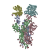
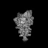
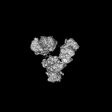
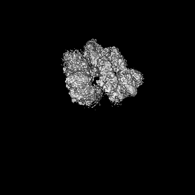
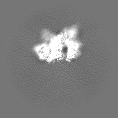
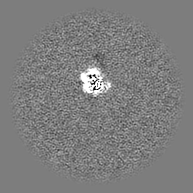
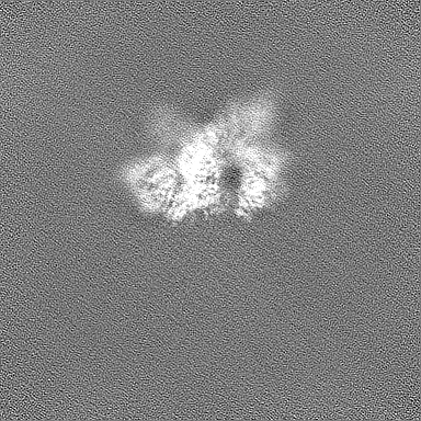
Journal: PLoS Pathog / Year: 2024 Title: Structure-guided in vitro evolution of nanobodies targeting new viral variants. Authors: Gang Ye / Fan Bu / Ruangang Pan / Alise Mendoza / Ge Yang / Benjamin Spiller / Brian E Wadzinski / Lanying Du / Stanley Perlman / Bin Liu / Fang Li / Abstract: A major challenge in antiviral antibody therapy is keeping up with the rapid evolution of viruses. Our research shows that nanobodies - single-domain antibodies derived from camelids - can be rapidly ...A major challenge in antiviral antibody therapy is keeping up with the rapid evolution of viruses. Our research shows that nanobodies - single-domain antibodies derived from camelids - can be rapidly re-engineered to combat new viral strains through structure-guided in vitro evolution. Specifically, for viral mutations occurring at nanobody-binding sites, we introduce randomized amino acid sequences into nanobody residues near these mutations. We then select nanobody variants that effectively bind to the mutated viral target from a phage display library. As a proof of concept, we used this approach to adapt Nanosota-3, a nanobody originally identified to target the receptor-binding domain (RBD) of early Omicron subvariants, making it highly effective against recent Omicron subvariants. Remarkably, this adaptation process can be completed in less than two weeks, allowing drug development to keep pace with viral evolution and provide timely protection to humans.
In the structure databanks used in Yorodumi, some data are registered as the other names, "COVID-19 virus" and "2019-nCoV". Here are the details of the virus and the list of structure data.
Jan 31, 2019. EMDB accession codes are about to change! (news from PDBe EMDB page)
EMDB accession codes are about to change! (news from PDBe EMDB page)
The allocation of 4 digits for EMDB accession codes will soon come to an end. Whilst these codes will remain in use, new EMDB accession codes will include an additional digit and will expand incrementally as the available range of codes is exhausted. The current 4-digit format prefixed with “EMD-” (i.e. EMD-XXXX) will advance to a 5-digit format (i.e. EMD-XXXXX), and so on. It is currently estimated that the 4-digit codes will be depleted around Spring 2019, at which point the 5-digit format will come into force.
The EM Navigator/Yorodumi systems omit the EMD- prefix.
Related info.:Q: What is EMD? / ID/Accession-code notation in Yorodumi/EM Navigator
Yorodumi is a browser for structure data from EMDB, PDB, SASBDB, etc.
This page is also the successor to EM Navigator detail page, and also detail information page/front-end page for Omokage search.
The word "yorodu" (or yorozu) is an old Japanese word meaning "ten thousand". "mi" (miru) is to see.
Related info.:EMDB / PDB / SASBDB / Comparison of 3 databanks / Yorodumi Search / Aug 31, 2016. New EM Navigator & Yorodumi / Yorodumi Papers / Jmol/JSmol / Function and homology information / Changes in new EM Navigator and Yorodumi
 Movie
Movie Controller


 Open data
 Basic information
 Map data
 Sample
 Keywords
 Function and homology information
 Severe acute respiratory syndrome coronavirus 2 /
 Authors
 United States, 3 items
  Citation
 Structure visualization
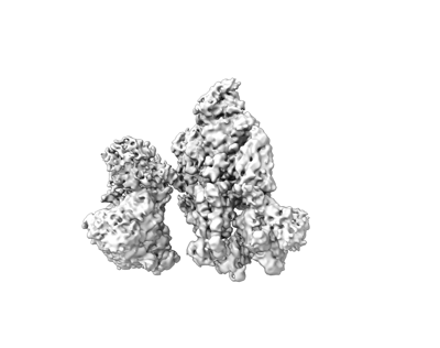
 Downloads & links
 emd_43832.png
 http://ftp.pdbj.org/pub/emdb/structures/EMD-43832
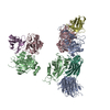


 Z (Sec.)
 Y (Row.)
 X (Col.)


 Sample components
 Homo sapiens (human)
 Processing
 Electron microscopy
 FIELD EMISSION GUN