[English] 日本語
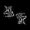
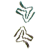
Yorodumi- EMDB-42294: Structure of recombinantly assembled murine alpha-synuclein fibrils -
+
Open data
-
Basic information
| Entry | ![]() | |||||||||
|---|---|---|---|---|---|---|---|---|---|---|
| Title | Structure of recombinantly assembled murine alpha-synuclein fibrils | |||||||||
Map data | ||||||||||
Sample |
| |||||||||
Keywords | synuclein / Parkinson's disease / neurodegeneration / amyloid / fibril / PROTEIN FIBRIL | |||||||||
| Function / homology | Function and homology informationneutral lipid metabolic process / regulation of acyl-CoA biosynthetic process / mitochondrial membrane organization / regulation of glutamate secretion / PKR-mediated signaling / regulation of macrophage activation / positive regulation of neurotransmitter secretion / regulation of neurotransmitter secretion / regulation of locomotion / platelet alpha granule membrane ...neutral lipid metabolic process / regulation of acyl-CoA biosynthetic process / mitochondrial membrane organization / regulation of glutamate secretion / PKR-mediated signaling / regulation of macrophage activation / positive regulation of neurotransmitter secretion / regulation of neurotransmitter secretion / regulation of locomotion / platelet alpha granule membrane / membrane organization / mitochondrial ATP synthesis coupled electron transport / regulation of reactive oxygen species metabolic process / arachidonate binding / negative regulation of dopamine uptake involved in synaptic transmission / negative regulation of norepinephrine uptake / response to desipramine / positive regulation of SNARE complex assembly / positive regulation of hydrogen peroxide catabolic process / supramolecular fiber / negative regulation of chaperone-mediated autophagy / positive regulation of protein localization to cell periphery / negative regulation of exocytosis / dopamine biosynthetic process / negative regulation of dopamine metabolic process / response to iron(II) ion / negative regulation of platelet-derived growth factor receptor signaling pathway / SNARE complex assembly / negative regulation of thrombin-activated receptor signaling pathway / dopamine metabolic process / Lewy body / negative regulation of microtubule polymerization / synaptic vesicle priming / synaptic vesicle transport / regulation of norepinephrine uptake / transporter regulator activity / phospholipid metabolic process / positive regulation of inositol phosphate biosynthetic process / protein complex oligomerization / regulation of dopamine secretion / positive regulation of receptor recycling / regulation of neuronal synaptic plasticity / cuprous ion binding / positive regulation of exocytosis / nuclear outer membrane / dynein complex binding / synaptic transmission, dopaminergic / synaptic vesicle exocytosis / response to magnesium ion / positive regulation of endocytosis / negative regulation of serotonin uptake / kinesin binding / positive regulation of synaptic transmission / cysteine-type endopeptidase inhibitor activity / regulation of presynapse assembly / synaptic vesicle endocytosis / alpha-tubulin binding / adult locomotory behavior / beta-tubulin binding / phospholipase binding / behavioral response to cocaine / cellular response to fibroblast growth factor stimulus / response to type II interferon / inclusion body / fatty acid metabolic process / response to interleukin-1 / Hsp70 protein binding / cytoplasmic vesicle membrane / excitatory postsynaptic potential / positive regulation of release of sequestered calcium ion into cytosol / cellular response to copper ion / axon terminus / enzyme inhibitor activity / glutathione metabolic process / regulation of microtubule cytoskeleton organization / SNARE binding / regulation of long-term neuronal synaptic plasticity / protein tetramerization / protein sequestering activity / receptor internalization / phosphoprotein binding / microglial cell activation / tubulin binding / protein destabilization / ferrous iron binding / synapse organization / phospholipid binding / tau protein binding / neuron apoptotic process / long-term synaptic potentiation / enzyme activator activity / positive regulation of inflammatory response / terminal bouton / actin cytoskeleton / synaptic vesicle / synaptic vesicle membrane / presynapse / growth cone / response to lipopolysaccharide / cellular response to oxidative stress Similarity search - Function | |||||||||
| Biological species | ![]() | |||||||||
| Method | helical reconstruction / cryo EM / Resolution: 3.1 Å | |||||||||
Authors | Zhou Y / Sokratian A | |||||||||
| Funding support | United States, 1 items
| |||||||||
Citation | Journal: Sci Adv / Year: 2024Title: Mouse α-synuclein fibrils are structurally and functionally distinct from human fibrils associated with Lewy body diseases. Authors: Arpine Sokratian / Ye Zhou / Meltem Tatli / Kevin J Burbidge / Enquan Xu / Elizabeth Viverette / Sonia Donzelli / Addison M Duda / Yuan Yuan / Huizhong Li / Samuel Strader / Nirali Patel / ...Authors: Arpine Sokratian / Ye Zhou / Meltem Tatli / Kevin J Burbidge / Enquan Xu / Elizabeth Viverette / Sonia Donzelli / Addison M Duda / Yuan Yuan / Huizhong Li / Samuel Strader / Nirali Patel / Lauren Shiell / Tuyana Malankhanova / Olivia Chen / Joseph R Mazzulli / Lalith Perera / Henning Stahlberg / Mario Borgnia / Alberto Bartesaghi / Hilal A Lashuel / Andrew B West / ![]() Abstract: The intricate process of α-synuclein aggregation and fibrillization holds pivotal roles in Parkinson's disease (PD) and multiple system atrophy (MSA). While mouse α-synuclein can fibrillize in ...The intricate process of α-synuclein aggregation and fibrillization holds pivotal roles in Parkinson's disease (PD) and multiple system atrophy (MSA). While mouse α-synuclein can fibrillize in vitro, whether these fibrils commonly used in research to induce this process or form can reproduce structures in the human brain remains unknown. Here, we report the first atomic structure of mouse α-synuclein fibrils, which was solved in parallel by two independent teams. The structure shows striking similarity to MSA-amplified and PD-associated E46K fibrils. However, mouse α-synuclein fibrils display altered packing arrangements, reduced hydrophobicity, and heightened fragmentation sensitivity and evoke only weak immunological responses. Furthermore, mouse α-synuclein fibrils exhibit exacerbated pathological spread in neurons and humanized α-synuclein mice. These findings provide critical insights into the structural underpinnings of α-synuclein pathogenicity and emphasize a need to reassess the role of mouse α-synuclein fibrils in the development of related diagnostic probes and therapeutic interventions. | |||||||||
| History |
|
-
Structure visualization
| Supplemental images |
|---|
-
Downloads & links
-EMDB archive
| Map data | emd_42294.map.gz | 1.3 MB | EMDB map data format | |
|---|---|---|---|---|
| Header (meta data) | emd-42294-v30.xml emd-42294.xml | 19.1 KB 19.1 KB | Display Display | EMDB header |
| FSC (resolution estimation) | emd_42294_fsc.xml | 9.1 KB | Display | FSC data file |
| Images | emd_42294.png | 65.7 KB | ||
| Filedesc metadata | emd-42294.cif.gz | 6.4 KB | ||
| Others | emd_42294_half_map_1.map.gz emd_42294_half_map_2.map.gz | 49.6 MB 49.6 MB | ||
| Archive directory | http://ftp.pdbj.org/pub/emdb/structures/EMD-42294 ftp://ftp.pdbj.org/pub/emdb/structures/EMD-42294 | HTTPS FTP |

-Related structure data
| Related structure data | ![]() 8uieMC ![]() 9ewvC M: atomic model generated by this map C: citing same article ( |
|---|---|
| Similar structure data | Similarity search - Function & homology F&H Search |
-
Links
| EMDB pages | EMDB (EBI/PDBe) / EMDataResource |
|---|
-
Map
| File | Download / File: emd_42294.map.gz / Format: CCP4 / Size: 8 MB / Type: IMAGE STORED AS FLOATING POINT NUMBER (4 BYTES) | ||||||||||||||||||||||||||||||||||||
|---|---|---|---|---|---|---|---|---|---|---|---|---|---|---|---|---|---|---|---|---|---|---|---|---|---|---|---|---|---|---|---|---|---|---|---|---|---|
| Projections & slices | Image control
Images are generated by Spider. | ||||||||||||||||||||||||||||||||||||
| Voxel size | X=Y=Z: 1.08 Å | ||||||||||||||||||||||||||||||||||||
| Density |
| ||||||||||||||||||||||||||||||||||||
| Symmetry | Space group: 1 | ||||||||||||||||||||||||||||||||||||
| Details | EMDB XML:
|
-Supplemental data
-Half map: #2
| File | emd_42294_half_map_1.map | ||||||||||||
|---|---|---|---|---|---|---|---|---|---|---|---|---|---|
| Projections & Slices |
| ||||||||||||
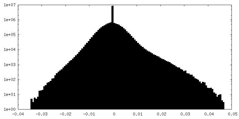
| Density Histograms |
-Half map: #1
| File | emd_42294_half_map_2.map | ||||||||||||
|---|---|---|---|---|---|---|---|---|---|---|---|---|---|
| Projections & Slices |
| ||||||||||||
| Density Histograms |
-
Sample components
-Entire : alpha-synuclein
| Entire | Name: alpha-synuclein |
|---|---|
| Components |
|
-Supramolecule #1: alpha-synuclein
| Supramolecule | Name: alpha-synuclein / type: complex / ID: 1 / Parent: 0 / Macromolecule list: all |
|---|---|
| Source (natural) | Organism: ![]() |
-Macromolecule #1: Alpha-synuclein
| Macromolecule | Name: Alpha-synuclein / type: protein_or_peptide / ID: 1 / Number of copies: 12 / Enantiomer: LEVO |
|---|---|
| Source (natural) | Organism: ![]() |
| Molecular weight | Theoretical: 14.501185 KDa |
| Recombinant expression | Organism: ![]() |
| Sequence | String: MDVFMKGLSK AKEGVVAAAE KTKQGVAEAA GKTKEGVLYV GSKTKEGVVH GVTTVAEKTK EQVTNVGGAV VTGVTAVAQK TVEGAGNIA AATGFVKKDQ MGKGEEGYPQ EGILEDMPVD PGSEAYEMPS EEGYQDYEPE A UniProtKB: Alpha-synuclein |
-Experimental details
-Structure determination
| Method | cryo EM |
|---|---|
Processing | helical reconstruction |
| Aggregation state | filament |
-
Sample preparation
| Concentration | 0.2 mg/mL | |||||||||||||||
|---|---|---|---|---|---|---|---|---|---|---|---|---|---|---|---|---|
| Buffer | pH: 7.4 Component:
Details: Phosphate-buffered saline (PBS) | |||||||||||||||
| Grid | Model: Quantifoil R1.2/1.3 / Material: GOLD / Mesh: 300 / Support film - Material: GOLD / Support film - topology: HOLEY ARRAY / Support film - Film thickness: 50 / Pretreatment - Type: PLASMA CLEANING / Pretreatment - Time: 75 sec. | |||||||||||||||
| Vitrification | Cryogen name: ETHANE / Chamber humidity: 90 % / Chamber temperature: 293.15 K / Instrument: LEICA EM GP |
-
Electron microscopy
| Microscope | FEI TITAN KRIOS |
|---|---|
| Image recording | Film or detector model: GATAN K3 (6k x 4k) / Number grids imaged: 2 / Average electron dose: 60.0 e/Å2 |
| Electron beam | Acceleration voltage: 300 kV / Electron source: FIELD EMISSION GUN |
| Electron optics | Illumination mode: FLOOD BEAM / Imaging mode: BRIGHT FIELD / Nominal defocus max: 2.5 µm / Nominal defocus min: 0.5 µm |
| Sample stage | Cooling holder cryogen: NITROGEN |
| Experimental equipment | ![]() Model: Titan Krios / Image courtesy: FEI Company |
Movie
Controller
About Yorodumi


Keywords
Authors
United States, 1 items
Citation


Z (Sec.)
Y (Row.)
X (Col.)


Processing
FIELD EMISSION GUN