nitrogenase / nitrogenase activity / bioluminescence / generation of precursor metabolites and energy / 4 iron, 4 sulfur cluster binding / ATP binding / metal ion binding Similarity search - Function
Nitrogenase iron protein NifH / NifH/frxC family / NifH/chlL conserved site / 4Fe-4S iron sulfur cluster binding proteins, NifH/frxC family / NifH/frxC family signature 2. / NifH/frxC family signature 1. / NIFH_FRXC family profile. / Green fluorescent protein, GFP / Green fluorescent protein-related / Green fluorescent protein ...Nitrogenase iron protein NifH / NifH/frxC family / NifH/chlL conserved site / 4Fe-4S iron sulfur cluster binding proteins, NifH/frxC family / NifH/frxC family signature 2. / NifH/frxC family signature 1. / NIFH_FRXC family profile. / Green fluorescent protein, GFP / Green fluorescent protein-related / Green fluorescent protein / Green fluorescent protein / P-loop containing nucleoside triphosphate hydrolase Similarity search - Domain/homology
National Institutes of Health/National Institute of General Medical Sciences (NIH/NIGMS)
GM045162
United States
National Institutes of Health/National Institute of General Medical Sciences (NIH/NIGMS)
GM143836-01
United States
Howard Hughes Medical Institute (HHMI)
United States
Citation
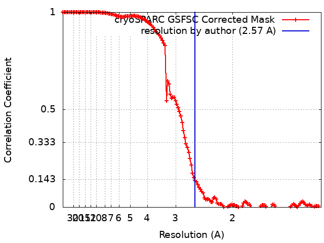
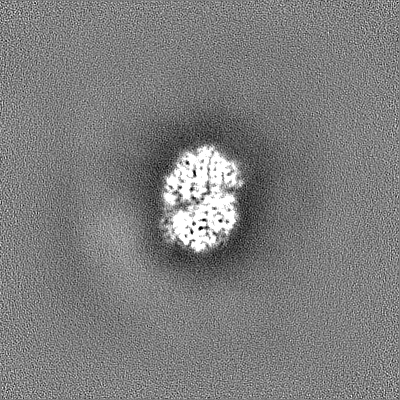
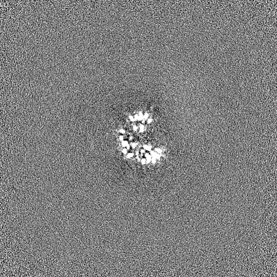
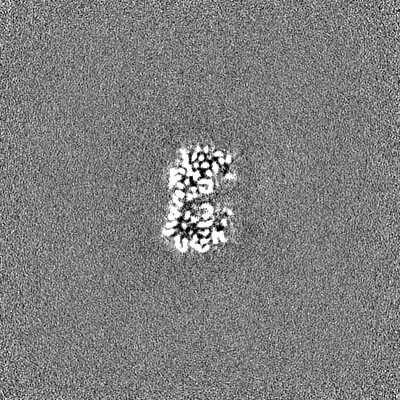
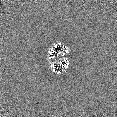
Journal: Nat Protoc / Year: 2024 Title: Anaerobic cryoEM protocols for air-sensitive nitrogenase proteins. Authors: Rebeccah A Warmack / Belinda B Wenke / Thomas Spatzal / Douglas C Rees / Abstract: Single-particle cryo-electron microscopy (cryoEM) provides an attractive avenue for advancing our atomic resolution understanding of materials, molecules and living systems. However, the vast ...Single-particle cryo-electron microscopy (cryoEM) provides an attractive avenue for advancing our atomic resolution understanding of materials, molecules and living systems. However, the vast majority of published cryoEM methodologies focus on the characterization of aerobically purified samples. Air-sensitive enzymes and microorganisms represent important yet understudied systems in structural biology. We have recently demonstrated the success of an anaerobic single-particle cryoEM workflow applied to the air-sensitive nitrogenase enzymes. In this protocol, we detail the use of Schlenk lines and anaerobic chambers to prepare samples, including a protein tag for monitoring sample exposure to oxygen in air. We describe how to use a plunge freezing apparatus inside of a soft-sided vinyl chamber of the type we routinely use for anaerobic biochemistry and crystallography of oxygen-sensitive proteins. Manual control of the airlock allows for introduction of liquid cryogens into the tent. A custom vacuum port provides slow, continuous evacuation of the tent atmosphere to avoid accumulation of flammable vapors within the enclosed chamber. These methods allowed us to obtain high-resolution structures of both nitrogenase proteins using single-particle cryoEM. The procedures involved can be generally subdivided into a 4 d anaerobic sample generation procedure, and a 1 d anaerobic cryoEM sample preparation step, followed by conventional cryoEM imaging and processing steps. As nitrogen is a substrate for nitrogenase, the Schlenk lines and anaerobic chambers described in this procedure are operated under an argon atmosphere; however, the system and these procedures are compatible with other controlled gas environments.
In the structure databanks used in Yorodumi, some data are registered as the other names, "COVID-19 virus" and "2019-nCoV". Here are the details of the virus and the list of structure data.
Jan 31, 2019. EMDB accession codes are about to change! (news from PDBe EMDB page)
EMDB accession codes are about to change! (news from PDBe EMDB page)
The allocation of 4 digits for EMDB accession codes will soon come to an end. Whilst these codes will remain in use, new EMDB accession codes will include an additional digit and will expand incrementally as the available range of codes is exhausted. The current 4-digit format prefixed with “EMD-” (i.e. EMD-XXXX) will advance to a 5-digit format (i.e. EMD-XXXXX), and so on. It is currently estimated that the 4-digit codes will be depleted around Spring 2019, at which point the 5-digit format will come into force.
The EM Navigator/Yorodumi systems omit the EMD- prefix.
Related info.:Q: What is EMD? / ID/Accession-code notation in Yorodumi/EM Navigator
Yorodumi is a browser for structure data from EMDB, PDB, SASBDB, etc.
This page is also the successor to EM Navigator detail page, and also detail information page/front-end page for Omokage search.
The word "yorodu" (or yorozu) is an old Japanese word meaning "ten thousand". "mi" (miru) is to see.
Related info.:EMDB / PDB / SASBDB / Comparison of 3 databanks / Yorodumi Search / Aug 31, 2016. New EM Navigator & Yorodumi / Yorodumi Papers / Jmol/JSmol / Function and homology information / Changes in new EM Navigator and Yorodumi
 Movie
Movie Controller

 Yorodumi
 Open data
 Basic information
 Map data
 Sample
 Keywords
 Function and homology information
 Azotobacter vinelandii DJ (bacteria) /
  Discosoma sp. LW-2004 (sea anemone)
 Authors
 United States, 3 items
  Citation
 Structure visualization
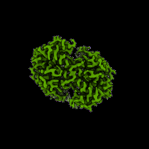
 Downloads & links
 emd_41151.png
 http://ftp.pdbj.org/pub/emdb/structures/EMD-41151
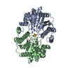


 Z (Sec.)
 Y (Row.)
 X (Col.)


 Sample components

 Processing
 Electron microscopy
 FIELD EMISSION GUN