Movie
Movie Controller


+ Open data

- Basic information
| Entry |  | |||||||||
|---|---|---|---|---|---|---|---|---|---|---|
| Title | CUS-3 Mature Virus Capsid | |||||||||
 Map data Map data | Single Particle Reconstruction of CUS3 Bacteriophage | |||||||||
 Sample Sample |
| |||||||||
 Keywords Keywords | Bacteriophage / Capsid / Virus | |||||||||
| Function / homology | Major capsid protein Gp5 / P22 coat protein - gene protein 5 / viral capsid / Putative coat protein Function and homology information Function and homology information | |||||||||
| Biological species |  Enterobacteria phage CUS-3 (virus) Enterobacteria phage CUS-3 (virus) | |||||||||
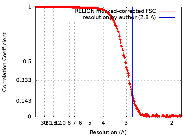
| Method | single particle reconstruction / cryo EM / Resolution: 2.8 Å | |||||||||
 Authors Authors | Whitehead III RD / Alexandrescu AT / White S / Teschke CM | |||||||||
| Funding support |  United States, 1 items United States, 1 items
| |||||||||
 Citation Citation | Journal: To Be Published Title: Not currently available Authors: Whitehead III RD / White S / Alexandrescu AT / Teschke CM | |||||||||
| History |
|
- Structure visualization
| Supplemental images |
|---|

- Downloads & links
-EMDB archive
| Map data | emd_40564.map.gz | 1.7 GB | EMDB map data format | |
|---|---|---|---|---|
| Header (meta data) | emd-40564-v30.xmlemd-40564.xml | 13.6 KB 13.6 KB | Display Display | EMDB header |
| FSC (resolution estimation) | emd_40564_fsc.xml | 28.1 KB | Display | FSC data file |
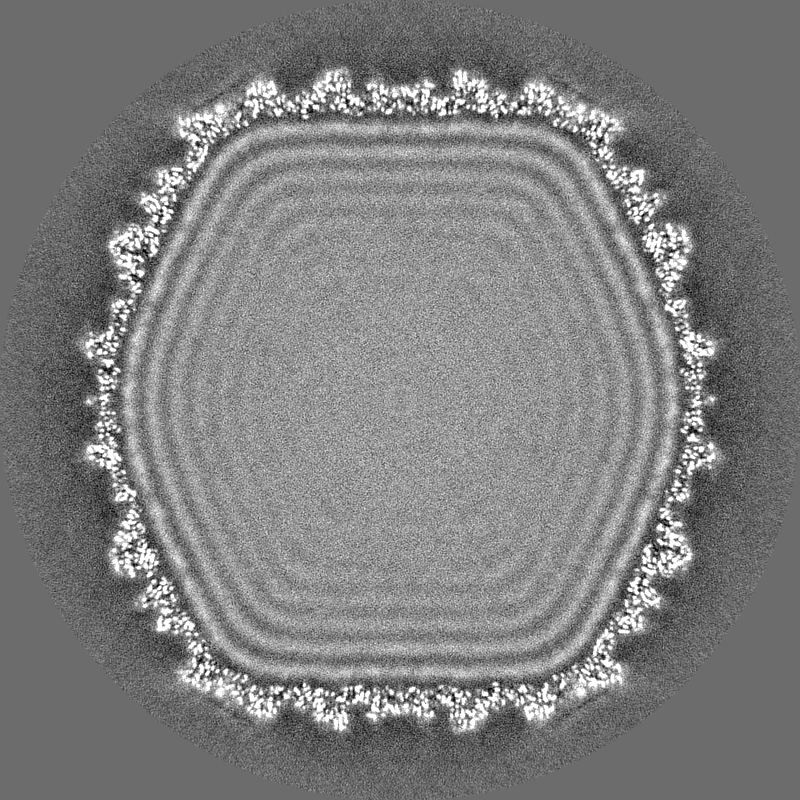
| Images |  emd_40564.png emd_40564.png | 117.4 KB | ||
| Filedesc metadata | emd-40564.cif.gz | 5.3 KB | ||
| Others | emd_40564_half_map_1.map.gzemd_40564_half_map_2.map.gz | 1.5 GB 1.5 GB | ||
| Archive directory |  http://ftp.pdbj.org/pub/emdb/structures/EMD-40564ftp://ftp.pdbj.org/pub/emdb/structures/EMD-40564 http://ftp.pdbj.org/pub/emdb/structures/EMD-40564ftp://ftp.pdbj.org/pub/emdb/structures/EMD-40564 | HTTPS FTP |
-Related structure data
| Related structure data |  8skgMC M: atomic model generated by this map C: citing same article ( |
|---|---|
| Similar structure data |
-Links
| EMDB pages | EMDB (EBI/PDBe) / EMDataResource |
|---|
-Map
| File | Download / File: emd_40564.map.gz / Format: CCP4 / Size: 1.9 GB / Type: IMAGE STORED AS FLOATING POINT NUMBER (4 BYTES) | ||||||||||||||||||||||||||||||||||||
|---|---|---|---|---|---|---|---|---|---|---|---|---|---|---|---|---|---|---|---|---|---|---|---|---|---|---|---|---|---|---|---|---|---|---|---|---|---|
| Annotation | Single Particle Reconstruction of CUS3 Bacteriophage | ||||||||||||||||||||||||||||||||||||
| Projections & slices | Image control
Images are generated by Spider. | ||||||||||||||||||||||||||||||||||||
| Voxel size | X=Y=Z: 0.935 Å | ||||||||||||||||||||||||||||||||||||
| Density |
| ||||||||||||||||||||||||||||||||||||
| Symmetry | Space group: 1 | ||||||||||||||||||||||||||||||||||||
| Details | EMDB XML:
|
 X (Sec.)
 Y (Row.)
 Z (Col.)


-Supplemental data
-Half map: Half Map 1
| File | emd_40564_half_map_1.map | ||||||||||||
|---|---|---|---|---|---|---|---|---|---|---|---|---|---|
| Annotation | Half Map 1 | ||||||||||||
| Projections & Slices |
| ||||||||||||
| Density Histograms |


-Half map: Half Map 2
| File | emd_40564_half_map_2.map | ||||||||||||
|---|---|---|---|---|---|---|---|---|---|---|---|---|---|
| Annotation | Half Map 2 | ||||||||||||
| Projections & Slices |
| ||||||||||||
| Density Histograms |


- Sample components
-Entire : Enterobacteria phage CUS-3
| Entire | Name: Enterobacteria phage CUS-3 (virus) |
|---|---|
| Components |
|
-Supramolecule #1: Enterobacteria phage CUS-3
| Supramolecule | Name: Enterobacteria phage CUS-3 / type: virus / ID: 1 / Parent: 0 / Macromolecule list: all / NCBI-ID: 539221 / Sci species name: Enterobacteria phage CUS-3 / Virus type: VIRION / Virus isolate: STRAIN / Virus enveloped: No / Virus empty: No |
|---|---|
| Virus shell | Shell ID: 1 / T number (triangulation number): 7 |
-Macromolecule #1: Putative coat protein
| Macromolecule | Name: Putative coat protein / type: protein_or_peptide / ID: 1 / Number of copies: 7 / Enantiomer: LEVO |
|---|---|
| Source (natural) | Organism: Enterobacteria phage CUS-3 (virus) |
| Molecular weight | Theoretical: 44.772359 KDa |
| Sequence | String: ANQLAKDLEI MFENYVEGFE AACVVSRNAK KFRPGDTAMQ RAGDVLYRPQ HYHMNIEEGL DLSSKTPTAL VQRLVPSVFK EPKNILYTL DAREMRDPEH KTEAGRAAGM RLAAQIDSDL ISMVTQRATN VITMADSTAG TQGRDLWNCA AGIDATMTAI G VPQGINRR ...String: ANQLAKDLEI MFENYVEGFE AACVVSRNAK KFRPGDTAMQ RAGDVLYRPQ HYHMNIEEGL DLSSKTPTAL VQRLVPSVFK EPKNILYTL DAREMRDPEH KTEAGRAAGM RLAAQIDSDL ISMVTQRATN VITMADSTAG TQGRDLWNCA AGIDATMTAI G VPQGINRR SFWNPFNYKD LAGELGHRAY AQGATLTAYE KAQIPPVASF DSYKTDISGR LPKGSTESLT VSGQPEHKVE AK DSNGMPV DNRQGTITVS ASGLQVGDAF TIAGVNSVHQ ITKDTTGQPQ VFRVLAVSGT TVTISPKILP VENTDVASRP YAN VDAKPA ESAAITILNK NAAPANLFWA DGSVELMYGK LAFPTGQGPQ VMTATTEQGA TLIMSYAFDH IKGVTTARFT TLYG CSVLV PEYTGIVIAG Q UniProtKB: Putative coat protein |
-Experimental details
-Structure determination
| Method | cryo EM |
|---|---|
 Processing Processing | single particle reconstruction |
| Aggregation state | particle |
-Sample preparation
| Buffer | pH: 7.4 |
|---|---|
| Grid | Model: C-flat-1.2/1.3 / Material: GOLD / Mesh: 300 / Pretreatment - Type: GLOW DISCHARGE |
| Vitrification | Cryogen name: ETHANE / Chamber humidity: 100 % / Chamber temperature: 277 K / Instrument: FEI VITROBOT MARK IV |
- Electron microscopy
| Microscope | FEI TITAN KRIOS |
|---|---|
| Image recording | Film or detector model: GATAN K3 (6k x 4k) / Average electron dose: 1.3075 e/Å2 |
| Electron beam | Acceleration voltage: 300 kV / Electron source:  FIELD EMISSION GUN FIELD EMISSION GUN |
| Electron optics | Illumination mode: FLOOD BEAM / Imaging mode: BRIGHT FIELD / Nominal defocus max: 2.5 µm / Nominal defocus min: 0.8 µm |
| Experimental equipment |  Model: Titan Krios / Image courtesy: FEI Company |