 Movie
Movie Controller


+ Open data

- Basic information
| Entry |  | |||||||||
|---|---|---|---|---|---|---|---|---|---|---|
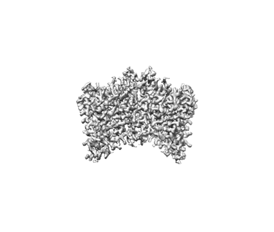
| Title | Structure of TaHKT2;1 in NaCl at 2.6 Angstroms resolution | |||||||||
 Map data Map data | ||||||||||
 Sample Sample |
| |||||||||
 Keywords Keywords | HKT / salt tolerance / ion selectivity / TRANSPORT PROTEIN | |||||||||
| Function / homology | : / Cation transporter / Cation transport protein / metal ion transport / monoatomic cation transmembrane transporter activity / : / plasma membrane / Uncharacterized protein Function and homology information Function and homology information | |||||||||
| Biological species |  | |||||||||
| Method | single particle reconstruction / cryo EM / Resolution: 2.6 Å | |||||||||
 Authors Authors | Wang J / Su N / Guo J | |||||||||
| Funding support |  China, 1 items China, 1 items
| |||||||||
 Citation Citation | Journal: Mol Plant / Year: 2024 Title: Structures and ion transport mechanisms of plant high-affinity potassium transporters. Authors: Jiangqin Wang / Yanping Luo / Fan Ye / Zhong Jie Ding / Shao Jian Zheng / Shuai Qiao / Yong Wang / Jiangtao Guo / Wei Yang / Nannan Su / Abstract: Plant high-affinity K transporters (HKTs) mediate Na and K uptake, maintain Na/K homeostasis, and therefore play crucial roles in plant salt tolerance. In this study, we present cryoelectron ...Plant high-affinity K transporters (HKTs) mediate Na and K uptake, maintain Na/K homeostasis, and therefore play crucial roles in plant salt tolerance. In this study, we present cryoelectron microscopy structures of HKTs from two classes, class I HKT1;1 from Arabidopsis thaliana (AtHKT1;1) and class II HKT2;1 from Triticum aestivum (TaHKT2;1), in both Na- and K-bound states at 2.6- to 3.0-Å resolutions. Both AtHKT1;1 and TaHKT2;1 function as homodimers. Each HKT subunit consists of four tandem domain units (D1-D4) with a repeated K-channel-like M-P-M topology. In each subunit, D1-D4 assemble into an ion conduction pore with a pseudo-four-fold symmetry. Although both TaHKT2;1 and AtHKT1;1 have only one putative Na ion bound in the selectivity filter with a similar coordination pattern, the two HKTs display different K binding modes in the filter. TaHKT2;1 has three K ions bound in the selectivity filter, but AtHKT1;1 has only two K ions bound in the filter, which has a narrowed external entrance due to the presence of a Ser residue in the first filter motif. These structures, along with computational, mutational, and electrophysiological analyses, enable us to pinpoint key residues that are critical for the ion selectivity of HKTs. The findings provide new insights into the ion selectivity and ion transport mechanisms of plant HKTs and improve our understanding about how HKTs mediate plant salt tolerance and enhance crop growth. | |||||||||
| History |
|
- Structure visualization
| Supplemental images |
|---|
- Downloads & links
-EMDB archive
| Map data | emd_37381.map.gz | 33.1 MB | EMDB map data format | |
|---|---|---|---|---|
| Header (meta data) | emd-37381-v30.xmlemd-37381.xml | 16.6 KB 16.6 KB | Display Display | EMDB header |
| Images |  emd_37381.png emd_37381.png | 42 KB | ||
| Filedesc metadata | emd-37381.cif.gz | 6.2 KB | ||
| Others | emd_37381_half_map_1.map.gzemd_37381_half_map_2.map.gz | 31.2 MB 30.3 MB | ||
| Archive directory |  http://ftp.pdbj.org/pub/emdb/structures/EMD-37381ftp://ftp.pdbj.org/pub/emdb/structures/EMD-37381 http://ftp.pdbj.org/pub/emdb/structures/EMD-37381ftp://ftp.pdbj.org/pub/emdb/structures/EMD-37381 | HTTPS FTP |
-Related structure data
| Related structure data | 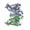 8w9tMC  8w9nC  8w9oC 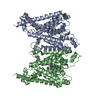 8w9vC M: atomic model generated by this map C: citing same article ( |
|---|---|
| Similar structure data |

-Links
| EMDB pages | EMDB (EBI/PDBe) / EMDataResource |
|---|
-Map
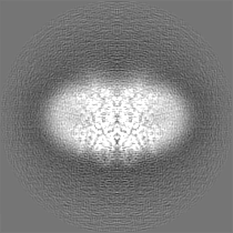
| File | Download / File: emd_37381.map.gz / Format: CCP4 / Size: 35.3 MB / Type: IMAGE STORED AS FLOATING POINT NUMBER (4 BYTES) | ||||||||||||||||||||||||||||||||||||
|---|---|---|---|---|---|---|---|---|---|---|---|---|---|---|---|---|---|---|---|---|---|---|---|---|---|---|---|---|---|---|---|---|---|---|---|---|---|
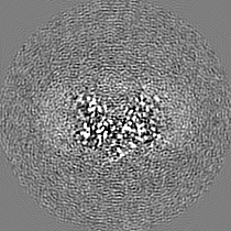
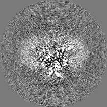
| Projections & slices | Image control
Images are generated by Spider. | ||||||||||||||||||||||||||||||||||||
| Voxel size | X=Y=Z: 0.93 Å | ||||||||||||||||||||||||||||||||||||
| Density |
| ||||||||||||||||||||||||||||||||||||
| Symmetry | Space group: 1 | ||||||||||||||||||||||||||||||||||||
| Details | EMDB XML:
|
 Z (Sec.)
 Y (Row.)
 X (Col.)


-Supplemental data
-Half map: #1
| File | emd_37381_half_map_1.map | ||||||||||||
|---|---|---|---|---|---|---|---|---|---|---|---|---|---|
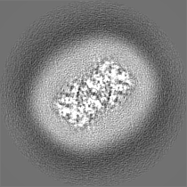
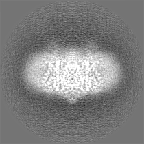
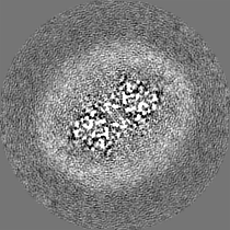
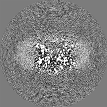
| Projections & Slices |
| ||||||||||||
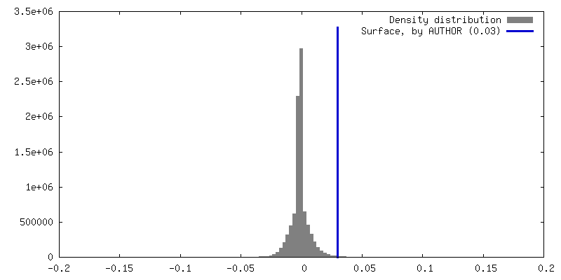
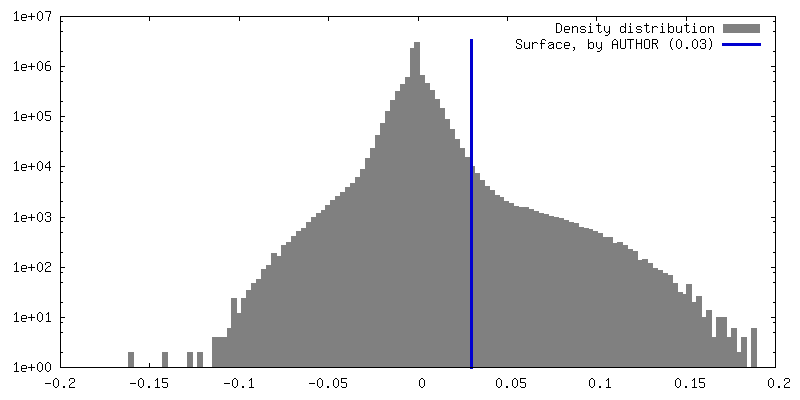
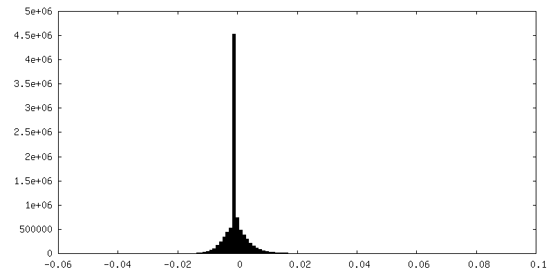
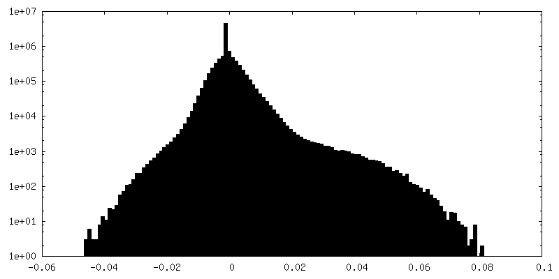
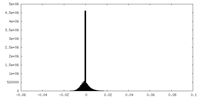
| Density Histograms |
-Half map: #2
| File | emd_37381_half_map_2.map | ||||||||||||
|---|---|---|---|---|---|---|---|---|---|---|---|---|---|
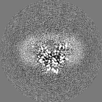
| Projections & Slices |
| ||||||||||||
| Density Histograms |


- Sample components
-Entire : Structure of TaHKT2;1 in NaCl at 2.6 Angstroms resolution
| Entire | Name: Structure of TaHKT2;1 in NaCl at 2.6 Angstroms resolution |
|---|---|
| Components |
|
-Supramolecule #1: Structure of TaHKT2;1 in NaCl at 2.6 Angstroms resolution
| Supramolecule | Name: Structure of TaHKT2;1 in NaCl at 2.6 Angstroms resolution type: complex / ID: 1 / Parent: 0 / Macromolecule list: #1 |
|---|---|
| Source (natural) | Organism: |
-Macromolecule #1: HKT2
| Macromolecule | Name: HKT2 / type: protein_or_peptide / ID: 1 / Number of copies: 2 / Enantiomer: LEVO |
|---|---|
| Source (natural) | Organism: |
| Molecular weight | Theoretical: 60.20059 KDa |
| Recombinant expression | Organism:  Homo sapiens (human) Homo sapiens (human) |
| Sequence | String: MHLFLTLVHS TMDRVKRFYQ DFIHIKLHSF SRISRYVVDS IVFIYRFVAL HVHPFWIQLS YFLAIAILGS VLLISLKPSN PEFSPPYID MLYLSTSALT VSGLSTVKME DLSSSQIVVL TLLMLVGGEI FVSLLGLMLR VNHQDMQDLP SVKISSVPVE L EVLDLANS ...String: MHLFLTLVHS TMDRVKRFYQ DFIHIKLHSF SRISRYVVDS IVFIYRFVAL HVHPFWIQLS YFLAIAILGS VLLISLKPSN PEFSPPYID MLYLSTSALT VSGLSTVKME DLSSSQIVVL TLLMLVGGEI FVSLLGLMLR VNHQDMQDLP SVKISSVPVE L EVLDLANS MALCDESQLE DASHAIPPKK CTELKRSRSV KCLGYVVFGY FAVIHVLGFV LVFLYITHVP TASAPLNKKG IN IVLFSLS VTVASCANAG LVPTNENMVI FSKNSGLLLL LSGQMLAGNT LFPLFLRLLV WFLGKLTKVK ELRLMTKNPE EVH FANLLP RLPTVFLSST VIGIVAAGVT LFCSVDWNSS VFDGLGSYQK TVNAFFMVVN ARHSGENSID CSLMSPAIVV LFIG MMYLP SSATFAPPSG DTKTTNENTK GKGKRGSLVQ NLAFSPLGCN IIFVIVACIT ERRRLRSDPL NFSTLNMIFE VISAY GNVG LSTGYSCSRL HQLHPEIICQ DMPYSFSGWW SDGGKFLLVL VMLYGRLKVF AVSTGKSWKV UniProtKB: Uncharacterized protein |
-Macromolecule #2: SODIUM ION
| Macromolecule | Name: SODIUM ION / type: ligand / ID: 2 / Number of copies: 2 |
|---|---|
| Molecular weight | Theoretical: 22.99 Da |
-Macromolecule #3: water
| Macromolecule | Name: water / type: ligand / ID: 3 / Number of copies: 4 / Formula: HOH |
|---|---|
| Molecular weight | Theoretical: 18.015 Da |
| Chemical component information |  ChemComp-HOH: |
-Experimental details
-Structure determination
| Method | cryo EM |
|---|---|
 Processing Processing | single particle reconstruction |
| Aggregation state | particle |
-Sample preparation
| Buffer | pH: 8 |
|---|---|
| Vitrification | Cryogen name: ETHANE |
- Electron microscopy
| Microscope | FEI TITAN KRIOS |
|---|---|
| Image recording | Film or detector model: GATAN K2 QUANTUM (4k x 4k) / Average electron dose: 52.0 e/Å2 |
| Electron beam | Acceleration voltage: 300 kV / Electron source:  FIELD EMISSION GUN FIELD EMISSION GUN |
| Electron optics | Illumination mode: FLOOD BEAM / Imaging mode: BRIGHT FIELD / Nominal defocus max: 1.6 µm / Nominal defocus min: 0.8 µm |
| Experimental equipment |  Model: Titan Krios / Image courtesy: FEI Company |
