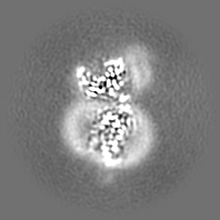
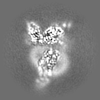
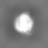
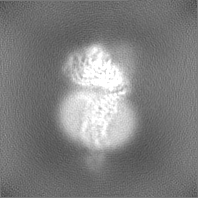
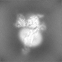
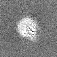
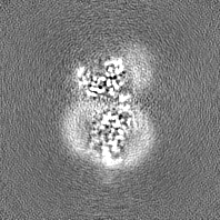
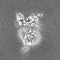
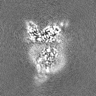
登録情報 データベース : EMDB / ID : EMD-35351タイトル Cryo-EM structure of CXCL8 bound C-X-C chemokine receptor 1 in complex with Gi heterotrimer sharpened map 複合体 : Cryo-EM structure of human chemokine receptor 1タンパク質・ペプチド : C-X-C chemokine receptor type 1タンパク質・ペプチド : Guanine nucleotide-binding protein G(i) subunit alpha-1タンパク質・ペプチド : Guanine nucleotide-binding protein G(I)/G(S)/G(T) subunit beta-1タンパク質・ペプチド : Guanine nucleotide-binding protein G(I)/G(S)/G(O) subunit gamma-2タンパク質・ペプチド : scFv16タンパク質・ペプチド : Interleukin-8 / / / / / 機能・相同性 分子機能 ドメイン・相同性 構成要素
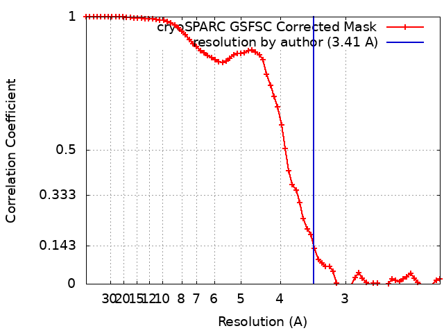
/ / / / / / / / / / / / / / / / / / / / / / / / / / / / / / / / / / / / / / / / / / / / / / / / / / / / / / / / / / / / / / / / / / / / / / / / / / / / / / / / / / / / / / / / / / / / / / / / / / / / / / / / / / / / / / / / / / / / / / / / / / / / / / / / / / / / / / / / / / / / / / / / / / / / / / / / / / / / / / / / / / 生物種 Homo sapiens (ヒト) / Mus musculus (ハツカネズミ)手法 / / 解像度 : 3.41 Å Ishimoto N / Park JH / Park SY 資金援助 1件 Organization Grant number 国 Not funded
ジャーナル : Nat Commun / 年 : 2023タイトル : Structural basis of CXC chemokine receptor 1 ligand binding and activation.著者 : Naito Ishimoto / Jae-Hyun Park / Kouki Kawakami / Michiko Tajiri / Kenji Mizutani / Satoko Akashi / Jeremy R H Tame / Asuka Inoue / Sam-Yong Park / 要旨 : Neutrophil granulocytes play key roles in innate immunity and shaping adaptive immune responses. They are attracted by chemokines to sites of infection and tissue damage, where they kill and ... Neutrophil granulocytes play key roles in innate immunity and shaping adaptive immune responses. They are attracted by chemokines to sites of infection and tissue damage, where they kill and phagocytose bacteria. The chemokine CXCL8 (also known as interleukin-8, abbreviated IL-8) and its G-protein-coupled receptors CXCR1 and CXCR2 are crucial elements in this process, and also the development of many cancers. These GPCRs have therefore been the target of many drug development campaigns and structural studies. Here, we solve the structure of CXCR1 complexed with CXCL8 and cognate G-proteins using cryo-EM, showing the detailed interactions between the receptor, the chemokine and Gαi protein. Unlike the closely related CXCR2, CXCR1 strongly prefers to bind CXCL8 in its monomeric form. The model shows that steric clashes would form between dimeric CXCL8 and extracellular loop 2 (ECL2) of CXCR1. Consistently, transplanting ECL2 of CXCR2 onto CXCR1 abolishes the selectivity for the monomeric chemokine. Our model and functional analysis of various CXCR1 mutants will assist efforts in structure-based drug design targeting specific CXC chemokine receptor subtypes. 履歴 登録 2023年2月10日 - ヘッダ(付随情報) 公開 2023年7月19日 - マップ公開 2023年7月19日 - 更新 2023年7月19日 - 現状 2023年7月19日 処理サイト : PDBj / 状態 : 公開
すべて表示 表示を減らす
 ムービー
ムービー コントローラー


 データを開く
 基本情報
基本情報
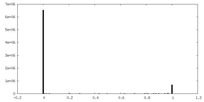
 マップデータ
 試料
試料 キーワード
キーワード 機能・相同性情報
 Homo sapiens (ヒト) /
 データ登録者
 引用
引用
 構造の表示
構造の表示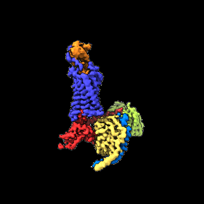
 ダウンロードとリンク
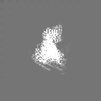
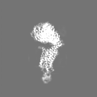
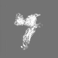
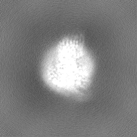
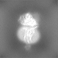
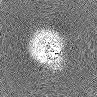
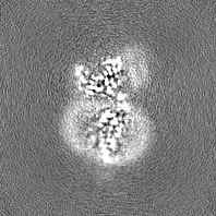
 emd_35351.png
 http://ftp.pdbj.org/pub/emdb/structures/EMD-35351


 Z
Z Y
Y X
X


 試料の構成要素
 Spodoptera frugiperda (ツマジロクサヨトウ)
 解析
解析 電子顕微鏡法
 FIELD EMISSION GUN