 Movie
Movie Controller


+ Open data

- Basic information
| Entry |  | |||||||||
|---|---|---|---|---|---|---|---|---|---|---|
| Title | Capsid structure of the Cyanophage P-SCSP1u | |||||||||
 Map data Map data | ||||||||||
 Sample Sample |
| |||||||||
 Keywords Keywords | Whole virus / Capsid / cyanophage / T7-like virus / VIRUS | |||||||||
| Biological species |  Prochlorococcus phage P-SCSP1u (virus) Prochlorococcus phage P-SCSP1u (virus) | |||||||||
| Method | single particle reconstruction / cryo EM / Resolution: 3.23 Å | |||||||||
 Authors Authors | Liu H / Dang S | |||||||||
| Funding support |  Hong Kong, 1 items Hong Kong, 1 items
| |||||||||
 Citation Citation | Journal: Nat Commun / Year: 2023 Title: Cryo-EM structure of cyanophage P-SCSP1u offers insights into DNA gating and evolution of T7-like viruses. Authors: Lanlan Cai / Hang Liu / Wen Zhang / Shiwei Xiao / Qinglu Zeng / Shangyu Dang / Abstract: Cyanophages, together with their host cyanobacteria, play important roles in marine biogeochemical cycles and control of marine food webs. The recently identified MPP-C (Marine Picocyanobacteria ...Cyanophages, together with their host cyanobacteria, play important roles in marine biogeochemical cycles and control of marine food webs. The recently identified MPP-C (Marine Picocyanobacteria Podovirus clade C) cyanophages, belonging to the T7-like podoviruses, contain the smallest genomes among cyanopodoviruses and exhibit distinct infection kinetics. However, understanding of the MPP-C cyanophage infection process is hindered by the lack of high-resolution structural information. Here, we report the cryo-EM structure of the cyanophage P-SCSP1u, a representative member of the MPP-C phages, in its native form at near-atomic resolution, which reveals the assembly mechanism of the capsid and molecular interaction of the portal-tail complex. Structural comparison of the capsid proteins of P-SCSP1u and other podoviruses with known structures provides insights into the evolution of T7-like viruses. Furthermore, our study provides the near-atomic resolution structure of portal-tail complex for T7-like viruses. On the basis of previously reported structures of phage T7, we identify an additional valve and gate to explain the DNA gating mechanism for the T7-like viruses. | |||||||||
| History |
|
- Structure visualization
| Supplemental images |
|---|

- Downloads & links
-EMDB archive
| Map data | emd_35174.map.gz | 227 MB |  EMDB map data format EMDB map data format | |
|---|---|---|---|---|
| Header (meta data) | emd-35174-v30.xmlemd-35174.xml | 14.2 KB 14.2 KB | Display Display | EMDB header |
| Images | 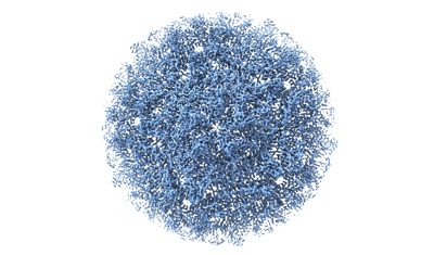 emd_35174.png emd_35174.png | 99.8 KB | ||
| Masks | emd_35174_msk_1.map | 244.1 MB | Mask map | |
| Filedesc metadata | emd-35174.cif.gz | 5.2 KB | ||
| Others | emd_35174_half_map_1.map.gzemd_35174_half_map_2.map.gz | 194.4 MB 194.4 MB | ||
| Archive directory |  http://ftp.pdbj.org/pub/emdb/structures/EMD-35174ftp://ftp.pdbj.org/pub/emdb/structures/EMD-35174 http://ftp.pdbj.org/pub/emdb/structures/EMD-35174ftp://ftp.pdbj.org/pub/emdb/structures/EMD-35174 | HTTPS FTP |
-Related structure data


-Links
| EMDB pages | EMDB (EBI/PDBe) / EMDataResource |
|---|
-Map
| File | Download / File: emd_35174.map.gz / Format: CCP4 / Size: 244.1 MB / Type: IMAGE STORED AS FLOATING POINT NUMBER (4 BYTES) | ||||||||||||||||||||||||||||||||||||
|---|---|---|---|---|---|---|---|---|---|---|---|---|---|---|---|---|---|---|---|---|---|---|---|---|---|---|---|---|---|---|---|---|---|---|---|---|---|
| Projections & slices | Image control
Images are generated by Spider. | ||||||||||||||||||||||||||||||||||||
| Voxel size | X=Y=Z: 0.85 Å | ||||||||||||||||||||||||||||||||||||
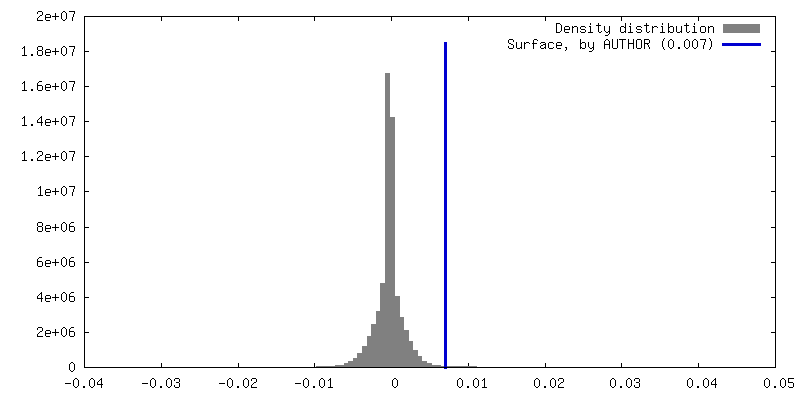
| Density |
| ||||||||||||||||||||||||||||||||||||
| Symmetry | Space group: 1 | ||||||||||||||||||||||||||||||||||||
| Details | EMDB XML:
|
 Z (Sec.)
 Y (Row.)
 X (Col.)


-Supplemental data
-Mask #1
| File | emd_35174_msk_1.map | ||||||||||||
|---|---|---|---|---|---|---|---|---|---|---|---|---|---|
| Projections & Slices |
| ||||||||||||
| Density Histograms |


-Half map: #2
| File | emd_35174_half_map_1.map | ||||||||||||
|---|---|---|---|---|---|---|---|---|---|---|---|---|---|
| Projections & Slices |
| ||||||||||||
| Density Histograms |


-Half map: #1
| File | emd_35174_half_map_2.map | ||||||||||||
|---|---|---|---|---|---|---|---|---|---|---|---|---|---|
| Projections & Slices |
| ||||||||||||
| Density Histograms |


- Sample components
-Entire : Prochlorococcus phage P-SCSP1u
| Entire | Name: Prochlorococcus phage P-SCSP1u (virus) |
|---|---|
| Components |
|
-Supramolecule #1: Prochlorococcus phage P-SCSP1u
| Supramolecule | Name: Prochlorococcus phage P-SCSP1u / type: virus / ID: 1 / Parent: 0 / Macromolecule list: all / NCBI-ID: 2914505 / Sci species name: Prochlorococcus phage P-SCSP1u / Virus type: VIRION / Virus isolate: STRAIN / Virus enveloped: No / Virus empty: No |
|---|---|
| Host (natural) | Organism:  Prochlorococcus (bacteria) Prochlorococcus (bacteria) |
-Macromolecule #1: The capsid protein(gp 19) of P-SCSP1u
| Macromolecule | Name: The capsid protein(gp 19) of P-SCSP1u / type: protein_or_peptide / ID: 1 / Number of copies: 7 / Enantiomer: LEVO |
|---|---|
| Source (natural) | Organism: Prochlorococcus phage P-SCSP1u (virus) |
| Molecular weight | Theoretical: 35.160438 KDa |
| Sequence | String: MANFTPSRLG LVNNTGTGVK DLFLKTFAGE VLSAFRKATI FEDLHTVRTI SSGKSAQFPI VGLSSTSYHS PGTQLTGNAI KHAEAVINI DDKLVSNVFI ADVDEAMNHY DVRSQYSVQM GNALAYTFDQ NVAAMIAQAA RTSTNPNTDL PGGTRIKILK S GTANTAAA ...String: MANFTPSRLG LVNNTGTGVK DLFLKTFAGE VLSAFRKATI FEDLHTVRTI SSGKSAQFPI VGLSSTSYHS PGTQLTGNAI KHAEAVINI DDKLVSNVFI ADVDEAMNHY DVRSQYSVQM GNALAYTFDQ NVAAMIAQAA RTSTNPNTDL PGGTRIKILK S GTANTAAA VAAVTGTDLA TALFSAAEQM DINNLPEEDR YCAIDPTNYY KLVQNTTVIN RDFGGRGAYA EGEVLKVAGI HI VKSNHLP KTNRSAATGE NNTYHANYTD NIGLVFNKQA VGTVKLMDLK MEQTGADIHA LYQGTFMVGS MMHGSGVLRP DCA IELYAA NS |
-Experimental details
-Structure determination
| Method | cryo EM |
|---|---|
 Processing Processing | single particle reconstruction |
| Aggregation state | particle |
-Sample preparation
| Buffer | pH: 7.4 |
|---|---|
| Vitrification | Cryogen name: ETHANE / Chamber humidity: 100 % |
- Electron microscopy
| Microscope | FEI TITAN KRIOS |
|---|---|
| Image recording | Film or detector model: GATAN K3 (6k x 4k) / Average exposure time: 4.5 sec. / Average electron dose: 50.0 e/Å2 |
| Electron beam | Acceleration voltage: 300 kV / Electron source:  FIELD EMISSION GUN FIELD EMISSION GUN |
| Electron optics | Illumination mode: FLOOD BEAM / Imaging mode: BRIGHT FIELD / Cs: 2.7 mm / Nominal defocus max: 2.5 µm / Nominal defocus min: 1.0 µm / Nominal magnification: 81000 |
| Sample stage | Cooling holder cryogen: NITROGEN |
| Experimental equipment |  Model: Titan Krios / Image courtesy: FEI Company |
-Image processing
| Startup model | Type of model: INSILICO MODEL |
|---|---|
| Final reconstruction | Resolution.type: BY AUTHOR / Resolution: 3.23 Å / Resolution method: FSC 0.143 CUT-OFF / Software - Name: cryoSPARC (ver. 2.15) / Number images used: 732864 |
| Initial angle assignment | Type: MAXIMUM LIKELIHOOD / Software - Name: cryoSPARC (ver. 2.15) |
| Final angle assignment | Type: MAXIMUM LIKELIHOOD / Software - Name: cryoSPARC (ver. 2.15) |
