 Movie
Movie Controller

[English] 日本語
 Yorodumi
- EMDB-34891: Cryo-EM structure of human high-voltage activated L-type calcium ... -
+ Open data

- Basic information
| Entry |  | |||||||||
|---|---|---|---|---|---|---|---|---|---|---|
| Title | Cryo-EM structure of human high-voltage activated L-type calcium channel CaV1.2 in complex with tetrandrine (TET) | |||||||||
 Map data Map data | ||||||||||
 Sample Sample |
| |||||||||
 Keywords Keywords | tet bound state / MEMBRANE PROTEIN | |||||||||
| Function / homology |  Function and homology information Function and homology informationvoltage-gated calcium channel activity involved in regulation of presynaptic cytosolic calcium levels / positive regulation of calcium ion transmembrane transport via high voltage-gated calcium channel / Presynaptic depolarization and calcium channel opening / regulation of membrane repolarization during action potential / membrane depolarization during atrial cardiac muscle cell action potential / calcium ion transmembrane transport via high voltage-gated calcium channel / Phase 2 - plateau phase / photoreceptor ribbon synapse / membrane depolarization during AV node cell action potential / signaling ...voltage-gated calcium channel activity involved in regulation of presynaptic cytosolic calcium levels / positive regulation of calcium ion transmembrane transport via high voltage-gated calcium channel / Presynaptic depolarization and calcium channel opening / regulation of membrane repolarization during action potential / membrane depolarization during atrial cardiac muscle cell action potential / calcium ion transmembrane transport via high voltage-gated calcium channel / Phase 2 - plateau phase / photoreceptor ribbon synapse / membrane depolarization during AV node cell action potential / signaling / membrane depolarization during bundle of His cell action potential / cell communication / L-type voltage-gated calcium channel complex / positive regulation of muscle contraction / NCAM1 interactions / calcium ion import / regulation of ventricular cardiac muscle cell membrane repolarization / positive regulation of calcium ion transport / cardiac muscle cell action potential involved in contraction / calcium ion transport into cytosol / regulation of calcium ion transmembrane transport via high voltage-gated calcium channel / Sensory processing of sound by inner hair cells of the cochlea / voltage-gated calcium channel complex / Mechanical load activates signaling by PIEZO1 and integrins in osteocytes / calcium ion transmembrane import into cytosol / regulation of heart contraction / neuromuscular junction development / Phase 0 - rapid depolarisation / regulation of calcium ion transport / regulation of heart rate by cardiac conduction / calcium ion import across plasma membrane / neuronal dense core vesicle / voltage-gated calcium channel activity / visual perception / presynaptic active zone membrane / T-tubule / muscle contraction / sarcoplasmic reticulum / protein localization to plasma membrane / Regulation of insulin secretion / regulation of membrane potential / postsynaptic density membrane / GABA-ergic synapse / calcium ion transmembrane transport / calcium channel activity / cellular response to amyloid-beta / actin filament binding / calcium ion transport / Adrenaline,noradrenaline inhibits insulin secretion / presynapse / chemical synaptic transmission / perikaryon / calmodulin binding / cilium / dendrite / extracellular exosome / nucleoplasm / metal ion binding / plasma membrane Similarity search - Function | |||||||||
| Biological species |  Homo sapiens (human) Homo sapiens (human) | |||||||||
| Method | single particle reconstruction / cryo EM / Resolution: 3.4 Å | |||||||||
 Authors Authors | Wei Y / Yu Z / Zhao Y | |||||||||
| Funding support |  China, 1 items China, 1 items
| |||||||||
 Citation Citation | Journal: Nat Commun / Year: 2024 Title: Structural bases of inhibitory mechanism of Ca1.2 channel inhibitors. Authors: Yiqing Wei / Zhuoya Yu / Lili Wang / Xiaojing Li / Na Li / Qinru Bai / Yuhang Wang / Renjie Li / Yufei Meng / Hao Xu / Xianping Wang / Yanli Dong / Zhuo Huang / Xuejun Cai Zhang / Yan Zhao / Abstract: The voltage-gated calcium channel Ca1.2 is essential for cardiac and vessel smooth muscle contractility and brain function. Accumulating evidence demonstrates that malfunctions of Ca1.2 are involved ...The voltage-gated calcium channel Ca1.2 is essential for cardiac and vessel smooth muscle contractility and brain function. Accumulating evidence demonstrates that malfunctions of Ca1.2 are involved in brain and heart diseases. Pharmacological inhibition of Ca1.2 is therefore of therapeutic value. Here, we report cryo-EM structures of Ca1.2 in the absence or presence of the antirheumatic drug tetrandrine or antihypertensive drug benidipine. Tetrandrine acts as a pore blocker in a pocket composed of S6, S6, and S6 helices and forms extensive hydrophobic interactions with Ca1.2. Our structure elucidates that benidipine is located in the D-D fenestration site. Its hydrophobic sidechain, phenylpiperidine, is positioned at the exterior of the pore domain and cradled within a hydrophobic pocket formed by S5, S6, and S6 helices, providing additional interactions to exert inhibitory effects on both L-type and T-type voltage gated calcium channels. These findings provide the structural foundation for the rational design and optimization of therapeutic inhibitors of voltage-gated calcium channels. | |||||||||
| History |
|
- Structure visualization
| Supplemental images |
|---|

- Downloads & links
-EMDB archive
| Map data | emd_34891.map.gz | 118 MB | EMDB map data format | |
|---|---|---|---|---|
| Header (meta data) | emd-34891-v30.xmlemd-34891.xml | 24 KB 24 KB | Display Display | EMDB header |
| FSC (resolution estimation) | emd_34891_fsc.xml | 10.5 KB | Display | FSC data file |
| Images | 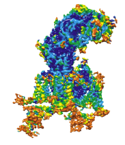 emd_34891.png emd_34891.png | 44.5 KB | ||
| Filedesc metadata | emd-34891.cif.gz | 9.1 KB | ||
| Others | emd_34891_half_map_1.map.gzemd_34891_half_map_2.map.gz | 116.1 MB 116.1 MB | ||
| Archive directory |  http://ftp.pdbj.org/pub/emdb/structures/EMD-34891ftp://ftp.pdbj.org/pub/emdb/structures/EMD-34891 http://ftp.pdbj.org/pub/emdb/structures/EMD-34891ftp://ftp.pdbj.org/pub/emdb/structures/EMD-34891 | HTTPS FTP |
-Related structure data
| Related structure data |  8hmaMC  8hlpC  8hmbC M: atomic model generated by this map C: citing same article ( |
|---|---|
| Similar structure data |


-Links
| EMDB pages | EMDB (EBI/PDBe) / EMDataResource |
|---|---|
| Related items in Molecule of the Month |


-Map
| File | Download / File: emd_34891.map.gz / Format: CCP4 / Size: 125 MB / Type: IMAGE STORED AS FLOATING POINT NUMBER (4 BYTES) | ||||||||||||||||||||||||||||||||||||
|---|---|---|---|---|---|---|---|---|---|---|---|---|---|---|---|---|---|---|---|---|---|---|---|---|---|---|---|---|---|---|---|---|---|---|---|---|---|
| Projections & slices | Image control
Images are generated by Spider. | ||||||||||||||||||||||||||||||||||||
| Voxel size | X=Y=Z: 1.04 Å | ||||||||||||||||||||||||||||||||||||
| Density |
| ||||||||||||||||||||||||||||||||||||
| Symmetry | Space group: 1 | ||||||||||||||||||||||||||||||||||||
| Details | EMDB XML:
|
 Z (Sec.)
 Y (Row.)
 X (Col.)


-Supplemental data
-Half map: #2
| File | emd_34891_half_map_1.map | ||||||||||||
|---|---|---|---|---|---|---|---|---|---|---|---|---|---|
| Projections & Slices |
| ||||||||||||
| Density Histograms |


-Half map: #1
| File | emd_34891_half_map_2.map | ||||||||||||
|---|---|---|---|---|---|---|---|---|---|---|---|---|---|
| Projections & Slices |
| ||||||||||||
| Density Histograms |


- Sample components
+Entire : cav1.2alpha2delta1beta2b
+Supramolecule #1: cav1.2alpha2delta1beta2b
+Macromolecule #1: Voltage-dependent L-type calcium channel subunit alpha
+Macromolecule #2: Isoform 2c of Voltage-dependent L-type calcium channel subunit beta-2
+Macromolecule #3: Voltage-dependent calcium channel subunit alpha-2/delta-1
+Macromolecule #4: HEXADECANE

+Macromolecule #5: 1,2-Distearoyl-sn-glycerophosphoethanolamine

+Macromolecule #6: 6,6',7,12-tetramethoxy-2,2'-dimethyl-1beta-3,4-didehydroberbaman
+Macromolecule #7: CALCIUM ION
+Macromolecule #8: 2-acetamido-2-deoxy-beta-D-glucopyranose

+Macromolecule #9: beta-D-mannopyranose

-Experimental details
-Structure determination
| Method | cryo EM |
|---|---|
 Processing Processing | single particle reconstruction |
| Aggregation state | particle |
-Sample preparation
| Buffer | pH: 7.5 |
|---|---|
| Vitrification | Cryogen name: ETHANE |
- Electron microscopy
| Microscope | FEI TITAN KRIOS |
|---|---|
| Image recording | Film or detector model: GATAN K2 SUMMIT (4k x 4k) / Average electron dose: 9.6 e/Å2 |
| Electron beam | Acceleration voltage: 300 kV / Electron source:  FIELD EMISSION GUN FIELD EMISSION GUN |
| Electron optics | Illumination mode: FLOOD BEAM / Imaging mode: BRIGHT FIELD / Nominal defocus max: 2.2 µm / Nominal defocus min: 1.2 µm |
| Experimental equipment |  Model: Titan Krios / Image courtesy: FEI Company |

