 Movie
Movie Controller


+ Open data

- Basic information
| Entry | 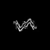 | ||||||||||||
|---|---|---|---|---|---|---|---|---|---|---|---|---|---|
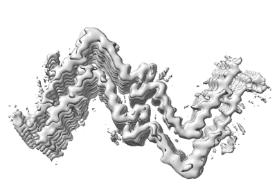
| Title | Structure of hIAPP-TF-type3 | ||||||||||||
 Map data Map data | hIAPP-WF-type3 | ||||||||||||
 Sample Sample |
| ||||||||||||
 Keywords Keywords | hIAPP / amylin / PROTEIN FIBRIL | ||||||||||||
| Function / homology |  Function and homology information Function and homology informationamylin receptor 3 signaling pathway / amylin receptor 2 signaling pathway / amylin receptor 1 signaling pathway / amylin receptor signaling pathway / Calcitonin-like ligand receptors / negative regulation of amyloid fibril formation / negative regulation of bone resorption / eating behavior / negative regulation of osteoclast differentiation / Regulation of gene expression in beta cells ...amylin receptor 3 signaling pathway / amylin receptor 2 signaling pathway / amylin receptor 1 signaling pathway / amylin receptor signaling pathway / Calcitonin-like ligand receptors / negative regulation of amyloid fibril formation / negative regulation of bone resorption / eating behavior / negative regulation of osteoclast differentiation / Regulation of gene expression in beta cells / positive regulation of cAMP/PKA signal transduction / bone resorption / negative regulation of protein-containing complex assembly / positive regulation of calcium-mediated signaling / osteoclast differentiation / sensory perception of pain / hormone activity / cell-cell signaling / amyloid-beta binding / G alpha (s) signalling events / positive regulation of MAPK cascade / positive regulation of apoptotic process / Amyloid fiber formation / receptor ligand activity / signaling receptor binding / neuronal cell body / apoptotic process / lipid binding / signal transduction / : / extracellular region / identical protein binding Similarity search - Function | ||||||||||||
| Biological species |  Homo sapiens (human) Homo sapiens (human) | ||||||||||||
| Method | helical reconstruction / Resolution: 3.3 Å | ||||||||||||
 Authors Authors | Li DG / Zhang XL | ||||||||||||
| Funding support |  China, 3 items China, 3 items
| ||||||||||||
 Citation Citation | Journal: iScience / Year: 2022 Title: A new polymorphism of human amylin fibrils with similar protofilaments and a conserved core. Authors: Dongyu Li / Xueli Zhang / Youwang Wang / Haonan Zhang / Kai Song / Keyan Bao / Ping Zhu / Abstract: Pancreatic amyloid deposits composed of a fibrillar form of the human islet amyloid polypeptide (hIAPP) are the pathological hallmark of type 2 diabetes (T2D). Although various cryo-EM structures of ...Pancreatic amyloid deposits composed of a fibrillar form of the human islet amyloid polypeptide (hIAPP) are the pathological hallmark of type 2 diabetes (T2D). Although various cryo-EM structures of polymorphic hIAPP fibrils were reported, the underlying polymorphic mechanism of hIAPP remains elusive. Meanwhile, the structure of hIAPP fibrils with all residues visible in the fibril core is not available. Here, we report the full-length structures of two different polymorphs of hIAPP fibrils, namely slim form (SF, dimer) and thick form (TF, tetramer), formed in a salt-free environment, which share a similar ζ-shaped protofilament but differ in inter-protofilament interfaces. In the absence of salt, electrostatic interactions were found to play a dominant role in stabilizing the fibril structure, suggesting an antagonistic effect between electrostatic and hydrophobic interactions in different salt concentrations environments. Our results shed light on understanding the mechanism of amyloid fibril polymorphism. | ||||||||||||
| History |
|
- Structure visualization
| Supplemental images |
|---|
- Downloads & links
-EMDB archive
| Map data | emd_33903.map.gz | 3.9 MB | EMDB map data format | |
|---|---|---|---|---|
| Header (meta data) | emd-33903-v30.xmlemd-33903.xml | 13.8 KB 13.8 KB | Display Display | EMDB header |
| FSC (resolution estimation) | emd_33903_fsc.xml | 9.4 KB | Display | FSC data file |
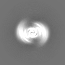
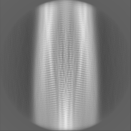
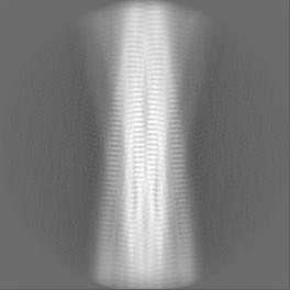
| Images |  emd_33903.png emd_33903.png | 67 KB | ||
| Filedesc metadata | emd-33903.cif.gz | 4.6 KB | ||
| Others | emd_33903_half_map_1.map.gzemd_33903_half_map_2.map.gz | 54.4 MB 54.4 MB | ||
| Archive directory |  http://ftp.pdbj.org/pub/emdb/structures/EMD-33903ftp://ftp.pdbj.org/pub/emdb/structures/EMD-33903 http://ftp.pdbj.org/pub/emdb/structures/EMD-33903ftp://ftp.pdbj.org/pub/emdb/structures/EMD-33903 | HTTPS FTP |
-Related structure data
| Related structure data |  7yl7MC  7ykwC  7yl0C  7yl3C M: atomic model generated by this map C: citing same article ( |
|---|---|
| Similar structure data |


-Links
| EMDB pages | EMDB (EBI/PDBe) / EMDataResource |
|---|---|
| Related items in Molecule of the Month |


-Map
| File | Download / File: emd_33903.map.gz / Format: CCP4 / Size: 70.2 MB / Type: IMAGE STORED AS FLOATING POINT NUMBER (4 BYTES) | ||||||||||||||||||||||||||||||||||||
|---|---|---|---|---|---|---|---|---|---|---|---|---|---|---|---|---|---|---|---|---|---|---|---|---|---|---|---|---|---|---|---|---|---|---|---|---|---|
| Annotation | hIAPP-WF-type3 | ||||||||||||||||||||||||||||||||||||
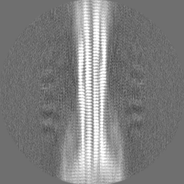
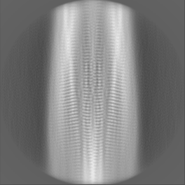
| Projections & slices | Image control
Images are generated by Spider. | ||||||||||||||||||||||||||||||||||||
| Voxel size | X=Y=Z: 1.04 Å | ||||||||||||||||||||||||||||||||||||
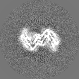
| Density |
| ||||||||||||||||||||||||||||||||||||
| Symmetry | Space group: 1 | ||||||||||||||||||||||||||||||||||||
| Details | EMDB XML:
|
 Z (Sec.)
 Y (Row.)
 X (Col.)


-Supplemental data
-Half map: #2
| File | emd_33903_half_map_1.map | ||||||||||||
|---|---|---|---|---|---|---|---|---|---|---|---|---|---|
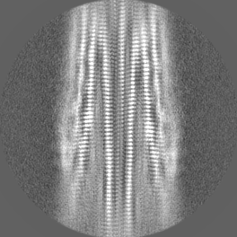
| Projections & Slices |
| ||||||||||||
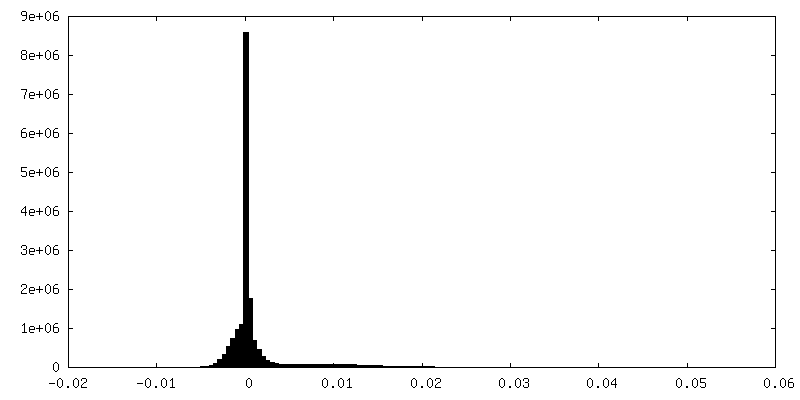
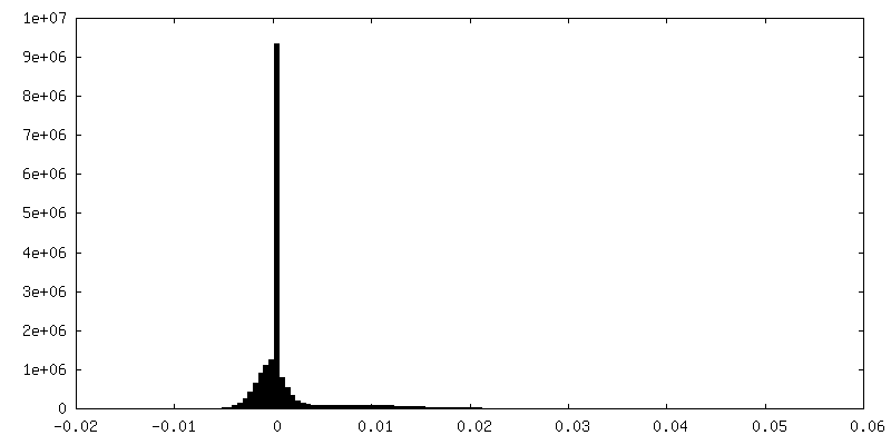
| Density Histograms |


-Half map: #1
| File | emd_33903_half_map_2.map | ||||||||||||
|---|---|---|---|---|---|---|---|---|---|---|---|---|---|
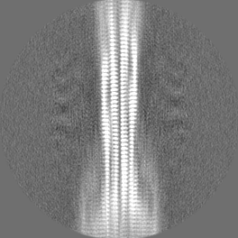
| Projections & Slices |
| ||||||||||||
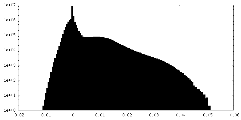
| Density Histograms |


- Sample components
-Entire : no
| Entire | Name: no |
|---|---|
| Components |
|
-Supramolecule #1: no
| Supramolecule | Name: no / type: organelle_or_cellular_component / ID: 1 / Parent: 0 / Macromolecule list: all |
|---|---|
| Source (natural) | Organism: Homo sapiens (human) / Synthetically produced: Yes |
-Macromolecule #1: Islet amyloid polypeptide
| Macromolecule | Name: Islet amyloid polypeptide / type: protein_or_peptide / ID: 1 / Number of copies: 12 / Enantiomer: LEVO |
|---|---|
| Source (natural) | Organism: Homo sapiens (human) |
| Molecular weight | Theoretical: 3.908319 KDa |
| Sequence | String: KCNTATCATQ RLANFLVHSS NNFGAILSST NVGSNT(TYC) UniProtKB: Islet amyloid polypeptide |
-Experimental details
-Structure determination
 Processing Processing | helical reconstruction |
|---|---|
| Aggregation state | helical array |
-Sample preparation
| Buffer | pH: 7 |
|---|
- Electron microscopy
| Microscope | FEI TITAN KRIOS |
|---|---|
| Image recording | Film or detector model: GATAN K2 SUMMIT (4k x 4k) / Detector mode: SUPER-RESOLUTION / Average exposure time: 3.0 sec. / Average electron dose: 60.0 e/Å2 |
| Electron beam | Acceleration voltage: 300 kV / Electron source:  FIELD EMISSION GUN FIELD EMISSION GUN |
| Electron optics | Illumination mode: FLOOD BEAM / Imaging mode: BRIGHT FIELD / Cs: 2.8 mm / Nominal defocus max: 1.5 µm / Nominal defocus min: 1.0 µm |
| Experimental equipment |  Model: Titan Krios / Image courtesy: FEI Company |
-Image processing
| Final reconstruction | Applied symmetry - Helical parameters - Δz: 2.35 Å Applied symmetry - Helical parameters - Δ&Phi: 179.41 ° Applied symmetry - Helical parameters - Axial symmetry: C1 (asymmetric) Resolution.type: BY AUTHOR / Resolution: 3.3 Å / Resolution method: FSC 0.143 CUT-OFF / Software - Name: RELION (ver. 3.0) / Number images used: 29587 |
|---|---|
| Startup model | Type of model: NONE |
| Final angle assignment | Type: NOT APPLICABLE / Software - Name: RELION (ver. 3.0) |
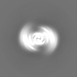
| FSC plot (resolution estimation) | 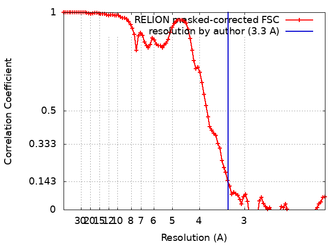 |
